. 2019 Sep 18;11(9):874.
doi: 10.3390/v11090874.

A Novel CXCR4 Targeting Protein SDF-1/54 as an HIV-1 Entry Inhibitor

Affiliations

A Novel CXCR4 Targeting Protein SDF-1/54 as an HIV-1 Entry Inhibitor

Suiyi Tan et al. Viruses. .

Abstract

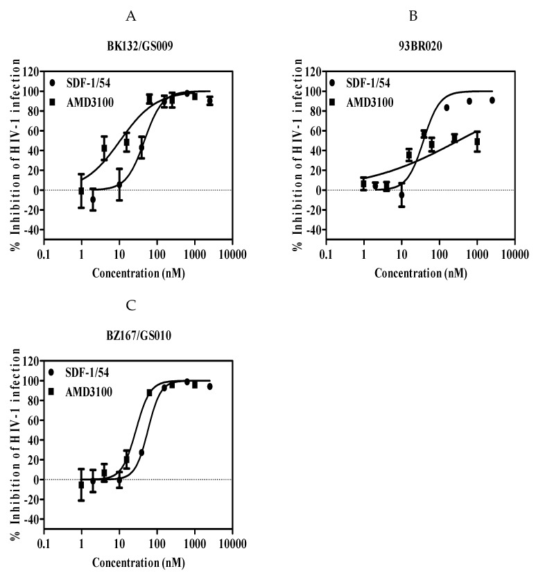
CXC chemokine receptor 4 (CXCR4) is a co-receptor for HIV-1 entry into target cells. Its natural ligand, the chemokine SDF-1, inhibits viral entry mediated by this receptor. However, the broad expression pattern of CXCR4 and its critical roles in various physiological and pathological processes indicate that the direct application of SDF-1 as an entry inhibitor might have severe consequences. Previously, we constructed an effective SDF-1 mutant, SDF-1/54, by deleting the α-helix of the C-terminal functional region of SDF-1. Of note, SDF-1/54 shows remarkable decreased chemotoxic ability, but maintains a similar binding affinity to CXCR4, suggesting SDF-1/54 might better serve as a CXCR4 inhibitor. Here, we found that SDF-1/54 exhibited potent antiviral activity against various X4 HIV-1 strains, including the infectious clone HIV-1 NL4-3, laboratory-adapted strain HIV-1 IIIB, clinical isolates and even drug-resistant strains. By using time-of-addition assay, non-infectious and infectious cell-cell fusion assay and CXCR4 internalization assay, we demonstrated SDF-1/54 is an HIV-1 entry inhibitor. A combination of SDF-1/54 with several antiretroviral drugs exhibited potent synergistic anti-HIV-1 activity. Moreover, SDF-1/54 was stable and its anti-HIV-1 activity was not significantly affected by the presence of seminal fluid, vaginal fluid simulant and human serum albumin. SDF-1/54 showed limited in vitro cytotoxicity to lymphocytes and vaginal epithelial cells. Based on these findings, SDF-1/54 could have a therapeutic potential as an HIV-1 entry inhibitor.

Keywords: CXCR4; HIV-1; SDF-1/54; entry inhibitor.


Conflict of interest statement

The authors declare no conflict of interest.

Figures

Figure 1
Figure 1
Antiviral potency of SDF-1/54 in a single-cycle viral infection. SDF-1/54-mediated inhibition of infection by infectious clone HIV-1 NL4-3 (A), laboratory-adapted HIV-1 IIIB (B) and HIV-1 SF162 (C) of TZM-bl cells. TZM-bl cells were pre-incubated with graded concentrations of SDF-1/54 for 15 min at 37 °C. Next, HIV-1 was added to the mixture. At 48 h post-infection, luciferase activity was detected. Average values (± SD) from three independent experiments are shown.
Figure 2
Figure 2
Antiviral potency of SDF-1/54 in multiple-cycle viral infection assays. SDF-1/54-mediated inhibition of infection by infectious clone HIV-1 NL4-3 (A) and laboratory-adapted HIV-1 IIIB (B) of MT-2 cells. MT-2 cells were pre-incubated with graded concentrations of inhibitor for 15 min at 37 °C. Next, mixtures were mixed with viruses. The culture medium was refreshed after overnight incubation. At 96 h post-infection, supernatant was collected and lyzed for p24 determination by ELISA and Western blotting (C). (D) SDF-1/54-mediated inhibition of infection by infectious clone HIV-1 NL4-3 in PBMC. Average values (±SD) from three independent experiments are shown.
Figure 3
Figure 3
SDF-1/54-mediated inhibition of infection by primary HIV-1 isolates of TZM-bl cells. Inhibition of the primary X4-tropic HIV-1 isolates included BK132/GS009 (A), 93BR020 (B) and BZ167/GS010 (C). TZM-bl cells were pre-incubated with graded concentrations of SDF-1/54 for 15 min at 37 °C. Next, primary HIV-1 isolates were added to the mixture. At 48 h post-infection, luciferase activity was detected. Average values (±SD) from three independent experiments are shown.
Figure 4
Figure 4
SDF-1/54 is effective against HIV-1 drug-resistant strains. SDF-1/54-mediated inhibition of infection by drug-resistant HIV-1 of TZM-bl cells which included viral strains resistant to T1144 (A,B) and T20 (C,D). TZM-bl cells were pre-incubated with graded concentrations of SDF-1/54 for 15 min at 37 °C. Then, drug-resistant HIV-1 was added to the mixture. At 48 h post-infection, luciferase activity was detected. Average values (±SD) from three independent experiments are shown.
Figure 5
Figure 5
Inhibition of HIV-1 entry by SDF-1/54 as determined by the time-of-addition assay. SDF-1/54 was added to MT-2 cells at different intervals after infection with HIV-1 IIIB. AZT and AMD3100 were included as controls. The final concentration was 800 nM for SDF-1/54, 100 nM for AZT and 400 nM for AMD3100. Averages (±SD) from three independent experiments are shown.
Figure 6
Figure 6
The inhibitory activity of SDF-1/54 against HIV-1-mediated cell–cell fusion. Effects of SDF-1/54 on the formation of syncytium with CHO-WT and MT-2 cells (A). Among them, the white arrow in the figure indicates the fusion bubble of cell fusion. Scale bar = 100 μm. (B) Dose-response curves and IC50 values of SDF-1/54 inhibiting cell–cell fusion. ADS-J1 was chosen as a positive control. (C) Cell–cell fusion between calcein-labeled H9/HIV-1 IIIB and MT-2 cells. Data are presented as mean ± SD.
Figure 6
Figure 6
The inhibitory activity of SDF-1/54 against HIV-1-mediated cell–cell fusion. Effects of SDF-1/54 on the formation of syncytium with CHO-WT and MT-2 cells (A). Among them, the white arrow in the figure indicates the fusion bubble of cell fusion. Scale bar = 100 μm. (B) Dose-response curves and IC50 values of SDF-1/54 inhibiting cell–cell fusion. ADS-J1 was chosen as a positive control. (C) Cell–cell fusion between calcein-labeled H9/HIV-1 IIIB and MT-2 cells. Data are presented as mean ± SD.
Figure 7
Figure 7
Capability of SDF-1/54 inducing CXCR4 internalization. MT-2 cells were treated with AMD3100 and SDF-1/54 for 2 h at 50 nM and CXCR4 on the cell surface was detected indirectly by PE-labeled CXCR4 antibody using flow cytometry. The level of CXCR4 on the cell surface is shown as mean fluorescence density, as described in Materials and Methods. MT-2 cells without any treatment were used as a control.
Figure 8
Figure 8
The effects of human SE-F, VFS and HSA on the anti-HIV-1 stability and activity of SDF-1/54. SDF-1/54 was incubated with PBS, VFS, SE-F and HSA (45mg/mL) for various time period. The remaining anti-viral activities of SDF-1/54 were tested (A). In addition, different concentrations of SDF-1/54 maintained antiviral activity in the presence or absence of SE, VFS and HSA (B), which were determined using p24 ELISA assay. Averages (± SD) from three independent experiments are shown.

References

    1. Joint United Nations Programme on HIV/AIDS (UNAIDS) Unaids Data 2018. UNAIDS; Geneva, Switzerland: 2018. [(accessed on 16 September 2019)]. Available online: http://www.unaids.org/sites/default/files/media_asset/unaids-data-2018_e....
    1. Cervia J.S., Smith M.A. Enfuvirtide (T-20): a novel human immunodeficiency virus type 1 fusion inhibitor. Clin. Infect. Dis. 2003;37:1102–1106. - PubMed
    1. Xu G.G., Guo J., Wu Y. Chemokine receptor CCR5 antagonist maraviroc: medicinal chemistry and clinical applications. Curr. Top. Med. Chem. 2014;14:1504–1514. doi: 10.2174/1568026614666140827143745. - DOI - PMC - PubMed
    1. Zhu T., Mo H., Wang N., Nam D.S., Cao Y., Koup R.A., Ho D.D. Genotypic and phenotypic characterization of HIV-1 patients with primary infection. Science. 1993;261:1179–1181. doi: 10.1126/science.8356453. - DOI - PubMed
    1. Schuitemaker H., Koot M., Kootstra N.A., Dercksen M.W., de Goede R.E., van Steenwijk R.P., Lange J.M., Schattenkerk J.K., Miedema F., Tersmette M. Biological phenotype of human immunodeficiency virus type 1 clones at different stages of infection: progression of disease is associated with a shift from monocytotropic to T–cell-tropic virus population. J. Virol. 1992;66:1354–1360. - PMC - PubMed

Publication types

LinkOut - more resources