. 2019 Sep 18;20(18):4634.
doi: 10.3390/ijms20184634.

FGF23-Mediated Activation of Local RAAS Promotes Cardiac Hypertrophy and Fibrosis

Affiliations

FGF23-Mediated Activation of Local RAAS Promotes Cardiac Hypertrophy and Fibrosis

Ineke Böckmann et al. Int J Mol Sci. .

Abstract

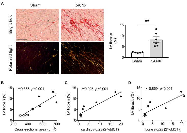
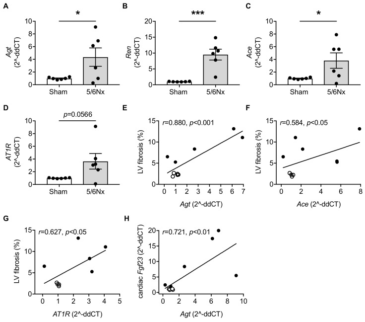
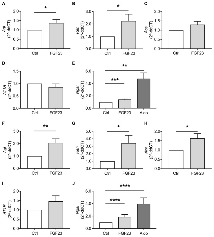
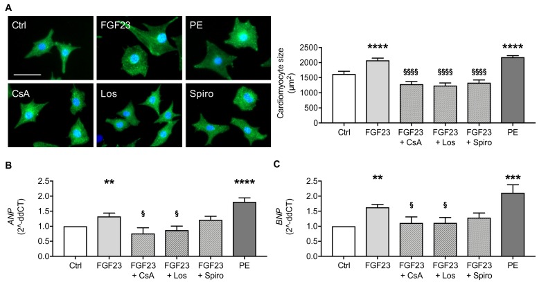
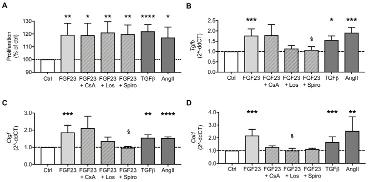
Patients with chronic kidney disease (CKD) are prone to developing cardiac hypertrophy and fibrosis, which is associated with increased fibroblast growth factor 23 (FGF23) serum levels. Elevated circulating FGF23 was shown to induce left ventricular hypertrophy (LVH) via the calcineurin/NFAT pathway and contributed to cardiac fibrosis by stimulation of profibrotic factors. We hypothesized that FGF23 may also stimulate the local renin-angiotensin-aldosterone system (RAAS) in the heart, thereby further promoting the progression of FGF23-mediated cardiac pathologies. We evaluated LVH and fibrosis in association with cardiac FGF23 and activation of RAAS in heart tissue of 5/6 nephrectomized (5/6Nx) rats compared to sham-operated animals followed by in vitro studies with isolated neonatal rat ventricular myocytes and fibroblast (NRVM, NRCF), respectively. Uremic rats showed enhanced cardiomyocyte size and cardiac fibrosis compared with sham. The cardiac expression of Fgf23 and RAAS genes were increased in 5/6Nx rats and correlated with the degree of cardiac fibrosis. In NRVM and NRCF, FGF23 stimulated the expression of RAAS genes and induced Ngal indicating mineralocorticoid receptor activation. The FGF23-mediated hypertrophic growth of NRVM and induction of NFAT target genes were attenuated by cyclosporine A, losartan and spironolactone. In NRCF, FGF23 induced Tgfb and Ctgf, which were suppressed by losartan and spironolactone, only. Our data suggest that FGF23-mediated activation of local RAAS in the heart promotes cardiac hypertrophy and fibrosis.

Keywords: cardiac fibrosis; chronic kidney disease; fibroblast growth factor 23; left ventricular hypertrophy; renin-angiotensin-aldosterone system.


Conflict of interest statement

The authors declare no conflict of interest.

Figures

Figure 1
Figure 1
The 5/6 nephrectomized (5/6Nx) rats develop left ventricular (LV) fibrosis, which correlates with fibroblast growth factor 23 (Fgf23) expression. (A) Representative bright field and polarized light microscopy images of picrosirius red-stained myocardial tissue of 5/6Nx and sham-operated rats and quantification of interstitial collagen fiber deposition demonstrating increased LV fibrosis in 5/6Nx rats (scale bar, 50 µm). (B–D) Pearson’s correlations of LV fibrosis with cross-sectional area of cardiomyocytes, and cardiac and bone Fgf23 mRNA expression as determined by quantitative real-time PCR using Gapdh as housekeeping gene. Clear dots, sham-operated rats; black dots, 5/6Nx rats. All values are shown as mean ± SEM; ** p < 0.01; n = 5–6 rats per group.
Figure 2
Figure 2
Here, 5/6 nephrectomy (5/6Nx) in rats induces cardiac expression of renin-angiotensin-aldosterone system (RAAS)-associated genes that correlates with left ventricular (LV) fibrosis. (A–C) Quantitative real-time PCR analyses in 5/6Nx rats show cardiac-specific induction of angiotensinogen (Agt), renin (Ren) and angiotensin converting enzyme (Ace), while (D) cardiac angiotensin II receptor type 1 (AT1R) mRNA expression is not significantly induced. (E–G) Pearson’s correlations of LV fibrosis with cardiac expression of Agt, Ace and AT1R, and (H) of cardiac Fgf23 with Agt mRNA expression. Clear dots, sham-operated rats; black dots, 5/6Nx rats. All values are shown as mean ± SEM; * p < 0.05, *** p < 0.001; n = 5–6 rats per group.
Figure 3
Figure 3
FGF23 induces RAAS-associated genes in neonatal rat ventricular myocytes (NRVM) and cardiac fibroblasts (NRCF) in vitro as shown by quantitative real-time PCR. (A–D) In NRVM, FGF23 treatment increases the mRNA expression of angiotensinogen (Agt) and renin (Ren), whereas the effects of FGF23 on angiotensin converting enzyme (Ace) and angiotensin II receptor type 1 (AT1R) expression levels are not statistically significant. (E) FGF23 and aldosterone (Aldo) induce the mRNA expression of neutrophil gelatinase-associated lipocalin (Ngal) in NRVM. (F–I) In NRCF, Agt, Ace and Ren mRNA expressions are enhanced after FGF23 treatment, but FGF23 does not significantly induce AT1R. (J) Ngal mRNA expression is elevated upon FGF23 and aldosterone stimulation. All values are shown as mean ± SEM; * p < 0.05, ** p < 0.01, *** p < 0.001, **** p < 0.0001 versus control; n = 6–8 independent cell isolations.
Figure 4
Figure 4
FGF23-induced hypertrophy in cultured NRVM is prevented by cyclosporine A (CsA), losartan (Los) and spironolactone (Spiro). (A) Representative immunofluorescent images of NRVM stained with sarcomeric α-actinin (green) and DAPI (blue) (scale bar, 50 µm), and quantification of NRVM size show significant induction of hypertrophic growth after treatment with FGF23, which is ameliorated by co-treatment with CsA, Los and Spiro. Phenylephrine (PE) served as positive control. (B,C) Quantitative real-time PCR analyses reveal FGF23-mediated increase of mRNA expression of prohypertrophic markers ANP and BNP, which is inhibited by CsA, Los and Spiro. Gapdh served as housekeeping gene. All values are shown as mean ± SEM; ** p < 0.01, *** p < 0.001, **** p < 0.0001 versus control; § p < 0.05, §§§§ p < 0.0001 versus FGF23; n = 5–9 independent cell isolations.
Figure 5
Figure 5
FGF23 stimulates the proliferation of NRCF and induces the expression of the profibrotic markers collagen 1 (Col1), transforming growth factor beta 1 (Tgfb) and connective tissue growth factor (Ctgf). (A) The proliferation of NRCF increases in response to stimulation with FGF23, which was not inhibited in the presence of cyclosporine A (CsA), losartan (Los) or spironolactone (Spiro) as demonstrated by MTS-based proliferation assay. (B–D) The mRNA expression of Tgfb, Ctgf and Col1 in NRCF is increased upon FGF23 stimulation as measured by quantitative real-time PCR. Co-treatment with Los and Spiro inhibits the induction of fibrotic markers by FGF23, while CsA has lesser effects. TGFβ and angiotensin II (AngII) served as positive controls. Horizontal dotted lines represent the level of controls. All values are shown as mean ± SEM; * p < 0.05, ** p < 0.01, *** p < 0.001, **** p < 0.0001 versus control; § p < 0.05 versus FGF23; n = 5–9 independent cell isolations.

References

    1. Hill N.R., Fatoba S.T., Oke J.L., Hirst J.A., O’Callaghan C.A., Lasserson D.S., Hobbs F.D.R. Global Prevalence of Chronic Kidney Disease—A Systematic Review and Meta-Analysis. PLoS ONE. 2016;11:e0158765. doi: 10.1371/journal.pone.0158765. - DOI - PMC - PubMed
    1. Thompson S., James M., Wiebe N., Hemmelgarn B., Manns B., Klarenbach S., Tonelli M. Cause of Death in Patients with Reduced Kidney Function. J. Am. Soc. Nephrol. 2015;26:2504–2511. doi: 10.1681/ASN.2014070714. - DOI - PMC - PubMed
    1. Go A.S., Chertow G.M., Fan D., McCulloch C.E., Hsu C.-Y. Chronic kidney disease and the risks of death, cardiovascular events, and hospitalization. N. Engl. J. Med. 2004;351:1296–1305. doi: 10.1056/NEJMoa041031. - DOI - PubMed
    1. Isakova T., Wahl P., Vargas G.S., Gutiérrez O.M., Scialla J., Xie H., Appleby D., Nessel L., Bellovich K., Chen J., et al. Fibroblast growth factor 23 is elevated before parathyroid hormone and phosphate in chronic kidney disease. Kidney Int. 2011;79:1370–1378. doi: 10.1038/ki.2011.47. - DOI - PMC - PubMed
    1. Pool L.R., Wolf M. FGF23 and Nutritional Metabolism. Annu. Rev. Nutr. 2017;37:247–268. doi: 10.1146/annurev-nutr-071816-064620. - DOI - PubMed

MeSH terms

Substances

LinkOut - more resources