. 2019 Nov 1;294(44):16465-16478.
doi: 10.1074/jbc.RA119.010105. Epub 2019 Sep 20.

The bacterial endoribonuclease RNase E can cleave RNA in the absence of the RNA chaperone Hfq

Affiliations

The bacterial endoribonuclease RNase E can cleave RNA in the absence of the RNA chaperone Hfq

Yu Mi Baek et al. J Biol Chem. .

Abstract

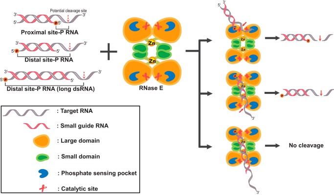
RNase E is a component of the RNA degradosome complex and plays a key role in RNA degradation and maturation in Escherichia coli RNase E-mediated target RNA degradation typically involves the RNA chaperone Hfq and requires small guide RNAs (sRNAs) acting as a seed by binding to short (7-12-bp) complementary regions in target RNA sequences. Here, using recombinantly expressed and purified proteins, site-directed mutagenesis, and RNA cleavage and protein cross-linking assays, we investigated Hfq-independent RNA decay by RNase E. Exploring its RNA substrate preferences in the absence of Hfq, we observed that RNase E preferentially cleaves AU-rich sites of single-stranded regions of RNA substrates that are annealed to an sRNA that contains a monophosphate at its 5'-end. We further found that the quaternary structure of RNase E is also important for complete, Hfq-independent cleavage at sites both proximal and distal to the sRNA-binding site within target RNAs containing monophosphorylated 5'-ends. Of note, genetic RNase E variants with unstable quaternary structure exhibited decreased catalytic activity. In summary, our results show that RNase E can degrade its target RNAs in the absence of the RNA chaperone Hfq. We conclude that RNase E-mediated, Hfq-independent RNA decay in E. coli requires a cognate sRNA sequence for annealing to the target RNA, a 5'-monophosphate at the RNA 5'-end, and a stable RNase E quaternary structure.

Keywords: RNA chaperone Hfq; RNA degradation; RNA processing; RNA turnover; RNase E; endoribonuclease; mRNA decay; protein expression; ribonuclease; small guide RNA (sRNA).


Conflict of interest statement

The authors declare that they have no conflicts of interest with the contents of this article

Figures

Figure 1.
Figure 1.
Cleavage of RNAs by RNase E in the absence or presence of Hfq. A, cleavage of RNAs with different chemical groups at the 5′-end (monophosphorylated ssRNAs, hydroxylated ssRNA, triphosphorylated ssRNA, and blunt-ended dsRNA). Top, monophosphorylated ssRNAs of two different lengths (25 and 47 nt for 25R1 and 47R0, respectively) were reacted with RNase E under different conditions (lanes a, b, c, and d; shown in table). Each reaction was performed by incubating 10 nm 32P-labeled RNA substrate in the absence (i.e. 0 min) or presence of RNase E (0.1 μm) for 30 min. Bottom, RNase E reactions were performed by incubating 10 nm 32P-labeled RNA substrates, such as triphosphorylated ssRNA (3P-47R0), 5′-end–hydroxylated ssRNA (HO-47R0), or blunt-ended duplex RNA (47R0/47cR0) with RNase E (0.1 μm) for increasing time (0 to 30 min). RNA cleavage products were resolved on 15% native PAGE and visualized using a phosphorimager. Asterisks represent 32P-labeled nucleotides. Lane c, size control for RNA of 47 and 25 nt. B, cleavage of sRNA (25R1)-guided target RNA (47R0) by RNase E in the absence or presence of Hfq. The partial duplex RNA (47R0/25R1, 10 nm) was incubated with RNase E (0.1 μm) in the absence or presence of Hfq (0.1 μm) for 0, 2, 4, 10, 20, and 40 min. RNA cleavage products were resolved on 15% native PAGE and visualized using a phosphorimager. Kinetic analysis results of cleaved RNA products, which were quantified using densitometry from the gel, were plotted and fitted to an exponential function. Mean values of triplicate independent experiments and S.D. (error bars) are shown. The difference between RNase E plus Hfq and RNase E alone was determined to be statistically insignificant by Student's t test (p = 0.079).
Figure 2.
Figure 2.
RNase E–mediated degradation of the single-stranded region in target RNA paired with 5′-monophosphorylated sRNA. A, RNase reaction of 47-mer target RNA paired with 5′-monophosphorylated sRNA by RNase E. The partial duplex RNA substrates (10 nm) were reacted with or without RNase E (0.1 μm) under different conditions (lanes a, b, c, and d; shown in table). RNase E reaction products were simultaneously resolved on 15% native and denaturing (12% polyacrylamide in 8 m urea) PAGE, in which 32P-labeled RNA were visualized using a phosphorimager. B, mapping of RNase E cleavage sites in the single-stranded region of the partial duplex RNA substrate. The 32P-labeled dsRNA substrate (30 nm, P47R0/P25R1 shown as RNA sequences) was incubated with or without RNase E (0.25 μm, lanes 6 and 7, respectively) at 37 °C for 30 min. For comparison as RNA ladders, the 5′-end 32P-labeled single-stranded RNA (30 nm; P47R0) was mixed with alkaline hydrolysis buffer at 25 °C for 10 or 5 min (lanes 1 and 2, respectively). RNase T1 (10, 5, or 1 unit; lanes 3, 4, and 5, respectively) was incubated with P47R0 RNA for 15 min. Cleavage sites after guanine residues are indicated in the RNA sequences (underlined Gs). All reactions were quenched with an equal volume of loading buffer and analyzed on 15% denaturing (8 m urea) PAGE and visualized using a phosphorimager. RNA fragments generated using RNase E cleavage at adenine residues in single-stranded regions are indicated with A and the nucleotide position with arrows. Dashed line, spliced part of two gel images for size controls (25- and 47-mer).
Figure 3.
Figure 3.
Cleavage of various partial duplex RNA substrates with different seed regions in sRNA (3′-overhang, 5′-overhang, and double overhang duplex RNAs). Various RNA substrates were reacted with RNase E: 3′-overhang dsRNA substrates (A), 5′-overhang dsRNA substrates (B), and double overhang dsRNA substrates (C). Reactions were performed by incubating 32P-labeled duplex RNA substrates (10 nm) in the presence or absence of RNase E (0.1 μm) at 37 °C for 30 min. Cleaved RNAs were simultaneously resolved on 15% native and denaturing (8 m urea) PAGE and visualized using a phosphorimager.
Figure 4.
Figure 4.
Degradation of target RNA by RNase E with 5′-monophosphorylated guide sRNA or sDNA. The RNase E reaction was performed using target RNA (47 nt, R0) annealed with 25-mer guide oligonucleotides: sRNA (A) or sDNA (C) (indicated with red lines). RNase E (0.1 μm) was incubated with 32P-labeled dsRNA or duplex RNA/DNA substrates (10 nm; asterisks represent 32P-labeled nucleotides) at 37 °C for 2, 4, 10, 20, 40, and 60 min. The reaction mixtures were resolved on 15% native PAGE and visualized using a phosphorimager. Kinetic analysis results of RNA cleavage product accumulation from A and C were plotted and fitted to an exponential function in B and D, respectively. Mean values of triplicate independent experiments and S.D. are shown.
Figure 5.
Figure 5.
Quaternary structure analysis and RNA cleavage activities of RNase E and RNase E mutants. A, size-exclusion chromatography profiles showing molecular mass distributions of WT and mutant RNase E proteins: RNase E (solid line), RNase EMT(499) (thick dashed line), and RNase EMT(C/S) (thin dashed line). The molecular weight of proteins was calculated using a standard curve obtained using size control proteins (inset graph). B, cleavage of partial duplex RNA substrates containing proximal or distal phosphate (HO47R0/P25R1 and P47R0/HO25R1, respectively) with RNase E and RNase E mutants. RNase E reactions were performed by incubating 32P-labeled 3′-overhang RNA duplex substrates (10 nm) with RNase E proteins (0.1 μm) at 37 °C for various incubation times (0, 2, 5, 10, 30, and 60 min). The reaction mixtures were resolved on 15% native PAGE and visualized using a phosphorimager. Kinetic analysis results of RNA cleavage product accumulation were plotted and fitted to an exponential function.
Figure 6.
Figure 6.
Suppression of target gene expression through target RNA decay with RNase E and 5′-monophosphorylated guide RNA in bacteria. A, time course analysis of GFP expression in E. coli cells transformed with GFP expression vector and transfected with GFP transcripts targeting or nontargeting (scrambled sequences irrespective of GFP transcript) sRNA. The GFP-transformed bacterial cells were transfected with 5 μm phosphorylated GFP target sRNA (p-GFP target sRNA) or nontarget control sRNA (p-nontarget sRNA) by electroporation. No transfection for negative control was included. The absorbance showing cell growth and GFP fluorescence was measured every 1 h, in which GFP fluorescence was normalized to cell growth absorbance. The resulting GFP fluorescence is shown in the graph as relative to the unity value at the starting point (time 0). Data are presented as the mean ± S.D. (error bars), n = 3. B, E. coli cells transformed with GFP expression vector were transfected with the GFP transcripts targeting phosphorylated (proximal or distal site) sRNAs or nontargeting sRNA by electroporation and further incubated for 6 h. GFP fluorescence was measured and represented as shown in A. C, levels of GFP mRNA in E. coli cells that were transfected with the GFP transcripts targeting phosphorylated (proximal or distal site) sRNA or nontargeting sRNA. Bacterial RNA was extracted and reverse-transcribed. Quantitative real-time PCR was performed for the quantification of GFP transcripts. D and E, time course analysis of GFP expression in the WT (D) or Hfq deletion (E) E. coli cells transformed with GFP expression vector and transfected with GFP transcripts targeting or nontargeting (scrambled sequences irrespective of GFP transcript) sRNAs. The experiment was conducted in the same manner as in A. F, levels of GFP mRNA in the WT and Hfq deletion E. coli cells that were transfected with the GFP transcripts targeting phosphorylated (proximal or distal site) sRNAs or nontargeting sRNA. The experiment was conducted in the same manner as in C. G, primer extension assay for the measurement of GFP mRNA. GFP mRNA synthesized in vitro (100 nm) was incubated with various concentrations (0, 0.1, 1, and 10 nm) of RNase E with or without GFP targeting (proximal) p-sRNA for 30 min. After the reaction, the GFP reverse primer DNAs annealed to GFP RNA were synthesized into complementary DNA using reverse transcriptase. Urea-PAGE and subsequent SYBR Gold staining were used to resolve the synthesized cDNA that reflects the amount of GFP mRNA remaining in the reaction. H, quantitative real-time PCR was used for monitoring the levels of the remaining target GFP RNA (100 nm) after RNA decay by RNase E. GFP mRNA synthesized in vitro (100 nm) was incubated with 200 nm targeting (proximal or distal site) sRNA or nontargeting sRNA. After the reaction, cDNA was synthesized and amplified for quantitative real-time PCR. I, RNA cleavage assay with long dsRNA (72- and 47-mer RNAs annealed). The partial duplex RNA (10 nm) was incubated with RNase E (100 nm) for 0, 2, 4, 10, 20, 40, and 60 min. For comparison, partial duplex RNA harboring 25 bp was also incubated with RNase E for 30 min as a positive control in the assay. RNA cleavage products were resolved on 15% native PAGE and visualized using a phosphorimager. Dashed line, spliced part of two gel images for comparison of RNA cleavage. Mean values of triplicate independent experiments and S.D. are shown. Statistical significance was determined using paired Student's t test (**, p < 0.01).
Figure 7.
Figure 7.
Models for RNase E cleavage with sRNA without the Hfq RNA chaperone. The quaternary structure of RNase E is assumed to be an intact dimer–dimer complex. RNase E can recognize and degrade target (partial duplex) RNA via the 5′-phosphate–sensing pocket and the RNA cleavage site in RNase E. When target RNA is base-paired with the proximal 5′-P sRNA, both the single-stranded RNA cleavage site and the 5′-phosphate–sensing pocket reside on the same dimer, enabling RNase E to cleave the target RNA near the 5′-P of sRNA. In the case of the distal 5′-P sRNA, target RNA base-paired with the distal 5′-P sRNA was recognized and degraded in separate dimeric sites. In contrast, RNA cleavage is not possible for target RNA with a longer duplex length exceeding the distance between the 5′-P–sensing site of a dimer and cleavage site of another dimer. The cleavage of RNA can occur both near to and distal from the 5′-P in RNA, depending on the alignment of the RNA cleavage site and the 5′-phosphate–sensing pocket on RNase E.

Similar articles

Cited by

References

    1. Chen H., Shiroguchi K., Ge H., and Xie X. S. (2015) Genome-wide study of mRNA degradation and transcript elongation in Escherichia coli. Mol. Syst. Biol. 11, 808 10.15252/msb.20159000 - DOI - PMC - PubMed
    1. Kushner S. R. (2002) mRNA decay in Escherichia coli comes of age. J. Bacteriol. 184, 4658–4665 10.1128/jb.184.17.4658-4665.2002 - DOI - PMC - PubMed
    1. Deutscher M. P. (2003) Degradation of stable RNA in bacteria. J. Biol. Chem. 278, 45041–45044 10.1074/jbc.R300031200 - DOI - PubMed
    1. Carpousis A. J. (2007) The RNA degradosome of Escherichia coli: an mRNA degrading machine assembled on RNase E. Annu. Rev. Microbiol. 61, 71–87 10.1146/annurev.micro.61.080706.093440 - DOI - PubMed
    1. Coburn G. A., Miao X., Briant D. J., and Mackie G. A. (1999) Reconstitution of a minimal RNA degradosome demonstrates functional coordination between a 3′ exonuclease and a DEAD-box RNA helicase. Genes Dev. 13, 2594–2603 10.1101/gad.13.19.2594 - DOI - PMC - PubMed

Publication types

MeSH terms

LinkOut - more resources