

Proteases as Secreted Exoproteins in Mycoplasmas from Ruminant Lungs and Their Impact on Surface-Exposed Proteins
- PMID: 31540994
- PMCID: PMC6856322
- DOI: 10.1128/AEM.01439-19
Proteases as Secreted Exoproteins in Mycoplasmas from Ruminant Lungs and Their Impact on Surface-Exposed Proteins
Abstract
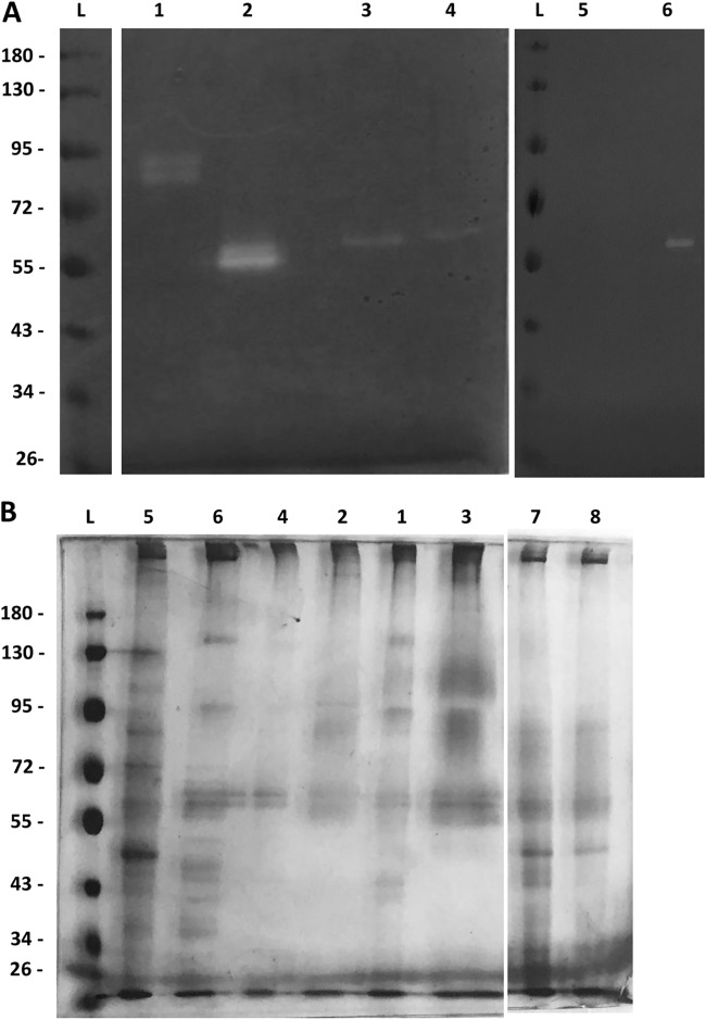
Many mycoplasma species are isolated from the ruminant lungs as either saprophytes or true pathogens. These wall-less bacteria possess a minimal genome and reduced metabolic capabilities. Accordingly, they rely heavily on their hosts for the supply of essential metabolites and, notably, peptides. Seven of 13 ruminant lung-associated Mycoplasma (sub)species were shown to possess caseinolytic activity when grown in rich media and assessed with a quantitative fluorescence test. For some species, this activity was detected in spent medium, an indication that proteases were secreted outside the mycoplasma cells. To identify these proteases, we incubated concentrated washed cell pellets in a defined medium and analyzed the supernatants by tandem mass spectrometry. Secreted-protease activity was detected mostly in the species belonging to the Mycoplasma mycoides cluster (MMC) and, to a lesser extent, in Mycoplasma bovirhinis Analyzing a Mycoplasma mycoides subsp. capri strain, chosen as a model, we identified 35 expressed proteases among 55 predicted coding genes, of which 5 were preferentially found in the supernatant. Serine protease S41, acquired by horizontal gene transfer, was responsible for the caseinolytic activity, as demonstrated by zymography and mutant analysis. In an M. capricolum mutant, inactivation of the S41 protease resulted in marked modification of the expression or secretion of 17 predicted surface-exposed proteins. This is an indication that the S41 protease could have a role in posttranslational cleavage of surface-exposed proteins and ectodomain shedding, whose physiological impacts still need to be explored.IMPORTANCE Few studies pertaining to proteases in ruminant mycoplasmas have been reported. Here, we focus on proteases that are secreted outside the mycoplasma cell using a mass spectrometry approach. The most striking result is the identification, within the Mycoplasma mycoides cluster, of a serine protease that is exclusively detected outside the mycoplasma cells and is responsible for casein digestion. This protease may also be involved in the posttranslational processing of surface proteins, as suggested by analysis of mutants showing a marked reduction in the secretion of extracellular proteins. By analogy, this finding may help increase understanding of the mechanisms underlying this ectodomain shedding in other mycoplasma species. The gene encoding this protease is likely to have been acquired via horizontal gene transfer from Gram-positive bacteria and sortase-associated surface proteases. Whether this protease and the associated ectodomain shedding are related to virulence has yet to be ascertained.
Keywords: Mycoplasma; exosecretion; posttranslational cleavage; proteases.
Copyright © 2019 American Society for Microbiology.
Figures

Similar articles
-
Highly dynamic genomic loci drive the synthesis of two types of capsular or secreted polysaccharides within the Mycoplasma mycoides cluster.Appl Environ Microbiol. 2015 Jan;81(2):676-87. doi: 10.1128/AEM.02892-14. Epub 2014 Nov 14. Appl Environ Microbiol. 2015. PMID: 25398856 Free PMC article.
-
Distinctive repertoire of contingency genes conferring mutation- based phase variation and combinatorial expression of surface lipoproteins in Mycoplasma capricolum subsp. capricolum of the Mycoplasma mycoides phylogenetic cluster.J Bacteriol. 2006 Jul;188(13):4926-41. doi: 10.1128/JB.00252-06. J Bacteriol. 2006. PMID: 16788201 Free PMC article.
-
Standardized analysis of nuclease activities in Mycoplasma species colonizing swine, poultry, and small ruminants.FEMS Microbiol Lett. 2025 Jan 10;372:fnaf057. doi: 10.1093/femsle/fnaf057. FEMS Microbiol Lett. 2025. PMID: 40489321
-
Secretion, processing and activation of bacterial extracellular proteases.Mol Microbiol. 1989 Dec;3(12):1825-31. doi: 10.1111/j.1365-2958.1989.tb00169.x. Mol Microbiol. 1989. PMID: 2695751 Review.
-
Taxonomy of the Mycoplasma mycoides cluster.Isr J Med Sci. 1987 Jun;23(6):632-5. Isr J Med Sci. 1987. PMID: 3312102 Review.
Cited by
-
Contagious Bovine and Caprine Pleuropneumonia: a research community's recommendations for the development of better vaccines.NPJ Vaccines. 2020 Jul 24;5(1):66. doi: 10.1038/s41541-020-00214-2. eCollection 2020. NPJ Vaccines. 2020. PMID: 32728480 Free PMC article. Review.
-
A toolbox for manipulating the genome of the major goat pathogen, Mycoplasma capricolum subsp. capripneumoniae.Microbiology (Reading). 2024 Jan;170(1):001423. doi: 10.1099/mic.0.001423. Microbiology (Reading). 2024. PMID: 38193814 Free PMC article.
-
Beware of Mycoplasma Anti-immunoglobulin Strategies.mBio. 2021 Dec 21;12(6):e0197421. doi: 10.1128/mBio.01974-21. Epub 2021 Nov 16. mBio. 2021. PMID: 34781733 Free PMC article. Review.
-
Genome-Wide Association Study of Nucleotide Variants Associated with Resistance to Nine Antimicrobials in Mycoplasma bovis.Microorganisms. 2022 Jul 6;10(7):1366. doi: 10.3390/microorganisms10071366. Microorganisms. 2022. PMID: 35889084 Free PMC article.
-
Comparative Secretome Analyses of Mycoplasma bovis Virulent and Attenuated Strains Revealed MbovP0145 as a Promising Diagnostic Biomarker.Front Vet Sci. 2021 Jun 18;8:666769. doi: 10.3389/fvets.2021.666769. eCollection 2021. Front Vet Sci. 2021. PMID: 34222397 Free PMC article.
References
-
- Nocard E, Roux E, Borrel A, Salimbeni AT, Dujardin-Beaumetz E. 1898. Le microbe de la péripneumonie. Ann Inst Pasteur (Paris) 12:240–262.
-
- Westberg J, Persson A, Holmberg A, Goesmann A, Lundeberg J, Johansson KE, Pettersson B, Uhlen M. 2004. The genome sequence of Mycoplasma mycoides subsp. mycoides SC type strain PG1T, the causative agent of contagious bovine pleuropneumonia (CBPP). Genome Res 14:221–227. doi:10.1101/gr.1673304. - DOI - PMC - PubMed
-
- Browning GF, Noormohammadi AH, Markham PF. 2014. Identification and characterization of virulence genes in mycoplasmas, p 77–90. In Browning GF, Citti C (ed), Mollicutes, molecular biology and pathogenesis. Caister Academic Press, Norfolk, United Kingdom.
MeSH terms
Substances
LinkOut - more resources
Full Text Sources
