. 2019 Sep 20;4(39):eaaw7083.
doi: 10.1126/sciimmunol.aaw7083.

Nucleoside-modified mRNA encoding HSV-2 glycoproteins C, D, and E prevents clinical and subclinical genital herpes

Affiliations

Nucleoside-modified mRNA encoding HSV-2 glycoproteins C, D, and E prevents clinical and subclinical genital herpes

Sita Awasthi et al. Sci Immunol. .

Abstract

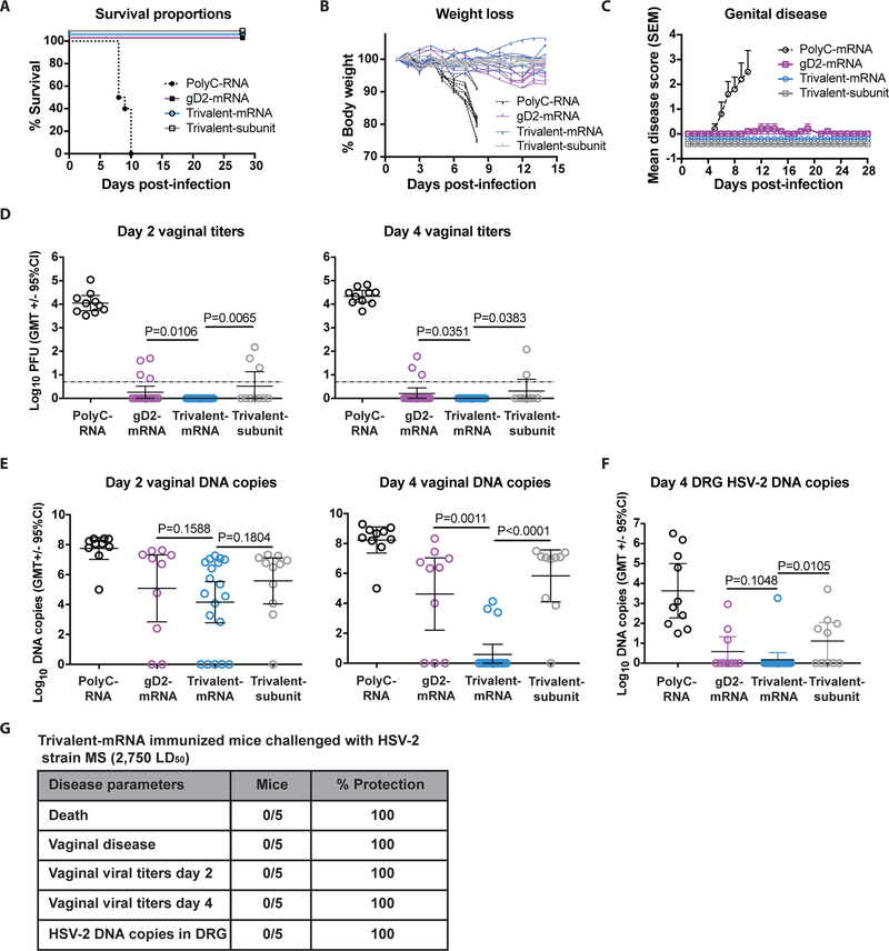
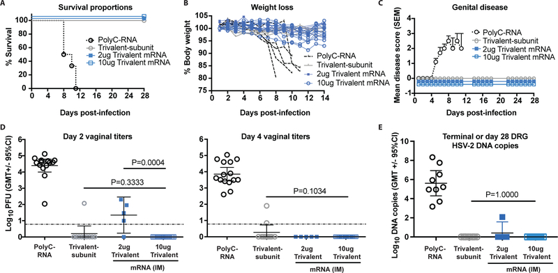
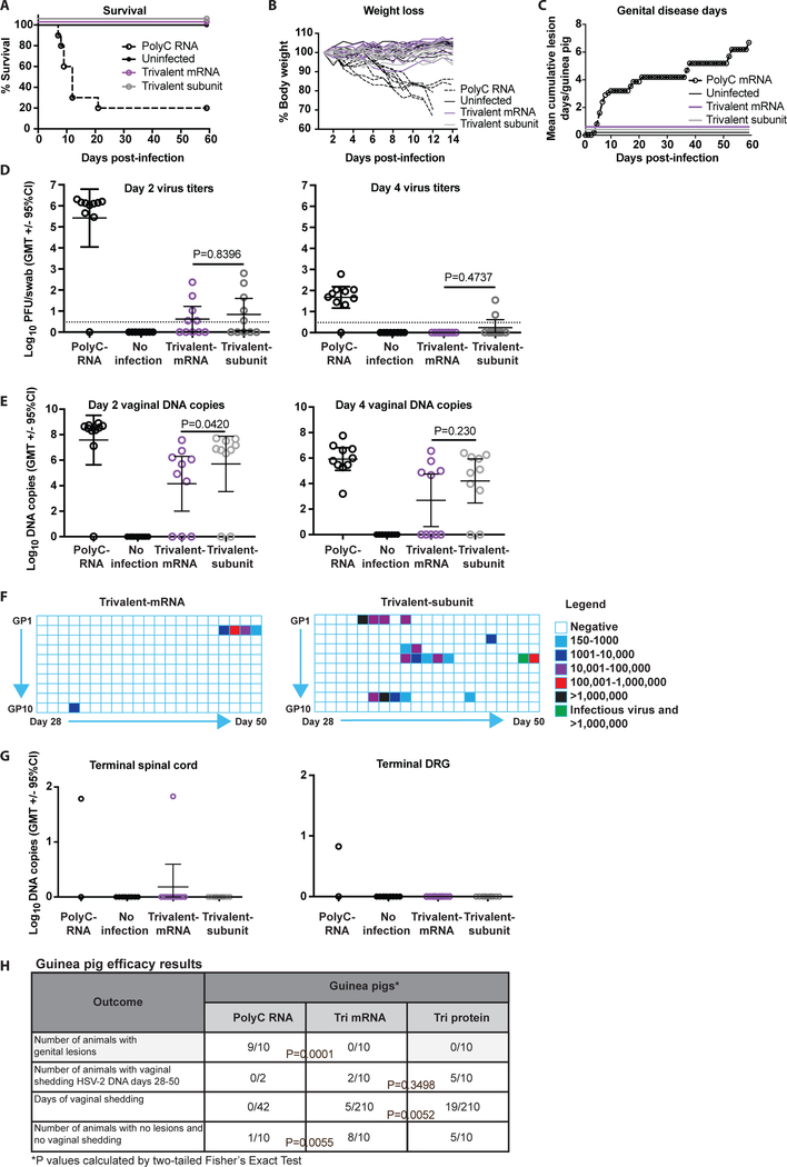
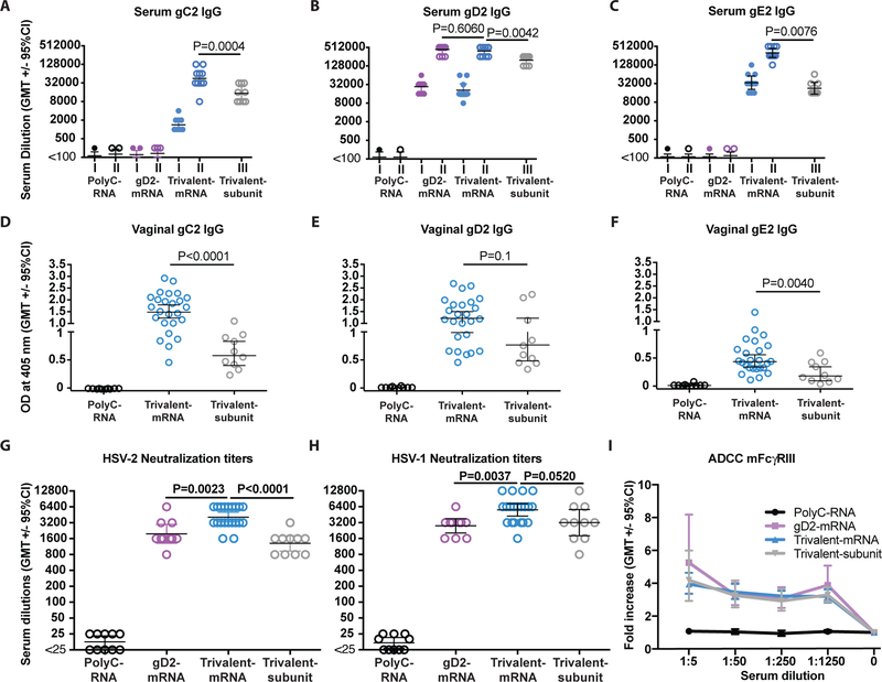
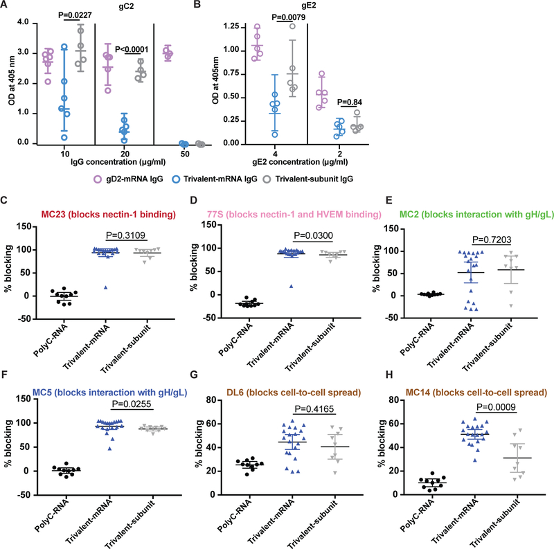
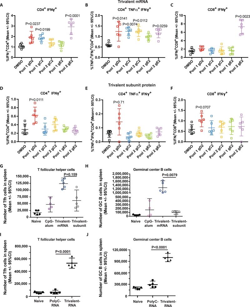
The goals of a genital herpes vaccine are to prevent painful genital lesions and reduce or eliminate subclinical infection that risks transmission to partners and newborns. We evaluated a trivalent glycoprotein vaccine containing herpes simplex virus type 2 (HSV-2) entry molecule glycoprotein D (gD2) and two immune evasion molecules: glycoprotein C (gC2), which binds complement C3b, and glycoprotein E (gE2), which blocks immunoglobulin G (IgG) Fc activities. The trivalent vaccine was administered as baculovirus proteins with CpG and alum, or the identical amino acids were expressed using nucleoside-modified mRNA in lipid nanoparticles (LNPs). Both formulations completely prevented genital lesions in mice and guinea pigs. Differences emerged when evaluating subclinical infection. The trivalent protein vaccine prevented dorsal root ganglia infection, and day 2 and 4 vaginal cultures were negative in 23 of 30 (73%) mice compared with 63 of 64 (98%) in the mRNA group (P = 0.0012). In guinea pigs, 5 of 10 (50%) animals in the trivalent subunit protein group had vaginal shedding of HSV-2 DNA on 19 of 210 (9%) days compared with 2 of 10 (20%) animals in the mRNA group that shed HSV-2 DNA on 5 of 210 (2%) days (P = 0.0052). The trivalent mRNA vaccine was superior to trivalent proteins in stimulating ELISA IgG antibodies, neutralizing antibodies, antibodies that bind to crucial gD2 epitopes involved in entry and cell-to-cell spread, CD4+ T cell responses, and T follicular helper and germinal center B cell responses. The trivalent nucleoside-modified mRNA-LNP vaccine is a promising candidate for human trials.


Figures

Figure 1.
Figure 1.. ID immunization with nucleoside-modified trivalent mRNA-LNP protects against genital HSV-2 in mice.
(a-f) BALB/c mice were immunized twice with PolyC RNA-LNP, gD2 nucleoside-modified mRNA-LNP, trivalent nucleoside-modified mRNA-LNP, or three times with trivalent subunit protein-CpG/alum and challenged one month after the final immunization intravaginally with 5×10³ HSV-2. (a) Survival curves: P<0.001 comparing PolyC RNA with each other group. P values were calculated by the log-rank test. (b) The mean weight loss each day post-infection of animals in the trivalent mRNA, trivalent subunit protein, or gD2 mRNA group was compared with PolyC RNA controls; P<0.0001. (c) Mean genital disease scores post-infection comparing trivalent mRNA, trivalent subunit protein or gD2 mRNA with PolyC RNA, P<0.0001. P value in (b) and (c) was calculated by two-way ANOVA followed by Tukey for significance. Error bars in (c) represent SEM. (d) Day 2 and day 4 vaginal titers. Dotted line indicates the limit of assay detection at 6.7 PFU/ml. (e) Day 2 and day 4 vaginal DNA copy number by qPCR. (f) Day 4 DRG HSV-2 DNA copy number by qPCR. P values in (d-f) are shown on the figures for the primary endpoint comparison between trivalent mRNA and trivalent protein or secondary endpoint comparison between trivalent mRNA and gD2 mRNA and were calculated by the two-tailed Fisher’s exact test. Sample size: (a-c and f), n=10 per group, except trivalent mRNA group, n=20 for (a-c) and n=19 for (f); (d), n=10 in PolyC RNA, n=20 in gD2 mRNA, n=39 in trivalent mRNA and n=10 in trivalent subunit protein; (e), n=10 per group except trivalent mRNA n=20. (g) Challenge of trivalent mRNA-immunized mice with 5×10⁴ PFU (2,750 LD50).
Figure 2.
Figure 2.. IM immunization with nucleoside-modified trivalent mRNA-LNP protects against genital HSV-2 in mice.
BALB/c mice were immunized as in Figure 1 except one group received 2μg trivalent mRNA. Mice were challenged one month after the final immunization intravaginally with 5×10³ HSV-2 (275 LD50). (a) Survival curves: P<0.0001 comparing PolyC RNA with each other group. P values were calculated by the log-rank test. (b) Weight loss of individual animals: P<0.0001 comparing PolyC RNA with the other three groups. (c) Genital disease: P<0.0001 comparing PolyC RNA with the other three groups. P values in (b-c) were calculated by two-way ANOVA followed by Tukey for significance. Sample size: (a-c), n=10 in PolyC RNA group for (a) and (c), n=5 for (b), n=10 in trivalent subunit protein, n=5 in 2μg trivalent mRNA, and n=10 in 10μg trivalent mRNA. Error bars in (c) represent SEM. P values in (d-e) are shown on the figures for the primary endpoint comparison between trivalent mRNA and trivalent protein or the comparison between 2μg and 10μg trivalent mRNA. Other P values are noted below. (d) Day 2 vaginal titers: comparing 10μg trivalent mRNA or trivalent subunit protein with PolyC RNA, P<0.0001; comparing 2μg trivalent mRNA with PolyC RNA, P=0.2500. Day 4 vaginal titers: comparing the number of animals with positive day 4 cultures in the 10μg trivalent mRNA, the 2μg trivalent mRNA or the trivalent subunit protein group with PolyC RNA, P<0.0001. Sample size: n=15 in PolyC RNA group, n=10 in trivalent subunit protein group, n=5 in the 2μg trivalent mRNA group and n=20 in the 10μg trivalent mRNA group. Dotted line indicates the limit of assay detection at 6.7 PFU/ml. (e) DRG HSV-2 DNA copy number at time of euthanasia of PolyC RNA group or on day 28 in the other groups; P<0.0001 comparing the number of animals with positive HSV-2 DNA copy number in the PolyC RNA with the 10μg trivalent mRNA or the trivalent subunit protein group; P=0.0050 comparing the number of animals with positive HSV-2 DNA copy in the PolyC RNA with the 2μg trivalent mRNA group. Other comparisons were not significant. Sample size: n=9 in the PolyC group, n=10 in the trivalent subunit protein group, n=5 in the 2μg trivalent mRNA group and n=10 in the 10μg trivalent mRNA group. P values for (d-e) were calculated by the two-tailed Fisher’s exact test.
Figure 3.
Figure 3.. ID immunization with nucleoside-modified trivalent mRNA-LNP protects against genital HSV-2 in guinea pigs.
(a-f) Guinea pigs were immunized three times monthly IM with 10μg PolyC RNA-LNP; ID with 20μg each trivalent nucleoside-modified mRNA-LNP; IM with 10μg each trivalent subunit protein-CpG/alum; or left unimmunized (n=10/group). One month after the final immunization, all immunized animals were infected intravaginally with 5×10⁵ HSV-2 (50 LD50), while unimmunized animals remained uninfected. (a) Survival curves: P<0.0001 comparing PolyC RNA with each other group. P values calculated by the log-rank test. (b) The mean weight loss each day post-infection of animals in the trivalent mRNA, trivalent subunit protein, or uninfected group was compared with PolyC RNA controls; P<0.0001. (c) Cumulative mean genital disease score per guinea pig is shown for each day post-infection. No animal developed genital lesions in the trivalent mRNA, trivalent subunit protein or uninfected group. Comparing trivalent mRNA, trivalent subunit protein or uninfected with PolyC RNA, P<0.0001. The P value in (b) and (c) was calculated by two-way ANOVA followed by Tukey for significance. (d-e) P values shown on the figures are for the primary endpoint comparison between trivalent mRNA and trivalent protein. Other P values are noted below. (d) Day 2 and day 4 vaginal titers: Day 2, P=0.0013 and day 4 P=0.0010 comparing PolyC RNA with trivalent mRNA. Dotted line indicates the limit of assay detection at 3.3 PFU/ml. (e) Day 2 and day 4 vaginal DNA copy number by qPCR. P value in (d) and (e) was calculated by the two-tailed Mann-Whitney, except the day 4 comparison between trivalent mRNA and trivalent subunit proteins was performed by the two-tailed Fisher’s exact test. (f) Vaginal shedding of HSV-2 DNA over 21 days from day 28–50 post-infection. One animal in the trivalent subunit protein group had replication competent virus recovered on a day HSV-2 DNA shedding was detected (marked in green). Comparing days of shedding in the trivalent mRNA and trivalent subunit protein group, P=0.0052, calculated by the two-tailed Fisher’s exact test. (g) HSV-2 DNA copy number/106 copies of GAPDH in spinal cord and DRG at the end of the experiment; n=2 in PolyC RNA and n=10 in the 3 other groups. (h) Table summarizing key results of guinea pig studies.
Figure 4.
Figure 4.. ELISA, neutralizing and ADCC antibody responses.
Mice were immunized ID with PolyC RNA-LNP, gD2 mRNA-LNP, 10μg trivalent mRNA-LNP, or IM with trivalent subunit protein-CpG/alum. Sera were obtained 4 weeks after the first and second immunizations in the mRNA-LNP groups and 4 weeks after the third immunization in the subunit protein group. P values are shown on the figures for the primary endpoint comparison between trivalent mRNA and trivalent protein or secondary endpoint comparison between trivalent mRNA and gD2 mRNA. Other P values are noted below. (a) Serum gC2 IgG: P=0.0058 comparing first and second trivalent mRNA immunization. (b) Serum gD2 IgG: P=0.6060 comparing final immunizations of trivalent mRNA with gD2 mRNA; P=0.0085 comparing first and second gD2 mRNA immunization; P=0.0056 comparing first and second trivalent mRNA immunization. (c) Serum gE2 IgG: P=0.0058 comparing first and second trivalent mRNA immunization. n=9 animals per group. (a-c) P values comparing first with second immunization were calculated by the two-tailed Wilcoxon signed-rank test. P values comparing final immunizations were performed by the two-tailed Mann-Whitney test. (d-f) Vaginal gC2 IgG, gD2 IgG or gE2 IgG ELISA titers: n=10 for PolyC and gD2 mRNA, n=25 for trivalent mRNA, n=10 for trivalent subunit protein. (g) HSV-2 strain MS neutralizing titers in the presence of 10% HSV-1/HSV-2 seronegative human serum as a source of complement. (h) HSV-1 strain NS neutralizing antibody titers in the presence of 10% seronegative human serum as a source of complement. n=10 in PolyC, n=10 in gD2, n=20 in trivalent mRNA and n=10 in trivalent subunit groups in (g-h). (d-h) P values comparing trivalent mRNA with trivalent subunit protein or with gD2 mRNA were calculated by the two-tailed Mann-Whitney test. (i) ADCC using sera from PolyC RNA, gD2 mRNA, trivalent mRNA and trivalent subunit mice. Results shown represent 5–6 separate sera tested per group. P<0.001 comparing each curve with PolyC RNA as calculated by two-way ANOVA with Tukey for significance.
Figure 5.
Figure 5.. Antibodies that block gC2 and gE2 immune evasion domains and that bind gD2 epitopes that mediate crucial functions.
P values are shown on the figures for the primary endpoint comparison between trivalent mRNA and trivalent protein. Other P values are noted below. (a) Antibodies produced by trivalent mRNA and trivalent subunit protein block C3b binding to gC2. Error bars represent geometric mean and 95% confidence intervals. Comparing trivalent mRNA with gD2 mRNA, P=0.0220 at 10μg/ml; P<0.0001 at 20 and 50μg/ml. Comparing trivalent subunit protein with gD2 mRNA, P<0.0001 at 50μg/ml; P not significant at other IgG concentrations. (b) Blocking gE2 binding to IgG Fc. Comparing trivalent mRNA with gD2 mRNA at 4μg/ml, P=0.0079. Comparing trivalent subunit protein with gD2 mRNA at 4μg/ml, P=0.1508; at 2μg/ml, P=0.0079. Results shown in (a-b) are geometric means with 95% CI. IgG was purified from pooled sera of 10 animals/group in (a-b). Each entry in (a-b) represents results from a single assay. P values in (a-b) were determined by the two-tailed Mann-Whitney test. (c-h) Sera were evaluated for blocking gD2 binding to prototype MAbs that recognize crucial gD2 epitopes. n= 10 for PolyC RNA, n=20 for trivalent mRNA, n=9 for trivalent subunit protein. Results in (c-g) represent means with 95% CI. P values comparing trivalent mRNA with trivalent subunit protein were calculated using the two-tailed Mann-Whitney test.
Figure 6.
Figure 6.. CD4+, CD8+, Tfh+ T cell responses and germinal center B cell responses.
Splenocytes for CD4+ and CD8+ T cell assays were pooled from 5 animals and the responses of 6 replicate wells are shown. (a) CD4+ T cell IFNγ responses in mice immunized with trivalent mRNA or (b) CD4+ polyfunctional IFNγ and TNFα responses in mice immunized with trivalent mRNA. (c) CD8+ T cell IFNγ responses in mice immunized with trivalent mRNA. (d) CD4+ T cell IFNγ responses in mice immunized with trivalent subunit protein or (e) CD4+ polyfunctional IFNγ and TNFα responses in mice immunized with trivalent subunit protein. (f) CD8+ T cell IFNγ responses in mice immunized with trivalent subunit protein. (a-f) Each peptide-stimulated CD4+ or CD8+ T cell response was compared with DMSO vehicle-treated controls. The CD4+ and CD8+ T cell assays for trivalent mRNA and trivalent subunit protein were run on separate days, which accounts for the variability in the DMSO control results. The percent of cytokine positive cells is based on the total number of CD4+ T cells counted in the trivalent mRNA and subunit protein assays. The mean number of CD4+ T cells counted in the trivalent mRNA experiments was 4680 ± 714, and in the trivalent subunit protein was 7850 ± 960. The mean number of CD8+ T cells counted in the trivalent mRNA experiments was 981 ± 232, and in the trivalent subunit protein 1118 ± 206. All values with P≤0.05 are shown in the figures. P values were calculated by the Kruskal-Wallis test with Dunn’s for multiple comparisons. (g-h) Tfh and GC B cell responses that include CpG/alum as a control. Splenocytes were harvested for Tfh and GC B cell assays from individual mice 10 days after a single immunization with CpG/alum alone, trivalent mRNA-LNP, trivalent subunit protein-CpG/alum, or no immunization. (g-h) P values are shown on the figures for the primary endpoint comparison between trivalent mRNA and trivalent protein. Other P values are noted below. (g) Tfh cells: P=0.532 comparing CpG/alum with trivalent subunit protein-CpG/alum. (h) Germinal center B cells: P=0.90 comparing CpG/alum with trivalent subunit protein. (g-h): n=5 animals per group, except n=4 for CpG/alum. P values were calculated using the two-tailed Mann-Whitney test. Error bars represent geometric means and 95% confidence intervals. (i-j) Tfh and GC B cell responses that include PolyC RNA-LNP as a control. Splenocytes were harvested for Tfh and GC B cell assays from individual mice 10 days after the second immunization with PolyC RNA-LNP, trivalent mRNA-LNP, or no immunization. P values are shown on the figures and were calculated by the two-tailed Mann-Whitney test. (i-j): n=5 animals per group.

References

    1. Looker KJ, Garnett GP, Schmid GP, An estimate of the global prevalence and incidence of herpes simplex virus type 2 infection. Bull World Health Organ 86, 805–812, A (2008). - PMC - PubMed
    1. Brown ZA, Selke S, Zeh J, Kopelman J, Maslow A, Ashley RL, Watts DH, Berry S, Herd M, Corey L, The acquisition of herpes simplex virus during pregnancy. N Engl J Med 337, 509–515 (1997). - PubMed
    1. Looker KJ, Magaret AS, May MT, Turner KM, Vickerman P, Newman LM, Gottlieb SL, First estimates of the global and regional incidence of neonatal herpes infection. Lancet Glob Health 5, e300–e309 (2017). - PMC - PubMed
    1. Kimberlin DW, Lin CY, Jacobs RF, Powell DA, Corey L, Gruber WC, Rathore M, Bradley JS, Diaz PS, Kumar M, Arvin AM, Gutierrez K, Shelton M, Weiner LB, Sleasman JW, de Sierra TM, Weller S, Soong SJ, Kiell J, Lakeman FD, Whitley RJ, Safety and efficacy of high-dose intravenous acyclovir in the management of neonatal herpes simplex virus infections. Pediatrics 108, 230–238 (2001). - PubMed
    1. Poole CL, Kimberlin DW, Antiviral Approaches for the Treatment of Herpes Simplex Virus Infections in Newborn Infants. Annu Rev Virol 5, 407–425 (2018). - PubMed

Publication types

MeSH terms

Substances