. 2019 Sep 20;10(1):4312.
doi: 10.1038/s41467-019-12280-9.

Homomeric GluA2(R) AMPA receptors can conduct when desensitized

Affiliations

Homomeric GluA2(R) AMPA receptors can conduct when desensitized

Ian D Coombs et al. Nat Commun. .

Abstract

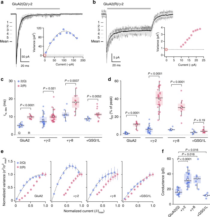
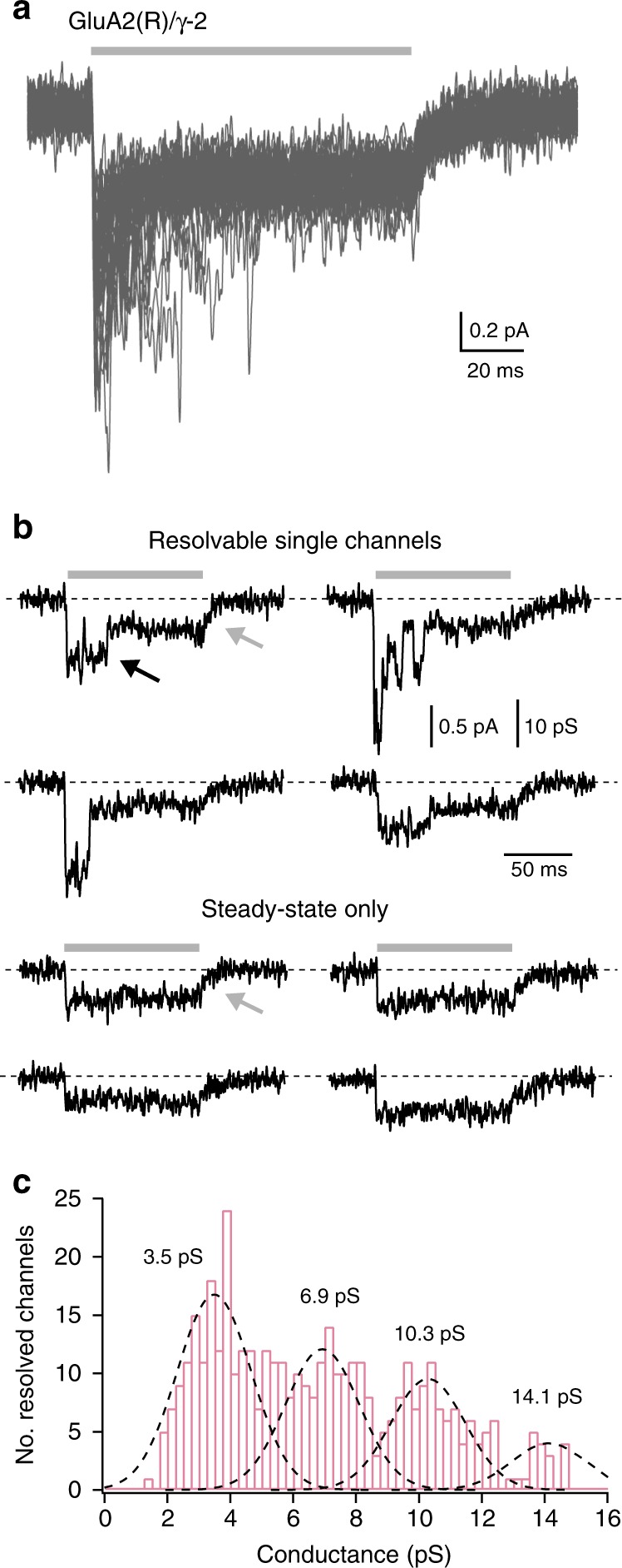
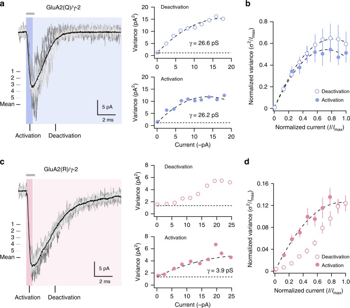
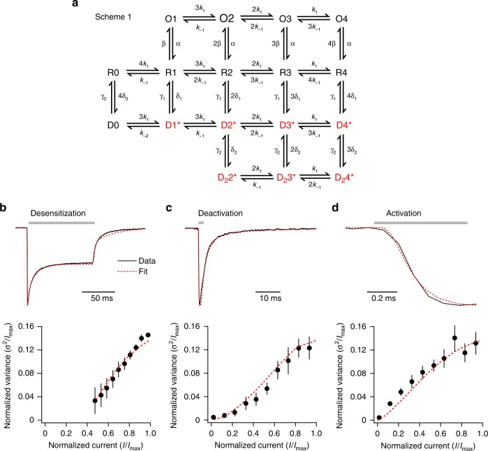
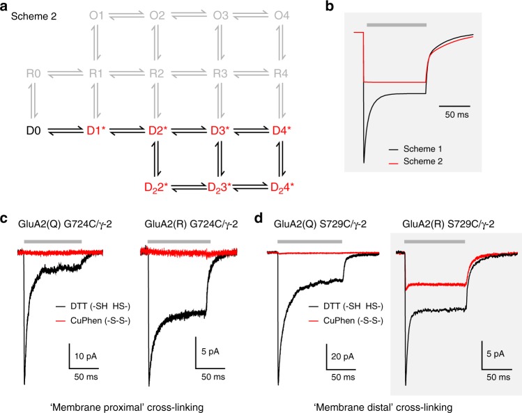
Desensitization is a canonical property of ligand-gated ion channels, causing progressive current decline in the continued presence of agonist. AMPA-type glutamate receptors (AMPARs), which mediate fast excitatory signaling throughout the brain, exhibit profound desensitization. Recent cryo-EM studies of AMPAR assemblies show their ion channels to be closed in the desensitized state. Here we present evidence that homomeric Q/R-edited AMPARs still allow ions to flow when the receptors are desensitized. GluA2(R) expressed alone, or with auxiliary subunits (γ-2, γ-8 or GSG1L), generates large fractional steady-state currents and anomalous current-variance relationships. Our results from fluctuation analysis, single-channel recording, and kinetic modeling, suggest that the steady-state current is mediated predominantly by conducting desensitized receptors. When combined with crystallography this unique functional readout of a hitherto silent state enabled us to examine cross-linked cysteine mutants to probe the conformation of the desensitized ligand binding domain of functioning AMPAR complexes.


Conflict of interest statement

The authors declare no competing interests.

Figures

Fig. 1
Fig. 1
Q/R editing affects the kinetics and variance of GluA2 currents. a Representative outside-out patch response (10 mM glutamate, 100 ms, –60 mV; gray bar) from a HEK293 cell transfected with GluA2(Q)/γ-2 (average current, black; five individual responses, grays). Inset: current–variance relationship (dotted line indicates background variance and red circle indicates expected origin). b As a, but for GluA2(R)/γ-2. Note that the data cannot be fitted with a parabolic relationship passing through the origin. c Pooled τw,des data for GluA2 alone (n= 12 Q-form and 9 R-form), GluA2/γ-2 (n= 21 and 27), GluA2/γ-8 (n= 7 and 10), and GluA2/GSG1L (n= 6 and 13). Box-and-whisker plots indicate the median (black line), the 25–75th percentiles (box), and the 10–90th percentiles (whiskers); filled circles are data from individual patches and open circles indicate means. Two-way ANOVA revealed an effect of Q/R editing (F1,97 = 111.34, P < 0.0001), an effect of auxiliary subunit type (F3,97 = 32.3, P < 0.0001) and an interaction (F3,97 = 2.84, P = 0.041). d Pooled data for Iss. Box-and-whisker plots and n numbers as in c. Two-way ANOVA indicated an effect of Q/R editing (F1,97 = 129.98, P < 0.0001), an effect of auxiliary subunit type (F3,97 = 58.30, P < 0.0001), and an interaction (F3,97 = 58.67, P < 0.0001). e Doubly normalized and averaged current–variance relationships (desensitizing current phase only) from GluA2(Q) and GluA2(R) expressed alone (n = 12 and 9), with γ-2 (n = 19 and 23), with γ-8 (n = 7 and 10), or with GSG1L (n = 6 and 13). Error bars are s.e.m.s. All Q-forms can be fitted with parabolic relationships passing through the origin, while R-forms cannot. f Pooled NSFA conductance estimates for GluA2(Q) alone, GluA2(Q)/γ-2, GluA2(Q)/γ-8, and GluA2(Q)/GSG1L (n = 12, 18, 7, and 6, respectively). Box-and-whisker plots as in c. Indicated P values are from Wilcoxon rank sum tests. Source data are provided as a Source Data file
Fig. 2
Fig. 2
GluA2(R)/γ-2 single-channel recordings. a GluA2(R)/γ-2 currents from an outside-out patch containing few channels (–60 mV). Forty consecutive applications of 10 mM glutamate (gray bar) are overlaid. b Individual responses exhibiting discrete channel openings superimposed on a persistent steady-state current (upper sweeps) or exhibiting only a persistent steady-state component (lower sweeps). Note the decay of the steady-state currents on glutamate removal (gray arrows) is much slower than the closure of resolved channels (black arrow). c Histogram of channel conductance for 392 discernible openings from six patches. The histogram is fit with the sum of four Gaussian curves (dashed lines) with a common standard deviation (1.2 pS), revealing four peaks (3.5, 6.9, 10.3, and 14.1 pS)
Fig. 3
Fig. 3
Estimated weighted mean conductance from NSFA of GluA2(R)/γ-2 activation. a Representative GluA2(Q)/γ-2 responses to 1 ms (gray bar) glutamate application (gray traces) with superimposed average (black trace). NSFA was performed on the activation phase (blue highlight; filled blue circles) and deactivation phase (light blue highlight; open blue circles) of the same records, yielding similar estimates of weighted mean conductance. b Pooled current–variance plots for the activation and deactivation of GluA2(Q)/γ-2 currents (n = 5). Error bars indicate s.e.m. c Representative GluA2(R)/γ-2 response to 1 ms glutamate application (as in a). NSFA was performed on the activation phase (pink highlight; filled pink circles) and deactivation phase (light pink highlight; open pink circles) of the same records. Current–variance relationship is non-parabolic for deactivation but parabolic for activation. d Pooled current–variance plots for the activation and deactivation of GluA2(R)/γ-2 currents (n = 8). Error bars indicate s.e.m.
Fig. 4
Fig. 4
Large steady-state GluA2(R)/γ-2 currents are observed even in conditions favoring desensitization. a Representative GluA2(R)/γ-2 current (–60 mV) evoked by 10 mM glutamate (gray bar) in the presence of 50 µM cyclothiazide (green bar). Note the minimal current decay when desensitization is inhibited (for pooled data ISS/Ipeak = 93.4 ± 1.6%, n = 6) (mean ± s.e.m. from n patches). b Representative glutamate-evoked currents from Q- and R-forms of GluA2 S754D/γ-2. Both forms exhibit very fast desensitization, but the R-form has an appreciable steady-state current. c Pooled data showing desensitization kinetics (τw,des) for wild-type (wt; n = 6 and 5) and mutant (S754D; n = 5 and 5) forms of GluA2(Q)/γ-2 and GluA2(R)/γ-2. Box-and-whisker plots as in Fig. 1c. Two-way ANOVA indicated an effect of Q/R editing (F1, 17 = 10.56, P= 0.0047), an effect of the mutation (F1, 17 = 43.19, P< 0.0001) but no interaction (F1, 17 = 2.63, P= 0.12). The mean difference between S754D and wild type was –8.1 ms (95% confidence interval, –10.9 to –6.0) for Q and –14.1 ms (95% confidence interval, –20.0 to –9.3) for R. d Pooled data (as in c) for recovery kinetics (τw,recov). Two-way ANOVA indicated no effect of Q/R editing (F1, 17 = 0.13, P= 0.72), an effect of the mutation (F1, 17 = 31.67, P< 0.0001) but no interaction (F1, 17 = 1.65, P= 0.22). The mean difference between S754D and wild type was 24.1 ms (95% confidence interval, 15.3 to 33.2) for Q and 38.7 ms (95% confidence interval, 23.7 to 53.9) for R. e Pooled data (as in c) for the fractional steady-state current (ISS). Two-way ANOVA indicated an effect of Q/R editing (F1, 17 = 65.37, P< 0.0001), an effect of the mutation (F1, 17 = 28.37, P< 0.0001), and an interaction (F1, 17 = 14.93, P= 0.0012). The mean difference in ISS (% of peak) between S754D and wild type was –7.7 (95% confidence interval, –11.2 to –5.2) for Q and –37.9 (95% confidence interval, –49.2 to –25.8) for R. Indicated P values are from Welch t-tests. Source data are provided as a Source Data file
Fig. 5
Fig. 5
A kinetic scheme including conducting desensitized states can mimic GluA2(R)/γ-2 behavior. a Scheme 1 is a modified form of a previously proposed kinetic model,. States which can conduct are red. Open states (O1–O4) and occupied desensitized states (D1*–D4*, D22*–D24*) have independent conductances that are occupancy-dependent. bd Global averaged GluA2(R)/γ-2 records (top) and variance data (bottom) for desensitization, deactivation, and activation (10 mM glutamate—gray bars). Using a single set of rate constants and conductances, the model closely mimics all six measures (dashed red lines): k1 = 1.3 ×  106 M–1 s–1, k−1 = 350 s–1, α = 3100 s–1, β = 1000 s–1, γ1 = 88 s–1, δ1 = 110 s–1, γ2 = 36 s–1, δ2 = 39 s–1, γ0 = 8 s–1, δ0 = 0.48 s–1, k−2 = 870 s–1, conductance of fully occupied open state (O4) = 3.9 pS, conductance of fully occupied desensitized state (D4* and D24*) = 670 fS. Conductances of partially occupied states were proportional to their occupancy (e.g. O3 = 0.75 × O4, O2 = 0.5 × O4, O1 = 0.25 × O4)
Fig. 6
Fig. 6
A model with access to only desensitized states predicts behavior of cross-linked GluA2(R) S729C/γ-2. a Scheme 2 is modified from Scheme 1 (Fig. 5a) and assumes that, following cross-linking, the receptor can occupy only desensitized states (excluded states are shown in gray). b Simulated responses to 10 mM glutamate (gray bar) using Scheme 1 to mimic the non-cross-linked condition and Scheme 2 to mimic the effect of cross-linking. c Representative currents at –60 mV activated by 10 mM glutamate (gray bar) from GluA2(Q) G724C/γ-2 and GluA2(R) G724C/γ-2 in DTT (black) or CuPhen (red). Note that, for both forms, currents are fully inhibited following cross-linking by 10 µM CuPhen. d Representative responses from individual patches demonstrate that following cross-linking by 10 µM CuPhen, GluA2(Q) S729C/γ-2 currents are inhibited, while GluA2(R) S729C/γ-2 currents show minimal desensitization and continue to display a large steady-state current, as predicted in a. Gray boxes (b and d) highlight the similarity of modeled currents and recorded GluA2(R) S729C/γ-2 currents
Fig. 7
Fig. 7
Cross-linked S729C LBD structures suggest a model of gating for desensitized GluA2(R). a Left, crystal structure of the dimeric GluA2 S729C ligand-binding core in the presence of NBQX, with the upper (D1, pale) and lower (D2, dark) lobes of each monomer (red and blue) distinguished by shading. The structure is viewed perpendicular to the axis between the Cα atoms of Pro632 (magenta spheres). Right, crystal structure of the GluA2 S729C ligand-binding core in the presence of glutamate (S729Cglu; PDB: 2I3W). The Pro632 separation seen in the presence of glutamate (right) is >3 Å greater than that seen with NBQX (left). b Cartoon representing possible conformations of the GluA2(R) LBD dimer and pore in our functional cross-linking recordings. Non-cross-linked GluA2(R) channels bind glutamate (gray spheres), closing the clamshell LBDs and opening the pore to the full open channel conductance. Desensitization does not fully close the pore. As determined in the presence of γ-2, cross-linking of the G724C mutant (yellow) does not allow the action of agonist binding to be communicated to the pore in any state, and disrupts the normal dimeric conformation of desensitized receptors. Cross-linking of the S729C mutant (yellow) is not compatible with the full open state, but the channel can adopt the normal desensitized conformation, meaning that (as with the non-cross-linked receptor) the pore is not closed

References

    1. Henley JM, Wilkinson KA. Synaptic AMPA receptor composition in development, plasticity and disease. Nat. Rev. Neurosci. 2016;17:337–350. doi: 10.1038/nrn.2016.37. - DOI - PubMed
    1. Huganir RL, Nicoll RA. AMPARs and synaptic plasticity: the last 25 years. Neuron. 2013;80:704–717. doi: 10.1016/j.neuron.2013.10.025. - DOI - PMC - PubMed
    1. Cathala L, Holderith NB, Nusser Z, DiGregorio DA, Cull-Candy SG. Changes in synaptic structure underlie the developmental speeding of AMPA receptor-mediated EPSCs. Nat. Neurosci. 2005;8:1310–1318. doi: 10.1038/nn1534. - DOI - PubMed
    1. Colquhoun D, Jonas P, Sakmann B. Action of brief pulses of glutamate on AMPA/kainate receptors in patches from different neurones of rat hippocampal slices. J. Physiol. 1992;458:261–287. doi: 10.1113/jphysiol.1992.sp019417. - DOI - PMC - PubMed
    1. Hestrin S. Activation and desensitization of glutamate-activated channels mediating fast excitatory synaptic currents in the visual cortex. Neuron. 1992;9:991–999. doi: 10.1016/0896-6273(92)90250-H. - DOI - PubMed

Publication types