

Video-rate multi-color structured illumination microscopy with simultaneous real-time reconstruction
- PMID: 31541134
- PMCID: PMC6754501
- DOI: 10.1038/s41467-019-12165-x
Video-rate multi-color structured illumination microscopy with simultaneous real-time reconstruction
Abstract
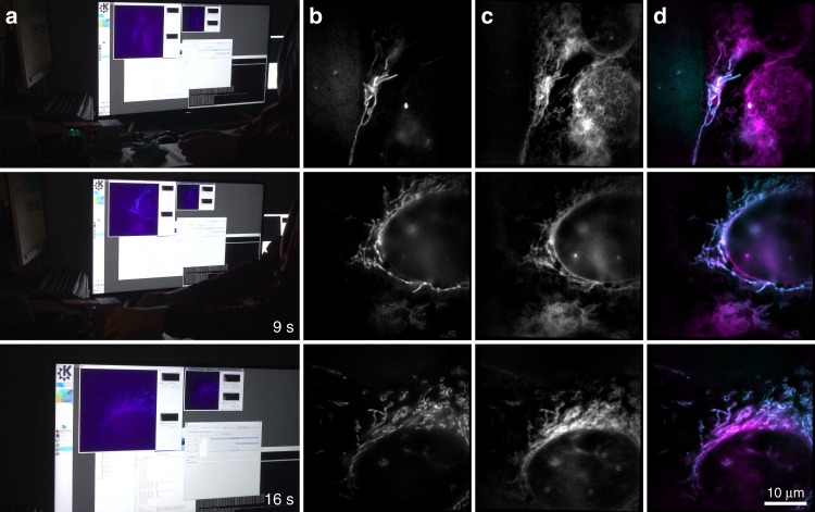
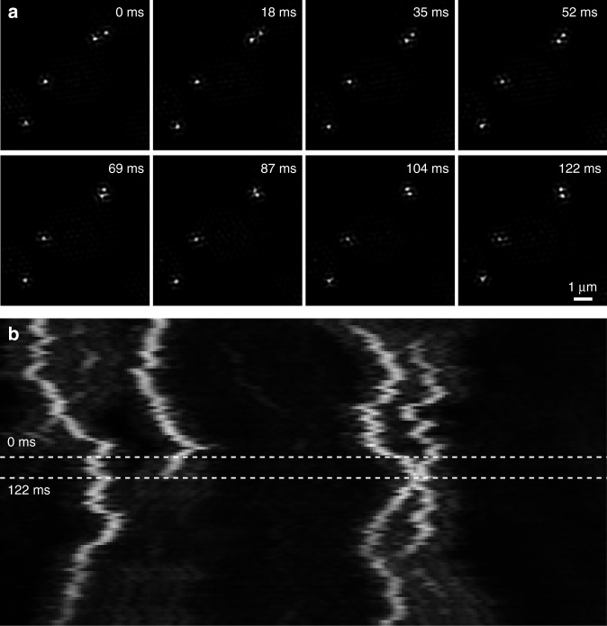
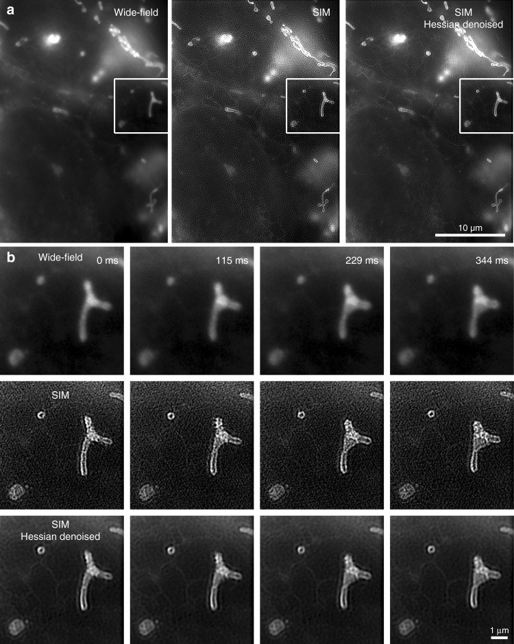
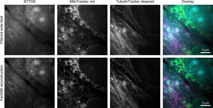
Super-resolved structured illumination microscopy (SR-SIM) is among the fastest fluorescence microscopy techniques capable of surpassing the optical diffraction limit. Current custom-build instruments are able to deliver two-fold resolution enhancement with high acquisition speed. SR-SIM is usually a two-step process, with raw-data acquisition and subsequent, time-consuming post-processing for image reconstruction. In contrast, wide-field and (multi-spot) confocal techniques produce high-resolution images instantly. Such immediacy is also possible with SR-SIM, by tight integration of a video-rate capable SIM with fast reconstruction software. Here we present instant SR-SIM by VIGOR (Video-rate Immediate GPU-accelerated Open-Source Reconstruction). We demonstrate multi-color SR-SIM at video frame-rates, with less than 250 ms delay between measurement and reconstructed image display. This is achieved by modifying and extending high-speed SR-SIM image acquisition with a new, GPU-enhanced, network-enabled image-reconstruction software. We demonstrate high-speed surveying of biological samples in multiple colors and live imaging of moving mitochondria as an example of intracellular dynamics.
Conflict of interest statement
The authors declare no competing interests.
Figures


Similar articles
-
Live, video-rate super-resolution microscopy using structured illumination and rapid GPU-based parallel processing.Microsc Microanal. 2011 Apr;17(2):191-6. doi: 10.1017/S1431927611000158. Epub 2011 Mar 9. Microsc Microanal. 2011. PMID: 21385522
-
High-speed TIRF and 2D super-resolution structured illumination microscopy with a large field of view based on fiber optic components.Opt Express. 2023 Aug 28;31(18):29156-29165. doi: 10.1364/OE.495353. Opt Express. 2023. PMID: 37710721
-
Imaging tissues and cells beyond the diffraction limit with structured illumination microscopy and Bayesian image reconstruction.Gigascience. 2019 Jan 1;8(1):giy126. doi: 10.1093/gigascience/giy126. Gigascience. 2019. PMID: 30351383 Free PMC article.
-
Recent advancements in structured-illumination microscopy toward live-cell imaging.Microscopy (Oxf). 2015 Aug;64(4):237-49. doi: 10.1093/jmicro/dfv034. Epub 2015 Jun 30. Microscopy (Oxf). 2015. PMID: 26133185 Review.
-
Faster, sharper, and deeper: structured illumination microscopy for biological imaging.Nat Methods. 2018 Dec;15(12):1011-1019. doi: 10.1038/s41592-018-0211-z. Epub 2018 Nov 26. Nat Methods. 2018. PMID: 30478322 Review.
Cited by
-
Structured illumination microscopy with noise-controlled image reconstructions.Nat Methods. 2021 Jul;18(7):821-828. doi: 10.1038/s41592-021-01167-7. Epub 2021 Jun 14. Nat Methods. 2021. PMID: 34127855 Free PMC article.
-
In vivo super-resolution of the brain - How to visualize the hidden nanoplasticity?iScience. 2022 Aug 17;25(9):104961. doi: 10.1016/j.isci.2022.104961. eCollection 2022 Sep 16. iScience. 2022. PMID: 36093060 Free PMC article. Review.
-
High-speed multicolor structured illumination microscopy using a hexagonal single mode fiber array.Biomed Opt Express. 2021 Feb 1;12(2):1181-1194. doi: 10.1364/BOE.416546. eCollection 2021 Feb 1. Biomed Opt Express. 2021. PMID: 33680566 Free PMC article.
-
Fast volumetric multifocus structured illumination microscopy of subcellular dynamics in living cells.Biomed Opt Express. 2024 Mar 11;15(4):2281-2292. doi: 10.1364/BOE.516261. eCollection 2024 Apr 1. Biomed Opt Express. 2024. PMID: 38633103 Free PMC article.
-
Multicolor structured illumination microscopy and quantitative control of polychromatic light with a digital micromirror device.Biomed Opt Express. 2021 Jun 1;12(6):3700-3716. doi: 10.1364/BOE.422703. eCollection 2021 Jun 1. Biomed Opt Express. 2021. PMID: 34221689 Free PMC article.
References
-
- Heintzmann R, Cremer CG. Laterally modulated excitation microscopy: improvement of resolution by using a diffraction grating. Proc. SPIE. 1999;3568:185–196. doi: 10.1117/12.336833. - DOI
Publication types
MeSH terms
LinkOut - more resources
Full Text Sources
Research Materials
