Comparative Study
. 2019 Sep 20;9(1):13643.
doi: 10.1038/s41598-019-50104-4.

Comparison of optical coherence tomography and high frequency ultrasound imaging in mice for the assessment of skin morphology and intradermal volumes

Affiliations
Comparative Study

Comparison of optical coherence tomography and high frequency ultrasound imaging in mice for the assessment of skin morphology and intradermal volumes

Kornelia Schuetzenberger et al. Sci Rep. .

Abstract

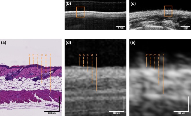
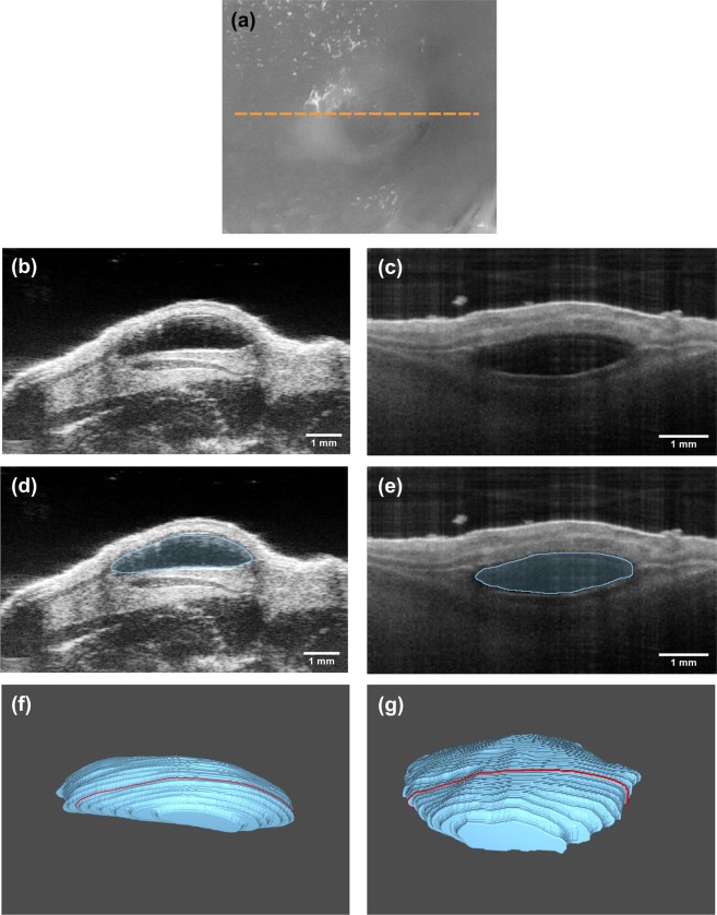
Optical coherence tomography (OCT) and high-frequency ultrasound (HFUS), two established imaging modalities in the field of dermatology, were evaluated and compared regarding their applicability for visualization of skin tissue morphology and quantification of murine intradermal structures. The accuracy and reproducibility of both methods were assessed ex vivo and in vivo using a standardized model for intradermal volumes based on injected soft tissue fillers. OCT revealed greater detail in skin morphology, allowing for detection of single layers due to the superior resolution. Volumetric data measured by OCT (7.9 ± 0.3 μl) and HFUS (7.7 ± 0.5 μl) were in good agreement and revealed a high accuracy when compared to the injected volume of 7.98 ± 0.8 µl. In vivo, OCT provided a higher precision (relative SD: 26% OCT vs. 42% HFUS) for the quantification of intradermal structures, whereas HFUS offered increased penetration depth enabling the visualization of deeper structures. A combination of both imaging technologies might be valuable for tumor assessments or other dermal pathologies in clinical settings.


Conflict of interest statement

The authors declare no competing interests.

Figures

Figure 1
Figure 1
Exemplary cross-sectional images of murine skin obtained by (b) OCT and (c) HFUS and corresponding (a) histology (H&E staining). Enlarged cross-sections as indicated by the orange rectangles are depicted in (d,e). Dashed orange lines in image (a,d,e) indicate the skin layers corresponding to: (a) epidermis, (b) papillary dermis, (c) reticular dermis, (d) subcutis, (e) sebaceous glands and hair follicles and (f) muscle. Skin layers with letters labeled with an asterisk are not distinguishable.
Figure 2
Figure 2
In vivo imaging of the murine soft tissue filler model, showing the same filler deposit imaged with OCT and HFUS. (a) Photograph of the dorsum of a mouse containing the soft tissue filler. The yellow line indicates the position of the cross-sectional image obtained by (b) HFUS and (c) OCT. The images of (d) HFUS and (e) OCT show examples of results of the automatic segmentation algorithm used for calculation of the volumes. Images (f,g) show 3D representation of intradermal deposit volumes as obtained from HFUS and OCT scanning data, respectively.
Figure 3
Figure 3
In vivo volumetric assessment of soft tissue fillers. (a) Bland-Altman plot, (b) box plot and (c) histogram comparing data obtained with OCT and HFUS.

References

    1. Olsen J, Holmes J, Jemec GB. Advances in optical coherence tomography in dermatology-a review. J. Biomed. Opt. 2018;23:1–10. doi: 10.1117/1.Jbo.23.4.040901. - DOI - PubMed
    1. Shahriari N, Grant-Kels JM, Rabinovitz H, Oliviero M, Scope A. In vivo reflectance confocal microscopy image interpretation for the dermatopathologist. J. Cutan. Pathol. 2018;45:187–197. doi: 10.1111/cup.13084. - DOI - PubMed
    1. Wollina U, Goldman A. Fillers for the improvement in acne scars. Clin. Cosmet. Investig. Dermatol. 2015;8:493–499. doi: 10.2147/ccid.S86478. - DOI - PMC - PubMed
    1. Jagdeo J, Ho D, Lo A, Carruthers A. A systematic review of filler agents for aesthetic treatment of HIV facial lipoatrophy (FLA) J. Am. Acad. Dermatol. 2015;73:1040–1054.e1014. doi: 10.1016/j.jaad.2015.08.040. - DOI - PubMed
    1. Koehler MJ, Lange-Asschenfeldt S, Kaatz M. Non-invasive imaging techniques in the diagnosis of skin diseases. Expert Opin. Med. Diagn. 2011;5:425–440. doi: 10.1517/17530059.2011.599064. - DOI - PubMed

Publication types