Review
. 2019 Oct;18(10):749-769.
doi: 10.1038/s41573-019-0041-4. Epub 2019 Sep 20.

Next-generation regulatory T cell therapy

Affiliations
Review

Next-generation regulatory T cell therapy

Leonardo M R Ferreira et al. Nat Rev Drug Discov. 2019 Oct.

Abstract

Regulatory T cells (Treg cells) are a small subset of immune cells that are dedicated to curbing excessive immune activation and maintaining immune homeostasis. Accordingly, deficiencies in Treg cell development or function result in uncontrolled immune responses and tissue destruction and can lead to inflammatory disorders such as graft-versus-host disease, transplant rejection and autoimmune diseases. As Treg cells deploy more than a dozen molecular mechanisms to suppress immune responses, they have potential as multifaceted adaptable smart therapeutics for treating inflammatory disorders. Indeed, early-phase clinical trials of Treg cell therapy have shown feasibility, tolerability and potential efficacy in these disease settings. In the meantime, progress in the development of chimeric antigen receptors and in genome editing (including the application of CRISPR-Cas9) over the past two decades has facilitated the genetic optimization of primary T cell therapy for cancer. These technologies are now being used to enhance the specificity and functionality of Treg cells. In this Review, we describe the key advances and prospects in designing and implementing Treg cell-based therapy in autoimmunity and transplantation.
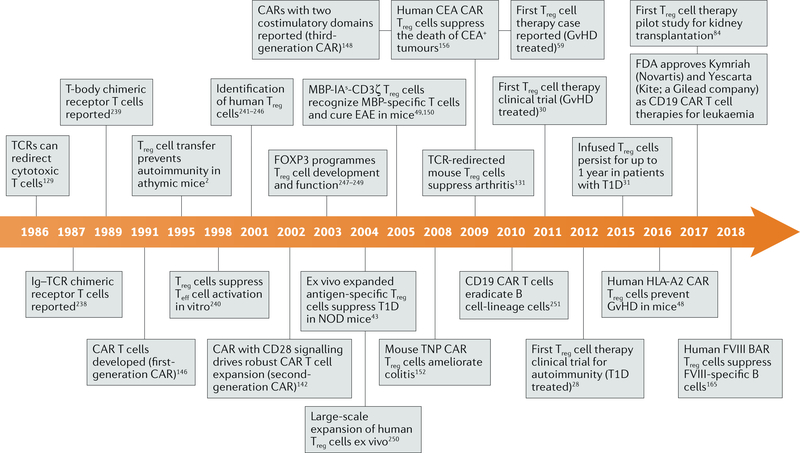

Figures

Fig. 1 |
Fig. 1 |. Registered clinical trials using regulatory T cells.
The number and status of registered clinical trials based on regulatory T cell (Treg cell) infusion were searched for in ClinicalTrials.gov and are summarized here (the numbers represent the data available in July 2019). The large circle shows the overall number of registered trials, divided into coloured segments that represent the proportion of these clinical trials with their status defined as not yet recruiting (blue), recruiting (green), active not recruiting (yellow), terminated or completed (pink), withdrawn or suspended (light grey) and unknown (dark grey). Unknown status corresponds to studies, the last known status of which was recruiting, not yet recruiting, or active, not recruiting, but that have passed their completion date but have not had their status verified within the past 2 years (http://clinicaltrials.gov). Small circles are categorized by indication: hematopoietic stem cell transplantation or graft-versus-host disease, solid organ transplantation and autoimmune disease.
Fig. 2 |
Fig. 2 |. Regulatory T cells as living drugs.
Four key properties are needed to successfully use regulatory T cells (Treg cells) as living drugs, summed up as the four S’s: suppression, specificity, stability and survival. In terms of suppression, Treg cells act through multiple suppression mechanisms (including IL-2 deprivation from the milieu, secretion of inhibitory cytokines and interactions with antigen-presenting cells (APCs)), which could be tailored to specific conditions or diseases by, for example, forcing the expression of specific transcription factors. Regarding specificity, it is possible to create Treg cells with a desired specificity using T cell receptor (TCR) gene transfer or artificial immune receptors, such as chimeric antigen receptors (CARs). Specificity can be made conditional by using synthetic Notch (SynNotch) receptors. Overexpressing transcription factors characteristic of T helper cell subsets can also enhance the specificity of Treg cells. With respect to stability, forkhead box protein P3 (FOXP3) expression is central to the lineage of Treg cells. Strategies to increase Treg cell stability include the ectopic expression of the transcription factors FOXP3, HELIOS and BACH2 or of a constitutively active form of signal transducer and activator of transcription 5 (STAT5-CA), as well as the ablation of carboxy terminus of Hsp70-interacting protein (CHIP), deleted in breast cancer gene 1 protein (DBC1) or protein kinase C-θ (PKCθ) to prevent the degradation of FOXP3. Finally, the survival of Treg cells depends on exogenous IL-2, metabolic requirements and tonic signalling mediated by the TCR and costimulatory molecules. Targeting Treg cell metabolic requirements or manipulating the phosphatidylinositol 3-OH kinase (PI3K)–AKT or JUN amino-terminal kinase 1 (JNK1) signalling pathways may increase Treg cell survival after infusion. MHC, major histocompatibility complex; RE, regulatory element.
Fig. 3 |
Fig. 3 |. Timeline of events in the development of regulatory T cell therapy.
A timeline of key developments leading to the use of regulatory T cell (Treg cell) therapy in the clinic. BAR, B cell-targeting antibody receptor; CAR, chimeric antigen receptor; CEA, carcinoembryonic antigen; EAE, experimental autoimmune encephalomyelitis; FOXP3, forkhead box protein P3; FVIII, factor VIIII; GvHD, graft-versus-host disease; HLA, human leukocyte antigen; IA, murine major histocompatibility complex class II molecule I-A; Ig, immunoglobulin; MBP, myelin basic protein; NOD, non-obese diabetic; T1D, type 1 diabetes; TCR, T cell receptor; Teff cell, effector T cell; TNP, 2,4,6-trinitrophenyl.
Fig. 4 |
Fig. 4 |. Redirecting regulatory T cells by engineering T cell receptors.
A tissue biopsy sample is taken from a patient with an autoimmune disease (step 1) and subjected to single-cell paired T cell receptor (TCR) sequencing to characterize the TCR repertoire of the regulatory T cells (Treg cells) or, alternatively, of the autoimmune T cells (step 2). On the basis of algorithms identifying conserved motifs and complementarity-determining regions, pathogenic epitopes are predicted (step 3) and selected TCR ultramer templates are synthesized (step 4). Using a non-viral approach, the ultramer and a Cas9 ribonucleoprotein complex targeting the endogenous TCR via homology repair are electroporated into peripheral blood-derived Treg cells (step 5). Edited Treg cells are further expanded in vitro (step 6) and finally reinfused into the patient (step 7).
Fig. 5 |
Fig. 5 |. Methods for delivering genetic material to engineer regulatory T cells.
a | The first FDA-approved method for delivering genetic material to engineer regulatory T cells uses retroviruses or lentiviruses, which are pseudodiploid single-stranded RNA viruses. Genomic integration of the gene cassette of interest is random, resulting in non-physiological gene regulation with often multiple copies of the gene per cell. The second FDA-approved method uses recombinant adenoassociated viruses (AAVs); these viruses persist inside the cell as a double-stranded DNA episome that is not integrated into the genome and will therefore be diluted over time in replicating cells. Many groups combine use of recombinant AAVs with use of CRISPR-based technologies, as the episome can serve as a homology repair (HR) template, an approach recently approved by the FDA for clinical trials. b | A third method, which is purely non-viral but not FDA approved yet, electroporates the desired cassette as double-stranded DNA or an ultramer (single-stranded DNA) template together with a Cas9–ribonucleoprotein (RNP) complex. Although this method leads to a single copy of the gene being precisely integrated at a desired location in the genome, it also typically results in lower modification efficiencies.

References

    1. Gershon RK. & Kondo K. Cell interactions in the induction of tolerance: the role of thymic lymphocytes. Immunology 18, 723–737 (1970). - PMC - PubMed
    1. Sakaguchi S, Sakaguchi N, Asano M, Itoh M & Toda M Immunologic self-tolerance maintained by activated T cells expressing IL-2 receptor alpha-chains (CD25). Breakdown of a single mechanism of self-tolerance causes various autoimmune diseases. J. Immunol. 155, 1151–1164 (1995). - PubMed
    1. Husebye ES, Anderson MS & Kampe O Autoimmune polyendocrine syndromes. N. Engl. J. Med. 378, 1132–1141 (2018). - PMC - PubMed
    1. Komatsu N et al. Pathogenic conversion of Foxp3+ T cells into TH17 cells in autoimmune arthritis. Nat. Med. 20, 62–68 (2014). - PubMed
    1. Zhou X et al. Instability of the transcription factor Foxp3 leads to the generation of pathogenic memory T cells in vivo. Nat. Immunol. 10, 1000–1007 (2009).

      This study reveals that Treg cells can become unstable, losing FOXP3 expression and converting into pathogenic T cells (‘ex-Treg cells’) in vivo.

Publication types

MeSH terms