. 2020 Mar;98(3):557-570.
doi: 10.1002/jnr.24535. Epub 2019 Sep 20.

Greater neurodegeneration and behavioral deficits after single closed head traumatic brain injury in adolescent versus adult male mice

Affiliations

Greater neurodegeneration and behavioral deficits after single closed head traumatic brain injury in adolescent versus adult male mice

Fernanda Guilhaume-Correa et al. J Neurosci Res. 2020 Mar.

Abstract

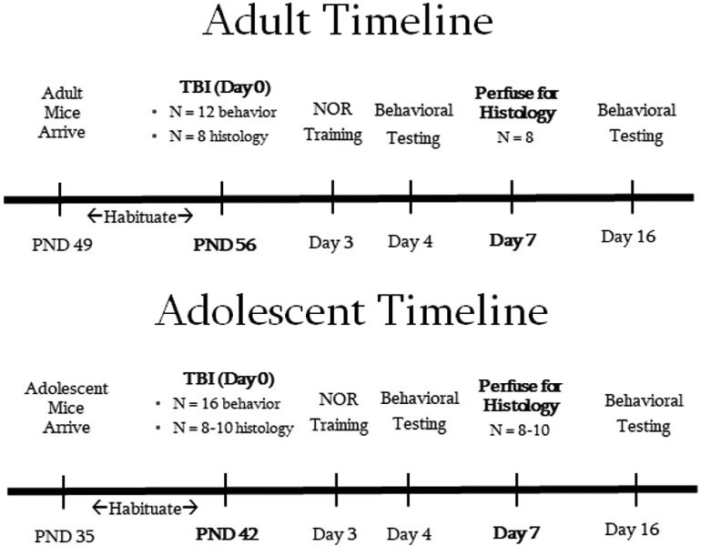
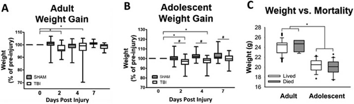
Traumatic brain injury (TBI) is a major public health concern affecting 2.8 million people per year in the United States, of whom about 1 million are children under 19 years old. Animal models of TBI have been developed and used in multiple ages of animals, but direct comparisons of adult and adolescent populations are rare. The current studies were undertaken to directly compare outcomes between adult and adolescent male mice, using a closed head, single-impact model of TBI. Six-week-old adolescent and 9-week-old adult male mice were subjected to mild-moderate TBI. Histological measures for neurodegeneration, gliosis, and microglial neuroinflammation, and behavioral tests of locomotion and memory were performed. Adolescent TBI mice have increased mortality (Χ2 = 20.72, p < 0.001) compared to adults. There is also evidence of hippocampal neurodegeneration in adolescents that is not present in adults. Hippocampal neurodegeneration correlates with histologic activation of microglia, but not with increased astrogliosis. Adults and adolescents have similar locomotion deficits after TBI that recover by 16 days postinjury. Adolescents have memory deficits as evidenced by impaired novel object recognition between 3-4 and 4-16 days postinjury (F1,26 = 5.23, p = 0.031) while adults do not. In conclusion, adults and adolescents within a close age range (6-9 weeks) respond to TBI differently. Adolescents are more severely affected by mortality, neurodegeneration, and inflammation in the hippocampus compared to adults. Adolescents, but not adults, have worse memory performance after TBI that lasts at least 16 days postinjury.

Keywords: RRID:AB_10013382; RRID:AB_10641962; RRID:AB_2307443; adolescent; memory; neurodegeneration; neuroinflammation; traumatic brain injury.


Conflict of interest statement

Conflict of Interest Statement

No competing financial interests exist.

Figures

Figure 1.
Figure 1.. Experimental timeline.
The timeline of the experimental procedures is summarized. There were four cohorts of animals (adult or adolescent for both histology and behavior). After arrival in the vivarium, mice were allowed to habituate to the animal facility for one week prior to beginning experimental procedures. TBI was performed on Day 0, and histology cohorts were sacrificed at postinjury day 7 (n = 8-10 per group). Behavioral testing was performed on the behavior cohorts through post-injury day 16 (n = 12-16 per group).
Figure 2.
Figure 2.. Weight gain and Mortality.
The graph illustrates weight gain and mortality in animals pooled from several experiments. By post-injury day 2, both (A) adult and (B) adolescent TBI animals lost weight compared to pre-injury, and were significantly below pre-injury weight through post-injury day 4. In contrast to adults, adolescent TBI animals were lighter than sham animals on days 2, 4, and 7. (C) Post-TBI mortality was much higher in adolescent mice than what we have observed using adult mice (described in the results), but there was not a significant difference in pre-injury weight between mice that died vs those that lived in adults or adolescents, though adults did weight significantly more than adolescents overall. (A and B) # p < 0.05 vs sham within time point, *weight lower than pre-injury within group. (C) * p < 0.05.
Figure 3.
Figure 3.. TBI causes somatic neurodegeneration in the adolescent hippocampus at 7 days post-mild/moderate TBI.
Histology using FJ-B staining was performed to evaluate neurodegeneration in the hippocampus. Representative photomicrographs, 10x and 20x magnification respectively, of adult sham (A-B) vs TBI (C-D) animals and adolescent sham (E-F) vs TBI (G-H) animals are shown. There is significant hippocampal somatic degeneration in adolescent TBI animals (I), particularly in and around the dentate gyrus, but not in shams or in adult TBI or sham mice. Background and luminescence of the FITC wavelength were matched to have the same background intensity and in order to identify structural features of the hippocampus. Scale bars represent 200 μm for 10x images and 100 μm for 20x images.
Figure 4.
Figure 4.. TBI is not associated with astrogliosis in adolescent or adult hippocampus.
There was no significant difference between TBI and sham in the hippocampus of adult (A) or adolescent (F) mice following a mild TBI in adults, and a mild-moderate TBI in adolescents. Representative photomicrographs of hippocampi in adult sham (B-C) and TBI (D-E) animals and adolescent sham (G-H) and TBI (I-J) animals at 10x and 20x magnifications. n.s. = not significant. Dotted lines indicate the granule cell layer of the dentate gyrus. Scale bars indicate 100 μm (50 μm for inset photos).
Figure 5.
Figure 5.. TBI per se does not lead to significant increase of reactive microglia in the hippocampus.
Tissues were stained for the microglial marker Iba-1, and expression in the hippocampus was examined in adult (A, C) and adolescent (B, D) mice. Representative photomicrographs from adult TBI (E) and sham (F) animals and from adolescent TBI (G) and sham (H) animals are shown at 5x and 20x magnifications. There was not a significant overall microglial activation in hippocampus of either age or condition group (as measured by increased soma perimeter and area). Scale bars indicate 100 μm (50 μm for the inset image).
Figure 6.
Figure 6.. There is increased microglial activation but not astrogliosis in hippocampus of animals with neuronal degeneration.
(A) There was not a significant correlation between positive FJ-B staining and increased GFAP. There was a significant correlation between positive FJ-B staining and increased microglial soma area (B) and perimeter (C).
Figure 7.
Figure 7.. Locomotor and anxiety-like behavior.
Neither adult (A-D) nor adolescent (E-H) sham animals show significant differences in behavior along the two time points except in time spent in the center of the open field. Compared to sham animals, both adolescent and adult TBI animals increase their center time and center frequency from Day 4 to 16, while only adolescent TBI mice increase distance traveled and mean velocity from Day 4 to 16. * p < 0.05 between DPI 4 and 16 within TBI; ** p < 0.05 between DPI 4 and 16 within sham; # p < 0.05 vs. sham within the timepoint.
Figure 8.
Figure 8.. Adolescent, but not adult, TBI mice have deficits in novel object recognition.
Data are presented as ratio of time spent interacting with the novel object to total interaction time. A ratio of 0.5 represents equal time interacting with each (represented by the dotted lines in the figure). (A) Adult TBI mice do not show deficits at either time point. (B) Adolescent TBI mice show deficits in novel object recognition at both time points. * p < 0.05 vs control group within the same time point.

Similar articles

Cited by

References

    1. Adelson PD, Dixon CE, Robichaud P, & Kochanek PM (1997). Motor and cognitive functional deficits following diffuse traumatic brain injury in the immature rat. J Neurotrauma, 14(2), 99–108. doi:10.1089/neu.1997.14.99 - DOI - PubMed
    1. Ahmed Z, Shaw G, Sharma VP, Yang C, McGowan E, & Dickson DW (2007). Actin-binding proteins coronin-1a and IBA-1 are effective microglial markers for immunohistochemistry. J Histochem Cytochem, 55(7), 687–700. doi:10.1369/jhc.6A7156.2007 - DOI - PubMed
    1. Ajao DO, Pop V, Kamper JE, Adami A, Rudobeck E, Huang L, … Badaut J (2012). Traumatic brain injury in young rats leads to progressive behavioral deficits coincident with altered tissue properties in adulthood. J Neurotrauma, 29(11), 2060–2074. doi:10.1089/neu.2011.1883 - DOI - PMC - PubMed
    1. Baratz R, Tweedie D, Wang JY, Rubovitch V, Luo W, Hoffer BJ, … Pick CG (2015). Transiently lowering tumor necrosis factor-alpha synthesis ameliorates neuronal cell loss and cognitive impairments induced by minimal traumatic brain injury in mice. J Neuroinflammation, 12, 45. doi:10.1186/s12974-015-0237-4 - DOI - PMC - PubMed
    1. Bevins RA, & Besheer J (2006). Object recognition in rats and mice: a one-trial non-matching-to-sample learning task to study 'recognition memory'. Nat Protoc, 1(3), 1306–1311. doi:10.1038/nprot.2006.205 - DOI - PubMed

Publication types