. 2019 Sep 21;151(11):114903.
doi: 10.1063/1.5121305.

Extension distribution for DNA confined in a nanochannel near the Odijk regime

Affiliations

Extension distribution for DNA confined in a nanochannel near the Odijk regime

Hui-Min Chuang et al. J Chem Phys. .

Abstract

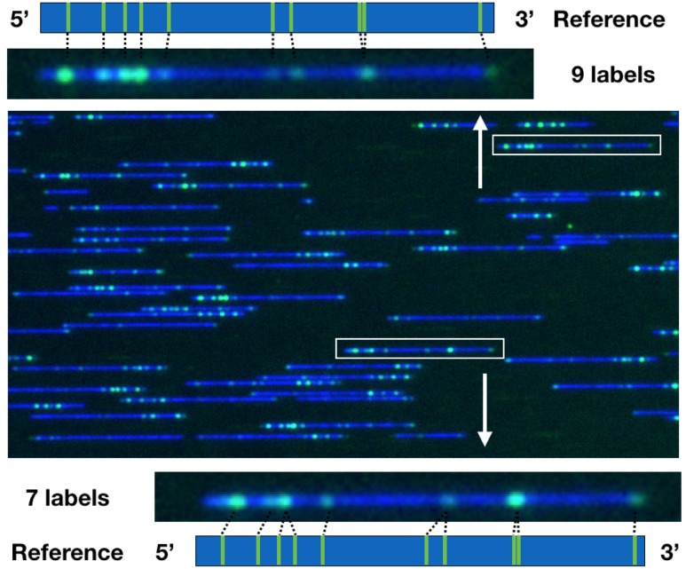
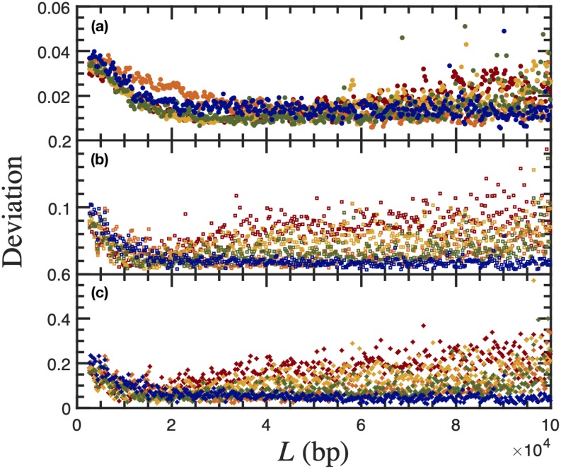
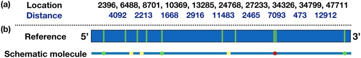
DNA confinement in a nanochannel typically is understood via mapping to the confinement of an equivalent neutral polymer by hard walls. This model has proven to be effective for confinement in relatively large channels where hairpin formation is frequent. An analysis of existing experimental data for Escherichia coli DNA extension in channels smaller than the persistence length, combined with an additional dataset for λ-DNA confined in a 34 nm wide channel, reveals a breakdown in this approach as the channel size approaches the Odijk regime of strong confinement. In particular, the predicted extension distribution obtained from the asymptotic solution to the weakly correlated telegraph model for a confined wormlike chain deviates significantly from the experimental distribution obtained for DNA confinement in the 34 nm channel, and the discrepancy cannot be resolved by treating the alignment fluctuations or the effective channel size as fitting parameters. We posit that the DNA-wall electrostatic interactions, which are sensible throughout a significant fraction of the channel cross section in the Odijk regime, are the source of the disagreement between theory and experiment. Dimensional analysis of the wormlike chain propagator in channel confinement reveals the importance of a dimensionless parameter, reflecting the magnitude of the DNA-wall electrostatic interactions relative to thermal energy, which has not been considered explicitly in the prevailing theories for DNA confinement in a nanochannel.


Figures

FIG. 1.
FIG. 1.
Comparison between the predictions of the telegraph model and experimental data in Ref. for the difference between the chain extension, X, relative to the average chain extension, ⟨X⟩ for L = 28 125 bp and two channel sizes: (a) D = 40 nm and (b) D = 51 nm. Reproduced with permission from A. B. Bhandari and K. D. Dorfman, Biomicrofluidics 13, 044110 (2019). Copyright 2019 AIP Publishing LLC.
FIG. 2.
FIG. 2.
Composite, false-color image of λ-DNA molecules with backbone (blue) and the Nt.BspQI nick sites (green) obtained in 34 nm nanochannels. The representative λ-DNA molecules (in the white rectangular boxes) have 9 (upper panel) and 7 (bottom panel) resolvable labels, respectively. The distances between the nearest pairs of labels are measured and compared to the reference. A detailed discussion on filtering data for contour length and the correlation coefficient between the image and the reference is provided in Sec. IV A.
FIG. 3.
FIG. 3.
Comparison between the predictions of the telegraph model in Eq. (4) and the experimental data of Reinhart et al. for (a) D = 40 nm, experiment; (b) D = 40 nm, theory; (c) D = 51 nm, experiment; and (d) D = 51 nm, theory. Panels (a) and (c) reproduced with permission from Reinhart et al., J. Chem. Phys. 142, 064902 (2015). Copyright 2015 AIP Publishing LLC.
FIG. 4.
FIG. 4.
The result of statistical tests for quantifying the degree of agreement between theory and experiments: (a) the rms error, (b) the Cramér-von Mises criterion, and (c) the Anderson-Darling criterion. Statistical data were obtained for 40 nm (red), 42 nm (orange), 43 nm (yellow), 49 nm (green), and 51 nm (blue) nanochannels using the probability distributions in Fig. 3 and Figs. S1–S3 of the supplementary material.
FIG. 5.
FIG. 5.
The result of statistical tests as a function of channel size by averaging the results in Fig. 4 from L = 20 kbp to L = 80 kbp for quantifying the degree of agreement between theory and experiments: the rms error (red), the Cramér-von Mises criterion (yellow), and the Anderson-Darling criterion (blue). The boxes span from 25% to 75% of the data, with the lines indicating the median value. For the comparison between theory and simulation, the dashed line alternating between red and yellow indicates the value of 0.02 which was obtained by the rms error and Cramér-von Mises criterion; the blue dashed line is at the value of 0.07 obtained by the Anderson-Darling criterion.
FIG. 6.
FIG. 6.
Scheme of λ-DNA reference with (a) the locations of all the nick sites counted from the 5′ end and the distance between every nearest pairs of nick sites and (b) a schematic molecule that illustrates the diffraction-limited spots. Due to the diffraction-limited optics, proximate nick sites can merge into a single label if the distance between pairs of nick sites is less than 1500–2500 bp. The pair of nick sites that always appear as a single label are labeled in red (the case of 8th and 9th nick sites); the nick sites that may be a single label due to fluctuations are labeled in yellow; all other resolvable nick sites are labeled in green.
FIG. 7.
FIG. 7.
Comparison between the predictions of telegraph model with σ0 = σOdijk (dotted lines) and the best fit for σ (solid lines) and the experimental data of λ-DNA (blue circles). The theoretical distributions were modeled at Deff = 36.5 nm (red), Deff = 34 nm (yellow), and Deff = 26.4 nm (green) nanochannels using the parameter values in Table II.
FIG. 8.
FIG. 8.
Plot of (a) the dimensionless wall interaction parameter β given by Eq. (15) and (b) the position z* at which the wall interaction potential βϕ(z) decays to 0.1kBT as a function of buffer ionic strength for channel sizes that are proximate to the Odijk regime. The dashed blue curve corresponds to D = lp, the solid black line corresponds to the ratio D = 0.62lp for a 34 nm channel and a 48 mM ionic strength, and the dashed-dotted gold line corresponds to D = 0.1lp. The black circles are the results of the analysis for the experiments of Sec. IV. Note that the ratio z*/D is the DNA-wall electrostatic interaction length due to a single wall relative to the entire channel size.

Similar articles

Cited by

References

    1. Daoud M. and de Gennes P. G., J. Phys. 38, 85 (1977).10.1051/jphys:0197700380108500 - DOI
    1. Odijk T., Macromolecules 16, 1340 (1983).10.1021/ma00242a015 - DOI
    1. Yang Y., Burkhardt T. W., and Gompper G., Phys. Rev. E 76, 011804 (2007).10.1103/physreve.76.011804 - DOI - PubMed
    1. Odijk T., Phys. Rev. E 77, 060901(R) (2008).10.1103/physreve.77.060901 - DOI - PubMed
    1. Burkhardt T. W., Yang Y., and Gompper G., Phys. Rev. E 82, 041801 (2010).10.1103/physreve.82.041801 - DOI - PubMed