

Neuronal Autophagy in Synaptic Functions and Psychiatric Disorders
- PMID: 31542152
- PMCID: PMC6986983
- DOI: 10.1016/j.biopsych.2019.07.018
Neuronal Autophagy in Synaptic Functions and Psychiatric Disorders
Abstract
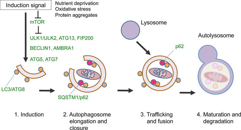
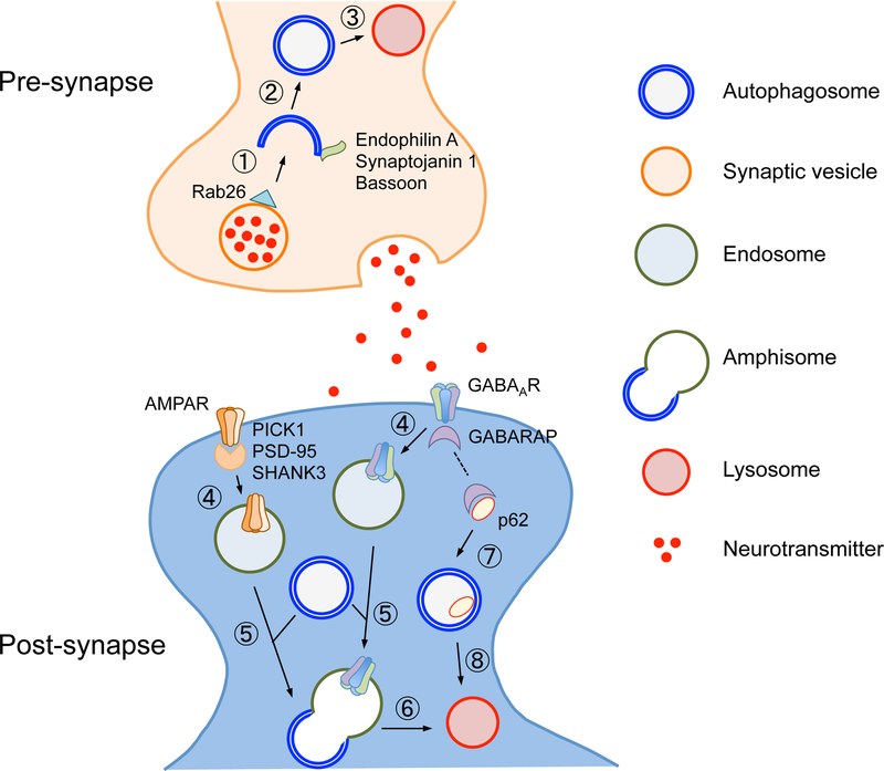
Homeostatic maintenance of physiological functions is fundamental to organismal well-being. Disruption or imbalance in homeostasis results in functional disturbances at molecular, cellular, and tissue levels, leading to manifestation as physical and mental illnesses. Homeostatic imbalance is caused by a range of pathophysiological mechanisms, including disrupted reduction-oxidation reactions, inflammatory responses, metabolic disturbances, or failure in quality control of cellular proteins and organelles. However, the roles for the protein/organelle quality control in the regulation of behaviors, in particular of cognitive processes, had not been well documented, until recent reports finally supported this concept. The frontline studies in neuroscience have revealed that synaptic components (e.g., synaptic proteins, organelles, neurotransmitters and their receptors) are selectively degraded by autophagy, a cellular recycling machinery implicated in surveillance and quality control of proteins and organelles responsible for the maintenance of cellular homeostasis. Apart from the canonical role of autophagy in supporting cell viability, synaptic autophagy appears to regulate synapse remodeling and plasticity. Consistently, emerging evidence suggests novel roles of autophagy in memory encoding, information processing, or cognitive functions. In this review, we overview recent progress in understanding the roles of neuronal autophagy in homeostatic maintenance of synaptic functions, with particular focus on how disruptions in these processes may contribute to the pathophysiology of psychiatric disorders.
Keywords: Aggregate; Autophagy; Cognition; Homeostasis; Psychiatric disorders; Synapse.
Copyright © 2019 Society of Biological Psychiatry. Published by Elsevier Inc. All rights reserved.
Conflict of interest statement
Disclosures
The authors report no biomedical financial interests or potential conflicts of interest.
Figures
Similar articles
-
Autophagy in neuronal physiology and disease.Curr Opin Pharmacol. 2021 Oct;60:133-140. doi: 10.1016/j.coph.2021.07.013. Epub 2021 Aug 17. Curr Opin Pharmacol. 2021. PMID: 34416525 Review.
-
Emerging Concepts and Functions of Autophagy as a Regulator of Synaptic Components and Plasticity.Cells. 2019 Jan 9;8(1):34. doi: 10.3390/cells8010034. Cells. 2019. PMID: 30634508 Free PMC article. Review.
-
Epigenetic regulation of autophagy in neuroinflammation and synaptic plasticity.Front Immunol. 2024 Feb 22;15:1322842. doi: 10.3389/fimmu.2024.1322842. eCollection 2024. Front Immunol. 2024. PMID: 38455054 Free PMC article. Review.
-
Mechanisms of homeostatic plasticity in the excitatory synapse.J Neurochem. 2016 Dec;139(6):973-996. doi: 10.1111/jnc.13687. Epub 2016 Jul 1. J Neurochem. 2016. PMID: 27241695 Review.
-
Molecular Mechanism and Regulation of Autophagy and Its Potential Role in Epilepsy.Cells. 2022 Aug 23;11(17):2621. doi: 10.3390/cells11172621. Cells. 2022. PMID: 36078029 Free PMC article. Review.
Cited by
-
Altered extracellular mRNA communication in postpartum depression is associated with decreased autophagy.Mol Psychiatry. 2022 Nov;27(11):4526-4535. doi: 10.1038/s41380-022-01794-2. Epub 2022 Sep 22. Mol Psychiatry. 2022. PMID: 36138128
-
The Role of Cathepsins in Memory Functions and the Pathophysiology of Psychiatric Disorders.Front Psychiatry. 2020 Jul 24;11:718. doi: 10.3389/fpsyt.2020.00718. eCollection 2020. Front Psychiatry. 2020. PMID: 32793006 Free PMC article. Review.
-
BDNF controls GABAAR trafficking and related cognitive processes via autophagic regulation of p62.Neuropsychopharmacology. 2022 Jan;47(2):553-563. doi: 10.1038/s41386-021-01116-0. Epub 2021 Aug 2. Neuropsychopharmacology. 2022. PMID: 34341497 Free PMC article.
-
Bipolar and schizophrenia risk gene AKAP11 encodes an autophagy receptor coupling the regulation of PKA kinase network homeostasis to synaptic transmission.Res Sq [Preprint]. 2025 Mar 13:rs.3.rs-6043477. doi: 10.21203/rs.3.rs-6043477/v1. Res Sq. 2025. PMID: 40162211 Free PMC article. Preprint.
-
Autophagy in Oligodendrocyte Lineage Cells Controls Oligodendrocyte Numbers and Myelin Integrity in an Age-dependent Manner.Neurosci Bull. 2025 Mar;41(3):374-390. doi: 10.1007/s12264-024-01292-1. Epub 2024 Sep 16. Neurosci Bull. 2025. PMID: 39283565
References
-
- Cannon WB (1932): The Wisdom of the Body. New York: W.W.Norton pp; 177–201.
-
- Barnham KJ, Masters CL, Bush AI (2004): Neurodegenerative diseases and oxidative stress. Nat Rev Drug Discov 3:205–214. - PubMed
Publication types
MeSH terms
Grants and funding
LinkOut - more resources
Full Text Sources
Medical
