. 2019 Oct:114:629-642.
doi: 10.1016/j.molimm.2019.09.011. Epub 2019 Sep 19.

Complement activation on neutrophils initiates endothelial adhesion and extravasation

Affiliations

Complement activation on neutrophils initiates endothelial adhesion and extravasation

Antonina Akk et al. Mol Immunol. 2019 Oct.

Abstract

Neutrophils are essential to the pathogenesis of many inflammatory diseases. In the autoantibody-mediated K/BxN model of inflammatory arthritis, the alternative pathway (AP) of complement and Fc gamma receptors (FcγRs) are required for disease development while the classical pathway is dispensable. The reason for this differential requirement is unknown. We show that within minutes of K/BxN serum injection complement activation (CA) is detected on circulating neutrophils, as evidenced by cell surface C3 fragment deposition. CA requires the AP factor B and FcγRs but not C4, implying that engagement of FcγRs by autoantibody or immune complexes directly triggers AP C3 convertase assembly. The absence of C5 does not prevent CA on neutrophils but diminishes the upregulation of adhesion molecules. In vivo two-photon microscopy reveals that CA on neutrophils is critical for neutrophil extravasation and generation of C5a at the site of inflammation. C5a stimulates the release of neutrophil proteases, which contribute to the degradation of VE-cadherin, an adherens junction protein that regulates endothelial barrier integrity. C5a receptor antagonism blocks the extracellular release of neutrophil proteases, suppressing VE-cadherin degradation and neutrophil transendothelial migration in vivo. These results elucidate the AP-dependent intravascular neutrophil-endothelial interactions that initiate the inflammatory cascade in this disease model but may be generalizable to neutrophil extravasation in other inflammatory processes.

Keywords: C5a receptor; Complement; Fc gamma receptor; Inflammation; Neutrophils; Two-photon microscopy.


Conflict of interest statement

Conflict of interest: JL is the inventor of patents and/or patent applications that describe the use of complement inhibitors for therapeutic purposes; the founder of Amyndas Pharmaceuticals, which is developing complement inhibitors (i.e., next generation compstatins) for clinical applications; and the inventor of the compstatin technology licensed to Apellis Pharmaceuticals (i.e., 4(1MeW)7W/POT-4/APL-1 and its PEGylated derivatives such as APL-2).The rest of the authors have declared that no conflict of interest exists.

Figures

Figure 1.
Figure 1.
In vivo K/BxN serum administration triggers intravascular complement activation. A) Wild type (WT) C57BL/6 mice were injected i.v. with K/BxN serum. After 10 or 20 min, blood was collected and white blood cells stained for C3 fragment deposition and CD11b, CD11a, PSGL-1 expression on Gr-1high cells. Quantitative assessment of C3, CD11b, CD11a, and PSGL-1 expression level on Gr-1high/Ly-6Ghigh cells is presented in Figure S1. B) In other experiments, C4−/−, FB−/−, FcγR−/−, and C5null mice were injected with K/BxN serum i.v. for 10 min, blood was collected and stained for C3 fragment deposition on Gr-1high cells. Histograms are representative of n = 4-9 animals per genotype.
Figure 2.
Figure 2.
K/BxN serum activates the alternative pathway of complement leading to C5a production ex vivo. A) Whole blood from different genotypes (WT, C4−/−, FB−/−, Pnull, and FcγR−/−) was obtained and stimulated ex vivo with heat inactivated (HI) K/BxN serum for 15 min and examined for C3 fragment deposition on Gr-1high neutrophils. Histograms are representative of at least 3 independent experiments. B) Plasma from stimulated whole blood was assayed for C5a. Values represent mean ± SEM, n = 3 mice per genotype. Statistical analysis was performed using one-way ANOVA.
Figure 3.
Figure 3.
Anti-GPI Ab recognizes an antigen on neutrophil cell surface. A) Peripheral white blood cells were stained for GPI (red) and Gr-1 (green, neutrophils), F4/80 (green, monocytes), CD19 (green, B cells), or CD3 (green, T cells). Colocalization appears yellow (see also Figure S6). Scale bar = 10 μm. B) Whole blood was stimulated with normal serum or HI K/BxN serum. Red blood cells were lysed and white blood cells were stained for MPO (green) and mouse IgG (red). DAPI stains nuclei blue. Scale bar = 5 μm.
Figure 4.
Figure 4.
Alternative pathway complement activation modulates neutrophil trafficking behavior at site of inflammation. A) Two-photon (2P) microscopy in the accelerated model of K/BxN arthritis. Within 15 min of s.c. K/BxN serum injection, a number of WT neutrophils were seen rolling, crawling then transmigrating (see supplemental video). Firmly adherent WT neutrophils exhibited a flattened morphology (seen at higher magnification in the far right, upper panel). L = length; W = width. In contrast, most FB−/− neutrophils retained a rounded morphology (far right, lower panel and supplemental video). The yellow arrow in the upper panels (WT) follows the same neutrophil after it transmigrated across the endothelial layer and meandered through the extracellular matrix. Red = neutrophils; green = FITC dextran; blue = collagen rich extracellular matrix. Note the disappearance of FITC dextran over time in WT animal indicating increased vascular permeability and enhanced clearance of the dextran. In contrast, FITC dextran outlining the blood vessel persisted in FB−/− animal. Scale bar = 20 μm. B) Enumeration of rolling (> 20 μm/min), crawling (< 10 μm/min), transmigrated neutrophils and neutrophil morphology (length:width ratio) in WT and FB−/− mice. At least 20 neutrophils were counted in 5 medium size blood vessels (20-50 μm in diameter) per animals and results presented as mean ± SEM (n = 3 animals per genotype). Statistical significance between phenotypes was determined by student’s t-test.
Figure 5.
Figure 5.
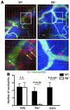
Alternative pathway complement activation promotes neutrophil transmigration. A) Acute inflammation was induced with s.c. injection of K/BxN serum. One hour after serum injection, 2P microscopy was performed on the inflamed paws to enumerate intravascular (white arrow), perivascular (green arrow) and extravascular (yellow arrow) neutrophils. Scale bar = 20 μm. Lower panels represent higher magnification. Red = neutrophils; green = FITC dextran; blue = collagen rich extracellular matrix. B) Enumerated number of intravascular, perivascular and extravascular neutrophils in WT versus FB−/− mice. Neutrophils were visualized and counted in at least 10 medium size blood vessels (20-50 μm in diameter) per animals and results presented as mean ± SEM (n = 3 animals per genotype). Statistical analysis was performed by one-way ANOVA.
Figure 6.
Figure 6.
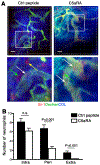
C5a receptor antagonist (C5aRA) blocks neutrophil transmigration. A) WT mice were administered C5aRA or control (Ctrl) peptide 10 min prior to s.c. K/BxN serum injection. Neutrophil trafficking to the inflamed paw was assessed by 2P microscopy 60 min after K/BxN injection. White arrow = intravascular neutrophils; green arrow = perivascular neutrophils; yellow arrow = extravascular neutrophils. Red = neutrophils; green = FITC dextran; blue = collagen rich extracellular matrix. Scale bar = 20 μm. B) The number of intravascular, perivascular and extravascular neutrophils in control peptide or C5aRA treated animals was enumerated. Neutrophils were visualized and counted in at least 10 medium size blood vessels (20-50 μm in diameter) per animal; results are presented as mean ± SEM (n = 3 animals per treatment condition). Statistical analysis was performed using one-way ANOVA.
Figure 7.
Figure 7.
In vitro C5a-induced degranulated neutrophil contents degrade VE-cadherin, generating intercellular gaps. A) C5a-induced degranulated neutrophil contents (30 ng of supernatant) were incubated with HUVEC monolayers. Loss of VE-cadherin (asterisks, left lower panel) and appearance of intercellular gaps (right lower panel) at time 0 (Media) and 30 min. VE-cadherin (red), F-actin (green), DAPI (blue). Scale bar = 30 μm. B) Quantification of intercellular gap formation over time, as detailed in Methods. C) Incubation of C5a-induced degranulated neutrophil supernatant with HUVECs led to VE-cadherin degradation. D) Neutrophil protease inhibitors, specifically NE inhibitor (NEi) or the combination of CGi, NEi, PR3i and MMPi (x4i), blocked VE-cadherin degradation. Actin served as control for protein loading. Statistical analysis was performed using one-way ANOVA.
Figure 8.
Figure 8.
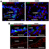
C5aRA blocks protease release and VE-cadherin degradation. A) Mice treated with control peptide or C5aRA were sacrificed 60 min after s.c. K/BxN serum injection. Their paw sections were stained for MPO (green), VE-cadherin (red) and DNA (DAPI). In mice treated with Ctrl peptide Z-stacks confocal images of blood vessels in inflamed paws revealed that in vivo neutrophils adherent to the endothelium displayed abundant extracellular MPO and were associated with areas of degraded VE-cadherin. C5aRA blocked degranulation and preserved adherens junctions. Scale bar = 25 μm. B) Higher magnification of inset areas in (A). Scale bar = 10 μm.
Figure 9.
Figure 9.
C5a-C5aR interaction induces endothelial F-actin cytoskeleton remodeling. A) Mice treated with control (Ctrl) peptide or C5aRA were sacrificed 60 min after s.c. K/BxN serum injection. Their paws were sectioned and stained for neutrophils (Gr-1, red), F-actin (green) and nuclei (DAPI). Scale bar = 100 μm. B) Integrity of the endothelial cytoskeleton was scored on a scale of 0 - 4 (see Methods). C) Density of microvascular F-actin fluorescent peaks was calculated using ImageJ software. D) The number of F-actin peaks per μm of endothelium was obtained from at least 10 medium size blood vessels per animals; results presented as mean ± SEM (n = 3 animals per treatment condition). Statistical significance between treatment group was determined by student’s t-test.

References

    1. Wipke BT & Allen PM (2001) Essential role of neutrophils in the initiation and progression of a murine model of rheumatoid arthritis. J Immunol 167(3):1601–1608. - PubMed
    1. Monach PA, et al. (2010) Neutrophils in a mouse model of autoantibody-mediated arthritis: critical producers of Fc receptor gamma, the receptor for C5a, and lymphocyte function-associated antigen 1. Arthritis Rheum 62(3):753–764. - PMC - PubMed
    1. Sadik CD, Kim ND, Iwakura Y, & Luster AD (2012) Neutrophils orchestrate their own recruitment in murine arthritis through C5aR and FcgammaR signaling. Proc Natl Acad Sci U S A 109(46):E3177–3185. - PMC - PubMed
    1. Ji H, et al. (2002) Arthritis critically dependent on innate immune system players. Immunity 16(2):157–168. - PubMed
    1. Grant EP, et al. (2002) Essential role for the C5a receptor in regulating the effector phase of synovial infiltration and joint destruction in experimental arthritis. J Exp Med 196(11):1461–1471. - PMC - PubMed

Publication types