Review
. 2019 Nov;90(11):1221-1233.
doi: 10.1136/jnnp-2017-317557. Epub 2019 Sep 21.

Understanding neurodegeneration after traumatic brain injury: from mechanisms to clinical trials in dementia

Affiliations
Review

Understanding neurodegeneration after traumatic brain injury: from mechanisms to clinical trials in dementia

Neil Sn Graham et al. J Neurol Neurosurg Psychiatry. 2019 Nov.

Abstract

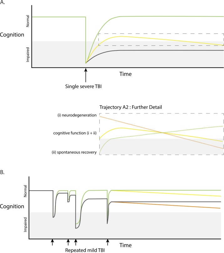
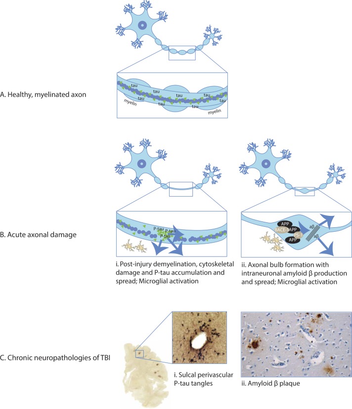
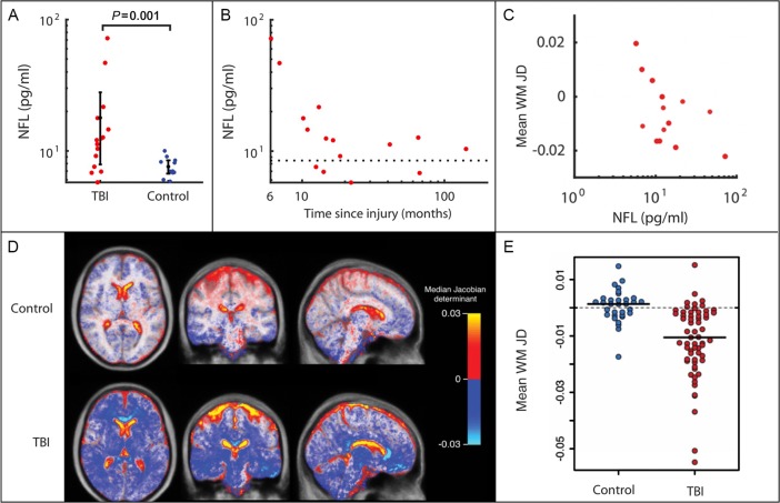
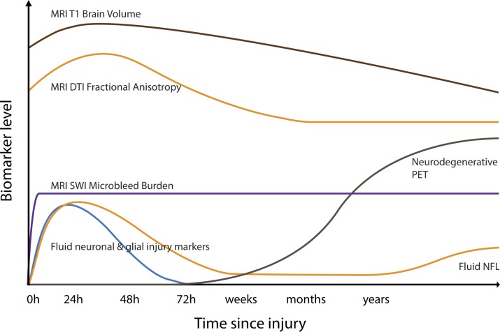
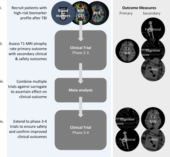
Traumatic brain injury (TBI) leads to increased rates of dementia, including Alzheimer's disease. The mechanisms by which trauma can trigger neurodegeneration are increasingly understood. For example, diffuse axonal injury is implicated in disrupting microtubule function, providing the potential context for pathologies of tau and amyloid to develop. The neuropathology of post-traumatic dementias is increasingly well characterised, with recent work focusing on chronic traumatic encephalopathy (CTE). However, clinical diagnosis of post-traumatic dementia is problematic. It is often difficult to disentangle the direct effects of TBI from those produced by progressive neurodegeneration or other post-traumatic sequelae such as psychiatric impairment. CTE can only be confidently identified at postmortem and patients are often confused and anxious about the most likely cause of their post-traumatic problems. A new approach to the assessment of the long-term effects of TBI is needed. Accurate methods are available for the investigation of other neurodegenerative conditions. These should be systematically employed in TBI. MRI and positron emission tomography neuroimaging provide biomarkers of neurodegeneration which may be of particular use in the postinjury setting. Brain atrophy is a key measure of disease progression and can be used to accurately quantify neuronal loss. Fluid biomarkers such as neurofilament light can complement neuroimaging, representing sensitive potential methods to track neurodegenerative processes that develop after TBI. These biomarkers could characterise endophenotypes associated with distinct types of post-traumatic neurodegeneration. In addition, they might profitably be used in clinical trials of neuroprotective and disease-modifying treatments, improving trial design by providing precise and sensitive measures of neuronal loss.

Keywords: acquired brain injury; cognition; dementia; image analysis; traumatic brain injury.


Conflict of interest statement

Competing interests: None declared.

Figures

Figure 1
Figure 1
Possible cognitive trajectories after traumatic brain injury (TBI). (A) Cognitive function in relation to single severe TBI (black arrow). Marked early deterioration in cognition which may recover fully (green colour), recover partially but subsequently deteriorate (progressive neurodegeneration, yellow colour), or recover partially leaving persistent non-progressive cognitive impairment (black colour). Further detail of trajectory A2 (dashed box) illustrating that overall cognitive function (yellow colour) may be influenced by a spontaneous recovery (green colour) and neurodegeneration (orange colour). (B) Cognitive function in relation to repeated mild TBI or ‘concussions’ (small black arrows). Possible trajectories include transient impairment in cognition associated with good recoveries and no progression (green colour), or late progressive neurodegeneration (yellow colour). TBI may be followed by incomplete recovery, without late progression (grey colour) or with late progressive deterioration (orange colour).
Figure 2
Figure 2
Acute neuropathologies and chronic neurodegeneration (A) Healthy, myelinated axon prior to traumatic brain injury (TBI). The box shows detail of the mid-segment of axon with central microtubules surrounded by tau with intact myelin sheath present. (B) Acute axonal damage with demyelination of the axon (panels i and ii). Tau pathology and demyelination of axon: (i) axonal injury causes cytoskeletal disruption, tau dissociation from microtubules and accumulation. Tau is aberrantly phosphorylated and may spread through extracellular, paracellular, transcellular and glymphatic mechanisms. Amyloid pathology: (ii) axonal damage causes formation of axonal bulbs/varicosities. Amyloid precursor protein (APP) accumulates with cleavage enzymes beta-site APP cleaving enzyme 1 (BACE-1) and presenilin 1 (PS-1). This produces amyloid beta which may spread to the surrounding structures following lysis of damaged neurons. Traumatic axonal damage stimulates local inflammatory response including microglial activation (panels i and ii). (C) Chronic neuropathologies. (i) Tau pathology: shearing forces during head injury localise to cortical sulcal depths causing microstructural damage, blood brain barrier disruption, axonopathy, astrogliopathy and inflammation. Sulcal perivascular localisation of P-tau neurofibrillary tangles is pathognomonic of chronic traumatic encephalopathy, visible on CP13 immunostaining. (ii) Amyloid pathology: amyloid beta plaques in a middle-aged woman who died many decades after TBI evident on immunohistochemical and thioflavine-S stains.
Figure 3
Figure 3
Quantifying neurodegeneration with brain atrophy and blood neurofilaments. (A) Plasma neurofilament light (NFL) levels plotted for moderate-severe traumatic brain injury (TBI) in the chronic phase and controls. Levels are significantly higher in patients with TBI than in controls. (B) NFL levels for moderate-severe TBI in the chronic phase plotted against time since injury (months). (C) Mean white matter (WM) Jacobian determinant (annualised JD rate) calculated over a 6-month scan–rescan interval in patients in the chronic phase after moderate-severe TBI, plotted against baseline plasma NFL level. (D) Spatial maps of average JD values in healthy controls and TBI patient groups. Marked progressive white matter atrophy is present after moderate-severe TBI (blue-white areas) with expansion of cerebrospinal fluid spaces (red-yellow areas) in comparison with minimal change in healthy controls. (E) Progressive atrophy of white matter following moderate-severe TBI. Scatter plot of JD rates of brain volume change in TBI compared with age-matched healthy volunteers, in white matter. A JD of 0 indicates no change in brain volume over the follow-up period.
Figure 4
Figure 4
Potential longitudinal biomarker trajectories following traumatic brain injury (TBI). Hypothecated trajectories of biomarkers after moderate/severe TBI. Brain volumes measured by volumetric MRI may initially increase due to oedema before progressively reducing and continuing to decline as a result of progressive neurodegeneration after injury. Fractional anisotropy, a measure of white matter integrity derived from diffusion tensor imaging (DTI), initially increases due to acute oedema, with a subacute reduction days–weeks later reflecting axonal damage. Cerebral microbleeds, a marker of diffuse vascular injury, appear rapidly after TBI and do not resolve. They are identified most sensitively with susceptibility weighted imaging (SWI). s106 Fluid neuronal and glial injury markers such as ubiquitin carboxy-terminal hydrolase L1, S100B, neuron-specific enolase, glial fibrillar acidic protein, amyloid and tau are briskly elevated after TBI. Neurofilament light levels (NFL) peak later and may be elevated in the chronic phase, correlating with progressive brain atrophy.s107 s108 PET, positron emission tomography.
Figure 5
Figure 5
Imaging traumatic brain injury (TBI) and post-traumatic neurodegeneration. (A) Progressive neurodegeneration is quantifiable using repeated T1 MRI used to generate atrophy rates over time. (B) Susceptibility weighted imaging (SWI) shows microbleeds in typical parafalcine distribution, typical of diffuse vascular injuries. (C) Diffusion MRI allows quantification of white matter integrity after axonal injury and provides a measure of diffuse axonal injury. (D) 11C-Pittsburgh compound B (PiB) positron emission tomography (PET) study shows amyloid deposition in a middle-aged woman several years after moderate-severe TBI. (E) 18F-AV1451 tau PET shows abnormal binding following TBI. (F) Persistent abnormal microglial activation on 11C-PBR28 translocator protein PET in a middle-aged man a decade after moderate-severe TBI, particularly in white matter regions. DTI, diffusion tensor imaging.
Figure 6
Figure 6
Biomarkers in clinical trials after traumatic brain injury (TBI). Stages for the evaluation of disease-modifying/neuroprotective treatment after TBI. (i) Recruitment of patients at high risk for neurodegeneration using baseline blood neurofilament light, diffusion tensor imaging abnormality (DTI) and positron emission tomography (PET) abnormality. (ii) Phase 2–3 trials powered to primary outcome measure of change in atrophy rate (using repeated T1 MRI) with secondary functional/cognitive/safety outcomes. (iii) Meta-analysis of phase 2–3 trials to clarify the relationship between the surrogate (T1 atrophy rate) and patient-centred outcomes. (iv) Late-stage phase 3–4 trials using primary functional or cognitive outcome. This may be a composite measure.

Similar articles

Cited by

References

    1. Maas AIR, Menon DK, Adelson PD, et al. . Traumatic brain injury: integrated approaches to improve prevention, clinical care, and research. Lancet Neurol 2017;16:987–1048. 10.1016/S1474-4422(17)30371-X - DOI - PubMed
    1. McMillan TM, Teasdale GM, Weir CJ, et al. . Death after head injury: the 13 year outcome of a case control study. J Neurol Neurosurg Psychiatry 2011;82:931–5. 10.1136/jnnp.2010.222232 - DOI - PubMed
    1. Scheid R, Walther K, Guthke T, et al. . Cognitive sequelae of diffuse axonal injury. Arch Neurol 2006;63:418–24. 10.1001/archneur.63.3.418 - DOI - PubMed
    1. Sariaslan A, Sharp DJ, D’Onofrio BM, et al. . Long-Term outcomes associated with traumatic brain injury in childhood and adolescence: a nationwide Swedish cohort study of a wide range of medical and social outcomes. PLoS Med 2016;13:e1002103 10.1371/journal.pmed.1002103 - DOI - PMC - PubMed
    1. Li Y, Li Y, Li X, et al. . Head injury as a risk factor for dementia and Alzheimer's disease: a systematic review and meta-analysis of 32 observational studies. PLoS One 2017;12:e0169650 10.1371/journal.pone.0169650 - DOI - PMC - PubMed

Publication types

MeSH terms