. 2019 Sep 22:25:7100-7114.
doi: 10.12659/MSM.917097.

Mechanisms of IFNalpha-1a-Induced Apoptosis in a Laryngeal Cancer Cell Line

Affiliations

Mechanisms of IFNalpha-1a-Induced Apoptosis in a Laryngeal Cancer Cell Line

Xiao-Lei Xin et al. Med Sci Monit. .

Abstract

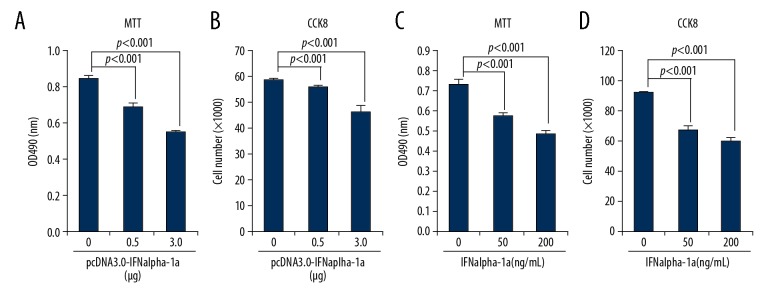
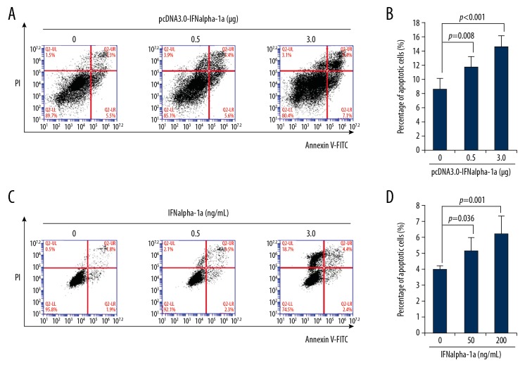
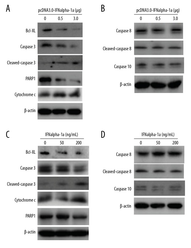
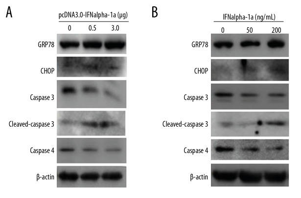
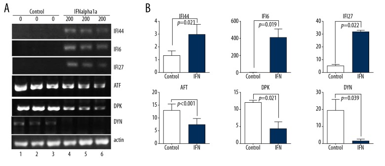
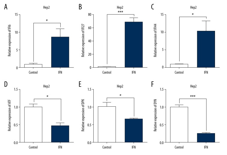
BACKGROUND Interferon alpha (IFNalpha) exerts its anti-proliferative effect on many human cancers. Among the 13 subtypes of human IFNalpha, IFNalpha-1 subtype has 2 variants, named IFNalpha-1a and IFNalpha-1b, that differ from each other in only 1 amino acid, at residue 114. However, the mechanism by which IFNalpha-1a mediates growth inhibition is still unclear. MATERIAL AND METHODS Human laryngeal carcinoma HEp2 cells were treated with IFNalpha-1a by either transient transfection or exogenous delivery. Western blot and RT-PCR analysis were carried out to assess apoptotic pathways active in IFNalpha-1a-treated HEp2 cells. Microarray analysis was conducted to uncover the differential gene expressions after IFNalpha-1a treatment. KEGG pathway enrichment analysis was also performed. RESULTS IFNalpha-1a markedly inhibited the proliferation and significantly promoted the apoptosis of HEp-2 cells. Mechanistic studies indicate that IFNalpha-1a-mediated cell apoptosis is directly linked to intrinsic and endoplasmic reticulum (ER) stress-related apoptosis, but is independent of extrinsic apoptosis. The top 40 differentially expressed genes discovered by microarray analysis included 20 upregulated genes (e.g., IFI6, IFI27, IFI44L, and MIR548X) and 20 downregulated genes (e.g., PRKDC, HIST1H3B, DYNC1H1, and HIST1H2AM). KEGG pathway enrichment analysis revealed that 4 out of 6 pathways are TP53-related. CONCLUSIONS We demonstrated a detailed mechanism involved in IFNalpha-1a-mediated anti-proliferation activity in human laryngeal carcinoma cells.


Conflict of interest statement

Conflicts of interest

None.

Figures

Figure 1
Figure 1
IFNalpha-1a inhibits the proliferation of laryngeal carcinoma HEp-2 cells. (A, C) 3-(4,5-dimethylthiazol-2-yl)-2,5-diphenyl tetrazolium bromide (MTT) analysis of the proliferation of HEp-2 cells. HEp-2 cells were either transiently transfected with increasing doses (0, 0.5, and 3 ug) of pcDNA3.0-IFNalpha-1a (A) or treated with increasing doses (0, 50, and 200 ng/mL) of recombinant human IFNalpha-1a (C). After 48-h incubation, the treated HEp-2 cells were collected and analyzed with MTT assay. The reaction products were measured at 490 nm with a microplate reader. Each value is represented as mean ±SD from 3 independent experiments. The results were considered to be significant if p≤0.05. (B, D) Cell counting kit 8 (CCK-8) analysis of cell proliferation. HEp-2 cells were either transiently transfected with increasing doses (0, 0.5, and 3 ug) of pcDNA3.0-IFNalpha-1a (B) or treated with increasing doses (0, 50, and 200 ng/mL) of recombinant human IFNalpha-1a (D). After 48-h incubation, the treated HEp-2 cells were collected and analyzed by CCK-8 assay. The reaction products were measured at 450 nm with a microplate reader. The number of viable cells for each dose was calculated against the standard curve. Each value is represented as mean ±SD from 3 independent experiments. The results were considered to be significant if p≤0.05.
Figure 2
Figure 2
IFNalpha-1a promotes the apoptosis of laryngeal carcinoma HEp-2 cells. (A) Flow cytometric analysis of the apoptosis of HEp-2 cells that were transiently transfected with pcDNA3.0-IFNalpha-1a. HEp-2 cells were first seeded onto a 12-well culture plate and transiently transfected with increasing doses (0, 0.5, and 3 ug) of pcDNA3.0-IFNalpha-1a. After 48-h incubation, the transfected HEp-2 cells were subjected to Annexin V/Propidium iodide (PI) double staining followed by flow cytometric analysis. (B) Quantitation of the apoptosis of the HEp-2 cells after transiently transfecting with pcDNA3.0-IFNalpha-1a. The transfected HEp2 cells in (A) were subjected to Annexin V/PI double staining followed by flow cytometric analysis. Each value is represented as mean ±SD from 3 independent experiments. After statistical analysis, results were considered to be significant if p≤0.05. (C) Flow cytometric analysis of the apoptosis of HEp-2 cells that were treated with recombinant human IFNalpha-1a. Hep-2 cells were seeded onto a 12-well culture plate and treated with increasing doses (0, 50, and 200 ng/mL) of recombinant human IFNalpha-1a. After 48-h culture, the cells were harvested and subjected to Annexin V/PI double staining followed by flow cytometric analysis. (D) Quantitation of the apoptosis of the HEp-2 cells after IFNalpha-1a treatment. The IFNalpha-1a treated HEp-2 cells were prepared as described in (C) and subjected to Annexin V/PI double staining followed by flow cytometric analysis. Each value is represented as mean ±SD from 3 independent experiments. After statistical analysis, results were considered to be significant if p≤0.05.
Figure 3
Figure 3
The responses to IFNalpha-1a by other cell lines. (A–C) Quantitation of the apoptosis of the HEK293T cells (A), HepG2 cells (B), and A549 cells (C) after transiently transfecting with increasing doses (0, 0.5, and 3 μg) of pcDNA3.0-IFNalpha-1a. The transfected cells were subjected to Annexin V/PI double staining followed by flow cytometric analysis. Each value is represented as mean ±SD from 3 independent experiments. After statistical analysis, results were considered to be significant if p≤0.05. (D–F) Quantitation of the apoptosis of the HEK293T cells (D), HepG2 cells (E), and A549 cells (F) after treatment with increasing doses (0, 50, and 200 ng/mL) of human recombinant IFNalpha-1a. The treated cells were subjected to Annexin V/PI double staining followed by flow cytometric analysis. Each value is represented as mean ±SD from 3 independent experiments. After statistical analysis, results were considered to be significant if p≤0.05.
Figure 4
Figure 4
IFNalpha-1a-mediated cell apoptosis in HEp-2 cells is associated with the intrinsic but not extrinsic apoptotic pathway. (A, C) Western blot analysis on the expressions of the key mediators involved in the intrinsic apoptotic pathway. HEp-2 cells were either transiently transfected with increasing doses (0, 0.5. and 3ug) of pcDNA3.0-IFNalpha-1a (A) or treated with increasing doses (0, 50, and 200 ng/mL) of recombinant human IFNalpha-1a (C). After 48-h incubation, the whole-cell lysates were prepared and probed with anti-Bcl-XL, anti-cytochrome c, anti-caspase 3, anti-cleaved caspase 3, and anti-PARP1 antibodies. The reaction products were subjected to Western blot analysis. β-actin gene expression served as internal control. (B, D) IFNalpha-1a does not activate the extrinsic apoptotic pathway. Hep-2 cells were either transiently transfected with increasing doses (0, 0.5, and 3 ug) of pcDNA3.0-IFNalpha-1a (B) or treated with increasing doses (0, 50, and 200 ng/mL) of recombinant human IFNalpha-1a (D). After 48-h incubation, the whole-cell lysates were prepared and probed with anti-caspase 8, anti-cleaved caspase 8, and anti-caspase 10 antibodies. The reaction products were subjected to Western blot analysis. β-actin gene expression served as an internal control.
Figure 5
Figure 5
IFNalpha-1a activates the endoplasmic reticulum stress-induced apoptosis. (A) Western blot analysis of the expressions of the key mediators associated with the endoplasmic reticulum stress-induced apoptosis. HEp-2 cells were either transiently transfected with increasing doses (0, 0.5, and 3 μg) of pcDNA3.0-IFNalpha-1a (A) or treated with increasing does (0, 50, and 200 ng/mL) of human recombinant IFNalpha-1a (B). After 48-h incubation, the whole-cell lysates (WCL) were prepared and probed with anti-caspase4, anti-GRP78, anti-CHOP, anti-caspase 3, and anti-cleaved caspase 3 antibodies. The reaction products were subjected to Western blot analysis. β-actin gene expression served as an internal control.
Figure 6
Figure 6
Confirmation of microarray analysis of IFNalpha-1a-responded gene expressions in HEp-2 cells by semi-quantitative RT-PCR analysis. (A) RT-PCR analysis of total RNAs from HEp-2 cells treated with or without IFNalpha-1a. The expressions of the 3 selected upregulated genes (IFI6, IFI27, and IFI44L) and 3 selected downregulated genes (ATF6 (ATF), PRKDC (DPK), and DYNC1H1 (DYN)) were subjected to RT-PCR analysis. The expression of β-actin gene was assayed as a loading control. (B) Quantitation of the results in (A). The relative band intensity was quantitated with Image J program in comparison with β-actin. The triplicated results of both IFNalpha-1a treated or untreated samples were subjected to statistical analysis. The results were considered to be significant if p≤0.05.
Figure 7
Figure 7
Confirmation of microarray analysis of IFNalpha-1a-responded gene expressions in HEp-2 cells by real-time qRT-PCR analysis. Real-time qRT-PCR was employed to detect the mRNA levels of IFI6 (A), IFI27 (B), IFI44L (C), ATF6 (ATF, D), PRKDC (DPK, E), and DYNC1H1 (DYN, F) on the samples isolated in Figure 6. Each value represents the mean ±SD from 3 reactions. The triplicated results of both IFNalpha-1a-treated or -untreated samples were subjected to statistical analysis. The symbols * and *** represent p values less than 0.05 and 0.001, respectively. The results were considered to be significant if p≤0.05.

Similar articles

Cited by

References

    1. Siegel RL, Miller KD, Jemal A. Cancer statistics, 2016. Cancer J Clin. 2016;66:7–30. - PubMed
    1. Kuper H, Boffetta P, Adami HO. Tobacco use and cancer causation: Association by tumour type. J Intern Med. 2002;252:206–24. - PubMed
    1. Boffetta P, Hashibe M. Alcohol and cancer. Lancet Oncol. 2006;7:149–56. - PubMed
    1. Sanchez Barrueco A, Gonzalez Galan F, Lora Pablos D, et al. HPV in larynx squamous cell carcinoma: New serotypes and survival study within 10-year follow-up. Otolaryngol Head Neck Surg. 2017;156:677–82. - PubMed
    1. Vietia D, Liuzzi J, Avila M, et al. Human papillomavirus detection in head and neck squamous cell carcinoma. Ecancermedicalscience. 2014;8:475. - PMC - PubMed