Case Reports
. 2020 Feb;9(1):24-29.
doi: 10.1007/s13730-019-00420-5. Epub 2019 Sep 21.

A case of female Fabry disease revealed by renal biopsy

Affiliations
Case Reports

A case of female Fabry disease revealed by renal biopsy

Sae Aratani et al. CEN Case Rep. 2020 Feb.

Abstract

Fabry disease (FD) is an X-linked inherited glycosphingolipid metabolism disorder, therefore, heterozygous female FD patients display highly variable clinical symptoms, disease severity, and pathological findings. This makes it very challenging to diagnosing female patients with FD. A 69-year-old Japanese female was introduced to the nephrologist for the evaluation of proteinuria. A renal biopsy was performed. Although the light microscopic examinations revealed that most of the glomeruli showed minor glomerular abnormalities, however, vacuolation was apparently found in the tubular epithelial cells. Immunofluorescence staining for globotriaosylceramide was positively detected in some podocytes and distal tubular epithelial cells. In addition, myelin-like structure (zebra body) was detected by electron microscopy. Pathological findings were most consistent with FD. Consequently, biochemical and genetic analysis confirmed the diagnosis of female FD. Enzyme replacement therapy was performed in conjunction with renin-angiotensin aldosterone system inhibitors and beta-blockers. The patient's family members received the analysis, and the same DNA missense mutation was detected in the patient's grandson. The enzyme replacement therapy was introduced to the grandson. The present case showed that renal biopsy can contribute towards a correct diagnosis for FD. Particularly, in female FD patients, careful examination of pathological changes is essential, for example, vacuolation of any type of renal cells may be a clue for the diagnosis.

Keywords: Enzyme replacement therapy; Fabry disease; Pathology; Renal biopsy; Vacuolation.


Conflict of interest statement

The authors have no competing interests to declare.

Figures

Fig. 1
Fig. 1
Family tree. Red color represents patients with Fabry disease, which was confirmed by genetic analysis. The patient’s mother (Case I-1) died of heart failure. The patient’s son (Case III/2) died due to sudden cardiac arrest. The patient’s daughter (Case III/3) was completely asymptomatic. The patient’s grandchild (Case IV/2) had developed neurological pain in his extremities, an early symptom of FD. He started ERT soon after the diagnosis was confirmed
Fig. 2
Fig. 2
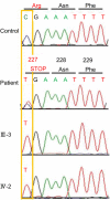
Genetic analysis. Genetic analysis was performed on the patient, the patient’s daughter (Case III/3), and the patient’s grandchild (Case IV/2). All showed the same DNA missense mutation in exon 5, c.[679C>T], which confirmed the diagnosis of FD
Fig. 3
Fig. 3
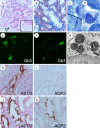
Renal pathology. The renal biopsy samples contained 22 glomeruli for the evaluation of light microscopy, two of which showed global sclerosis. Most of the glomeruli showed mild hypertrophy and minor glomerular abnormalities with mild mesangial expansion (a). Low levels of podocyte vacuolization were detected (white arrowhead in a). Tubular epithelial cell vacuolization occurred predominantly in the distal tubules (b) [a Periodic acid-Schiff (PAS) stain, × 600; b Masson trichrome stain, × 600]. Electron microscopy of toluidine blue-stained semi-thin sections revealed dark blue osmiophilic cells in podocytes, Bowman’s epithelial cells, and distal tubular epithelial cells (c, d). Immunofluorescence staining for Gb3 was positively detected in glomerular and tubular cells (e, f). Electron microscopy revealed myelin-like structures in the podocytes (g). Immunohistochemistry for AE1/3 and AQP2 detected distal tubular epithelial cells and principal cells in collecting tubules, respectively. Serial sections were stained with AE1/3 and AQP2, and counter stained with hematoxylin and eosin. Varying levels of vacuolation were observed in each tubule. In the collecting tubules, both intercalated cells and principal cells showed vacuolation (hk × 600)
Fig. 4
Fig. 4
Clinical Course. Black circles and gray squares represent the clinical course of eGFR and proteinuria, respectively. eGFR estimated glomerular filtration rate, ERT enzyme replacement therapy, g/gCr gram/gram creatine, RAAS renin–angiotensin aldosterone system; and U-Pro urine-protein

Similar articles

Cited by

References

    1. Brady RO, Gal AE, Bradley RM, Martensson E, Warshaw AL, Laster L. Enzymatic defect in Fabry's disease: ceramidetrihexosidase deficiency. N Engl J Med. 1967;27:163–167. - PubMed
    1. Germain DP. Fabry disease. Orphanet J Rare Dis. 2010;5:30. doi: 10.1186/1750-1172-5-30. - DOI - PMC - PubMed
    1. Garman SC, Garboczi DN. The molecular defect leading to Fabry disease: structure of human alpha-galactosidase. J Mol Biol. 2004;337:319–335. doi: 10.1016/j.jmb.2004.01.035. - DOI - PubMed
    1. Wilcox WR, Oliveira JP, Hopkin RJ, Ortiz A, Banikazemi M, Feldt-Rasmussen U, Sims K, Waldek S, Pastores GM, Lee P, Eng CM, Marodi L, Stanford KE, Breunig F, Wanner C, Warnock DG, Lemay RM, Germain DP. Females with Fabry disease frequently have major organ involvement: lessons from the Fabry Registry. Mol Genet Metab. 2008;93:112–128. doi: 10.1016/j.ymgme.2007.09.013. - DOI - PubMed
    1. Banikazemi M, Bultas J, Waldek S, Wilcox WR, Whitley CB, McDonald M, Finkel R, Packman S, Bichet DG, Warnock DG, Desnick RJ. Agalsidase-beta therapy for advanced Fabry disease: a randomized trial. Ann Intern Med. 2007;146:77–86. doi: 10.7326/0003-4819-146-2-200701160-00148. - DOI - PubMed

Publication types

MeSH terms