

Prostatic epithelial cells and their high expressions of CKIP-1 affect the TGF-β1 expression levels which might reduce the scar formation in remodeling stage at prostatic urethral wounds after wound repair
- PMID: 31542883
- PMCID: PMC6957543
- DOI: 10.1007/s11255-019-02286-z
Prostatic epithelial cells and their high expressions of CKIP-1 affect the TGF-β1 expression levels which might reduce the scar formation in remodeling stage at prostatic urethral wounds after wound repair
Abstract
Objective: There are less scar formations in some wounds after wound repair. Our earlier study had shown that the amount of collagen fibers in canine prostatic urethra wound were less than in bladder neck wound after 2-μm laser resection of the prostate (TmLRP) and partial bladder neck mucosa at 4 weeks. The purpose of this study was to observe the amount of scar tissue and characterize the probable causes of "less scar healing" in prostatic urethra wound.
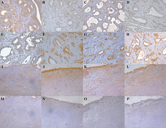
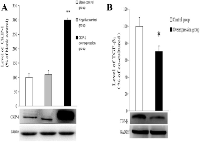
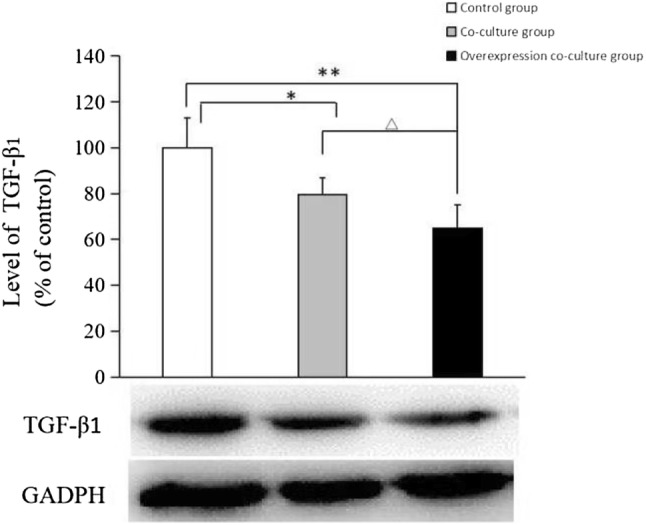
Methods: A total of 12 healthy adult male crossbred canines underwent resection of prostate and partial bladder neck mucosa using 2-μm laser. The prostatic urethra and bladder neck wound specimens were harvested at 3, 4, 8 and 12 weeks after operation, respectively. The histopathologic characteristics were observed by hematoxylin and eosin(HE)staining, and the expression of transforming growth factor-β1 (TGF-β1) and casein kinase-2 interacting protein-1 (CKIP-1) were examined by immunohistochemistry in prostatic urethra and bladder neck wound, respectively. Overexpressed CKIP-1 human prostate epithelial cells (BPH-1 cells) were established and the expression of TGF-β1 was detected by Western blotting. Furthermore, a non-contact co-culture system of BPH-1 cells and human fibroblast (HFF-1) cells was used to observe the effects of BPH-1 cell and their high CKIP-1 levels on the expression of TGF-β1 in HFF-1 in vitro.
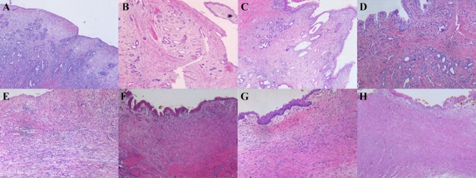
Results: The histology showed that there were a large number of prostatic epithelium and a small amount of scar tissue in prostatic urethra wound, while no epithelial cells and more scar tissue in bladder neck wound at 4, 8 and 12 weeks after repair. There were a higher expression level of TGF-β1 in prostate epithelial cells and fibroblasts and a lower expression level of CKIP-1 in prostate epithelial cells at 3 weeks after surgery in prostatic urethral wound. Compared to week 3, the TGF-β1 expression decreased both in prostate epithelial cells and fibroblasts at 4, 8 and 12 weeks in prostatic urethral wound (p < 0.05 or p < 0.01). The CKIP-1 expression increased in prostate epithelial cells at 4, 8 and 12 weeks compared to 3 weeks in prostatic urethra wound (p < 0.01). A higher TGF-β1 expression level of fibroblasts was observed in bladder neck wound at 3 weeks. And there was no significant change in the expression of TGF-β1 of fibroblasts in 3, 4, 8 and 12 weeks after operation in bladder neck wound. Both the prostate urethra and bladder neck wound fibroblasts showed weak expression of CKIP-1 and there was no significant change in 3, 4, 8 and 12 weeks. The vitro experiments showed that the TGF-β1 expression in BPH-1 cells with CKIP-1 overexpression decreased 25% compared with control group (p < 0.05). Furthermore, the expression of TGF-β1 in HFF-1 cells of co-cultured group decreased by 20% compared with Control group (p < 0.05); the expression of TGF-β1 in HFF-1 cells of overexpression co-culture group were reduced by 15% compared with co-cultured group (p < 0.01).
Conclusions: A large number of prostate epithelial cells in prostatic urethra wound may be one of the causes of less formation of scar tissue after repair. The prostate epithelial cells might reduce expression level of TGF-β1 by raising CKIP-1 expression and inhibit expression of TGF-β1 in peripheral fibroblasts at remodeling stage to reduce the excessive proliferation of fibrous cells and the excessive scar formation.
Keywords: CKIP-1; Less scar; Prostatic urethra wound; TGF-β1; Wound healing.
Conflict of interest statement
The authors declare that they have no conflict of interest.
Figures
References
-
- Yang P, Zhao J, Zhan L, et al. To explore the mechanism of repairing oral mucosa without scar healing. J Clin stomatol. 2012;28(11):699–701.
-
- Cao Y, Luo GH, Luo L, et al. Comparative study on re-epithelialization of canine prostatic urethra and bladder neck after 2-μm laser resection of the prostate. Chin J Urol. 2014;35(5):378–382.
MeSH terms
Substances
Grants and funding
LinkOut - more resources
Full Text Sources
Medical
Research Materials
