. 2019 Nov 4;29(21):3657-3668.e5.
doi: 10.1016/j.cub.2019.09.005. Epub 2019 Sep 19.

NODULE INCEPTION Recruits the Lateral Root Developmental Program for Symbiotic Nodule Organogenesis in Medicago truncatula

Affiliations

NODULE INCEPTION Recruits the Lateral Root Developmental Program for Symbiotic Nodule Organogenesis in Medicago truncatula

Katharina Schiessl et al. Curr Biol. .

Abstract

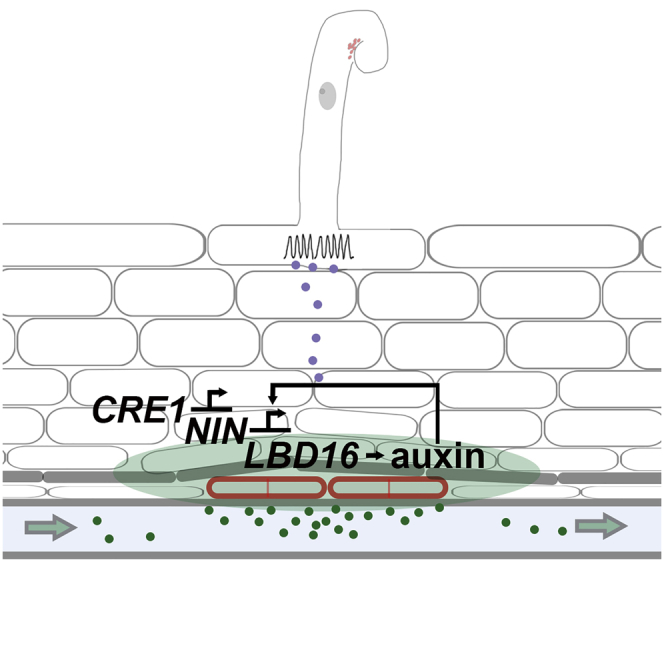
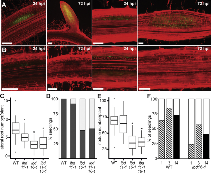
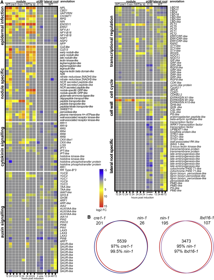
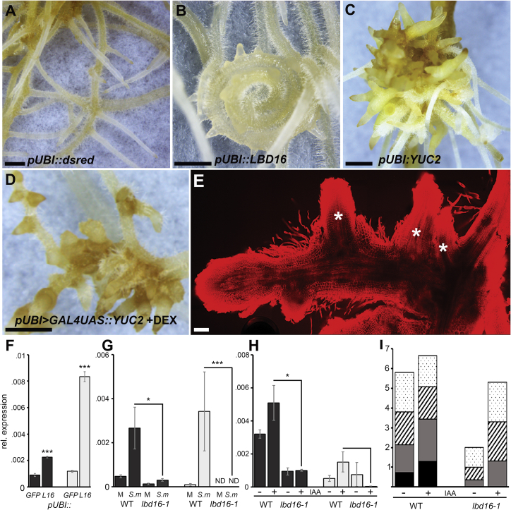
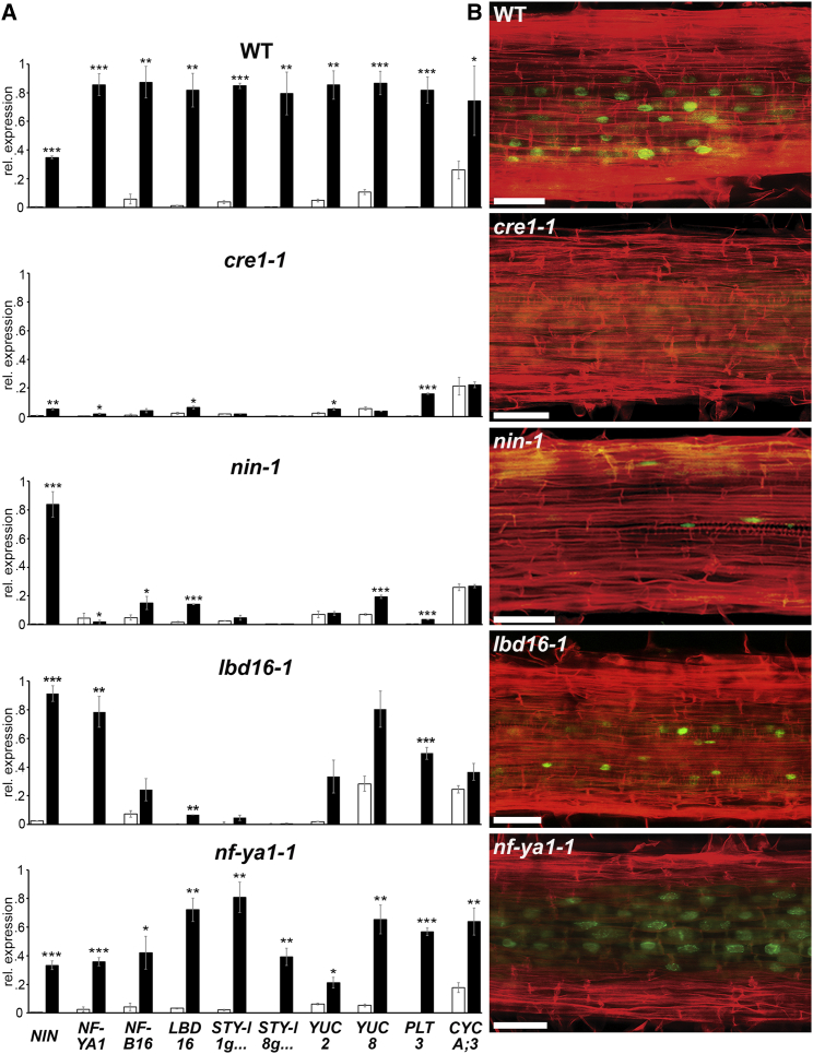
To overcome nitrogen deficiencies in the soil, legumes enter symbioses with rhizobial bacteria that convert atmospheric nitrogen into ammonium. Rhizobia are accommodated as endosymbionts within lateral root organs called nodules that initiate from the inner layers of Medicago truncatula roots in response to rhizobial perception. In contrast, lateral roots emerge from predefined founder cells as an adaptive response to environmental stimuli, including water and nutrient availability. CYTOKININ RESPONSE 1 (CRE1)-mediated signaling in the pericycle and in the cortex is necessary and sufficient for nodulation, whereas cytokinin is antagonistic to lateral root development, with cre1 showing increased lateral root emergence and decreased nodulation. To better understand the relatedness between nodule and lateral root development, we undertook a comparative analysis of these two root developmental programs. Here, we demonstrate that despite differential induction, lateral roots and nodules share overlapping developmental programs, with mutants in LOB-DOMAIN PROTEIN 16 (LBD16) showing equivalent defects in nodule and lateral root initiation. The cytokinin-inducible transcription factor NODULE INCEPTION (NIN) allows induction of this program during nodulation through activation of LBD16 that promotes auxin biosynthesis via transcriptional induction of STYLISH (STY) and YUCCAs (YUC). We conclude that cytokinin facilitates local auxin accumulation through NIN promotion of LBD16, which activates a nodule developmental program overlapping with that induced during lateral root initiation.

Keywords: CYTOKININ RESPONSE FACTOR; LATERAL ORGAN BOUNDARIES DOMAIN; Medicago truncatula; NODULE INCEPTION; YUCCA; auxin; endosymbiosis; lateral root/nodule organogenesis; nitrogen; rhizobia.


Conflict of interest statement

The authors declare no competing interests.

Figures

None
Graphical abstract
Figure 1
Figure 1
Lateral Roots and Nodules Show Overlapping Development (A and B) (A) Optical sections of lateral roots and (B) nodules hours (h) post induction. Red propidium iodide demarks cell walls and green EdU-labeled nuclei DNA replication. Arrowheads indicate vascular strands that in lateral roots are apparent by 72 hpi compared to nodules at 120–168 hpi. Scale bars:100 μm. See also Figure S1.
Figure 2
Figure 2
Lateral Roots and Nodules Show Overlap in Gene Expression (A) Heatmap showing selected genes induced during lateral root and nodule development with fold changes ≥±1.5; p < 0.05. Expression depicts log2 fold changes. (B) Correlation heatmap depicting the overlap between genes differentially expressed during lateral root and nodule organogenesis over a time course of development. See also Figures S1 and S2 and Data S1.
Figure 3
Figure 3
LATERAL ORGAN BOUNDARIES and YUCCAs Are Expressed during Root and Nodule Primordium Initiation and Development Expression patterns of YUC2, YUC8, LBD11, and LBD16 during nodule and lateral root development visualized by GUS staining (blue). Rhizobial-expressed LacZ is stained magenta. Scale bars: 100 μm. See also Figure S3 and Data S2A and S2B.
Figure 4
Figure 4
Lateral Root and Nodule Number Are Reduced in lbd16 (A and B) Optical sections of lateral roots and nodules in (A) wild-type (WT) and (B) lbd16-1 at 24 or 72 hpi. Scale bars: 50 μm. (C) Lateral root number in 14-day-old seedlings. Boxplots show median (thick line), second to third quartiles (box), minimum and maximum ranges (lines), and outliers (single points). A one-way Kruskal-Wallis rank-sum test showed that lateral root number is dependent on genotype; asterisks indicate significantly different (95% confidence) means compared with WT. n = 56 (WT), 58 (lbd11-1), 64 (lbd16-1), and 66 (lbd11lbd16). (D) Percentage of gravi-stimulated seedlings with ≥1 lateral roots (dark gray) or 0 lateral roots (white) in the bend 5 dpi in WT (n = 25), lbd11-1 (n = 60), lbd16-1 (n = 53), and lbd11lbd16 (n = 60) showing significant reduction in the number of emerging lateral roots in lbd16-1 and lbd11lbd16 compared to WT. p = 0.166, 7.401e−07, and 1.413e−06 respectively; Fisher’s exact test. (E) Nodule number 21 days post S. meliloti inoculation. n = 13 (WT), 15 (lbd11-1), 14 (lbd16-1), and 14 (lbd11lbd16). A normal distribution allowed a one-way ANOVA test revealing the mean number of nodules differed significantly between genotypes; asterisks indicate significantly different (95% confidence) means compared to WT. (F) Percentage of seedlings with ≥1 primordia (hashed), ≥1 emerged nodule (black), or no structure (white) developing at the spot inoculation site at 1, 3, and 14 days post inoculation (dpi; n = 116 WT and 142 lbd16-1). lbd16-1 showed significantly different rates of initiation of primordia or nodules at all time points: p = 0.003 (1 dpi), 0.022 (3 dpi), and 2.084e−04 (14 dpi); Fisher’s exact test. See also Figures S3 and S4 and Data S2A.
Figure 5
Figure 5
The Impact of CRE1, NIN, and LBD16 on Nodulation-Associated Gene Expression (A) Heatmap of selected genes (as in Figure 2; see also Figure S2 for gene identifiers and Data S1) in WT (jemalong), cre1-1, and nin-1 root sections at 12 and 24 h and WT (R108), lbd16-1 (ls), and lbd11lbd16 (ld) at 24 h post S. meliloti spot inoculation; response to LBD11 (11) and LBD16 (16) overexpression in 3-week-old hairy roots compared to control roots and during lateral root induction. Expression represents log2 fold changes. (B) Pairwise comparisons of all differentially expressed genes dependent on cre1-1 (blue), nin-1 (red), and lbd16-1 (purple). cre1-1 and nin-1 comparisons were to WT jemalong and lbd16-1 to WT R108. See also Figures 2, S2, and S5 and Data S1.
Figure 6
Figure 6
Overexpression of LBD16 or YUC2 Is Sufficient to Promote Root Primordia Formation (A–E) Constitutive expression of (A) dsred (control), (B) LBD16, or (C) YUC2 under control of the LjUBI promoter in hairy roots. 44 out of 103 and 53 out of 57 transformed plants expressing pLjUBI:LBD16 and pLjUBI:YUC2, respectively, showed similar phenotypes as depicted in (B) and (C). (D and E) (D) Bright field image and (E) optical sections of propidium iodide-stained root structures expressing dexamethasone (Dex)-inducible YUC2 under control of the LjUBI promoter (pLjUBI > GAL4UAS::MtYUC2). 82 out of 167 plants transformed with pLjUBI > GAL4UAS::MtYUC2 showed ectopic primordium induction 2 weeks post Dex treatment as depicted in (D) and (E) and Figures S5C–S5G compared to 3 out of 147 Dex treated plants transformed with pLjUBI > GAL4UAS::GFP. Ectopic primordia in (E) are indicated with asterisks. Scale bars: (A–D) 1 mm and (E) 50 μm. (F–H). (F) Quantification of transcript levels by qRT-PCR of STY-like (Medtr1g023320) (dark gray bars) and STY-like (Medtr8g076620) (light gray bars) in hairy roots constitutively expressing LBD16 or GFP (pUBI::LBD16 or pUBI::GFP), (G) S. meliloti and mock spot inoculation, and (H) IAA (+) and mock (−) treatment in WT and lbd16-1 root sections at 24 hpi. Expression levels were measured by qRT-PCR and normalized to HH3. Statistical comparisons were performed as indicated. Values are the mean of 3 biological replicates ± SEM (Student’s t test; asterisks indicate statistical significance; p < 0.05, ∗∗p < 0.01, ∗∗∗p < 0.001). (I) Number of lateral root primordia in 3-day-old WT and lbd16-1 seedlings 24 h post IAA or mock treatment represented as means (n ≥ 11). The different stages of primordia development are indicated: dotted, stages I–II; striped, stage III; light gray, stages IV–V; black, emerged. Mock-treated lbd16-1 seedlings had significantly fewer primordia compared to WT (Student’s t test; p < 0.001). IAA treatment significantly increased primordia number in lbd16-1 seedlings (Student’s t test; p < 0.001), but not in WT. More stages IV–V and emerged primordia developed in WT than in lbd16-1 in both treatments (Student’s t test; p < 0.001). See also Figures S5 and S6.
Figure 7
Figure 7
NIN and LBD16 Mediate Auxin Regulators and Cell-Cycle Activation in Response to Cytokinin (A) Expression profiling on root segments treated with 100 nM (6-Benzylaminopurine) BAP for 24 h by qRT-PCR normalized to HH3. Statistical comparisons were performed between mock (white bars) and BAP (black bars). Values are the mean ΔCt values of three biological replicates normalized to the maximum value obtained for that gene within the ecotype. Data are presented ± SEM (Student’s t test; p < 0.05; ∗∗p < 0.01, ∗∗∗p < 0.001). (B) Representative optical sections (≥20 roots analyzed) in root segments (susceptibility zone) treated with 100 nM BAP. Red, cell walls; green, cell-cycle activation. Scale bars: 50 μm. See also Figures S6A–S6C.

References

    1. Oldroyd G.E., Murray J.D., Poole P.S., Downie J.A. The rules of engagement in the legume-rhizobial symbiosis. Annu. Rev. Genet. 2011;45:119–144. - PubMed
    1. Crespi M., Frugier F. De novo organ formation from differentiated cells: root nodule organogenesis. Sci. Signal. 2008;1:re11. - PubMed
    1. Plet J., Wasson A., Ariel F., Le Signor C., Baker D., Mathesius U., Crespi M., Frugier F. MtCRE1-dependent cytokinin signaling integrates bacterial and plant cues to coordinate symbiotic nodule organogenesis in Medicago truncatula. Plant J. 2011;65:622–633. - PubMed
    1. Liu J., Rutten L., Limpens E., van der Molen T., van Velzen R., Chen R., Chen Y., Geurts R., Kohlen W., Kulikova O. A Remote cis-Regulatory Region Is Required for NIN Expression in the Pericycle to Initiate Nodule Primordium Formation in Medicago truncatula. Plant Cell. 2019;31:68–83. - PMC - PubMed
    1. Marsh J.F., Rakocevic A., Mitra R.M., Brocard L., Sun J., Eschstruth A., Long S.R., Schultze M., Ratet P., Oldroyd G.E. Medicago truncatula NIN is essential for rhizobial-independent nodule organogenesis induced by autoactive calcium/calmodulin-dependent protein kinase. Plant Physiol. 2007;144:324–335. - PMC - PubMed

Publication types

MeSH terms