. 2019 Oct 8;13(4):590-598.
doi: 10.1016/j.stemcr.2019.08.008. Epub 2019 Sep 19.

Targeted Repair of p47-CGD in iPSCs by CRISPR/Cas9: Functional Correction without Cleavage in the Highly Homologous Pseudogenes

Affiliations

Targeted Repair of p47-CGD in iPSCs by CRISPR/Cas9: Functional Correction without Cleavage in the Highly Homologous Pseudogenes

Denise Klatt et al. Stem Cell Reports. .

Abstract

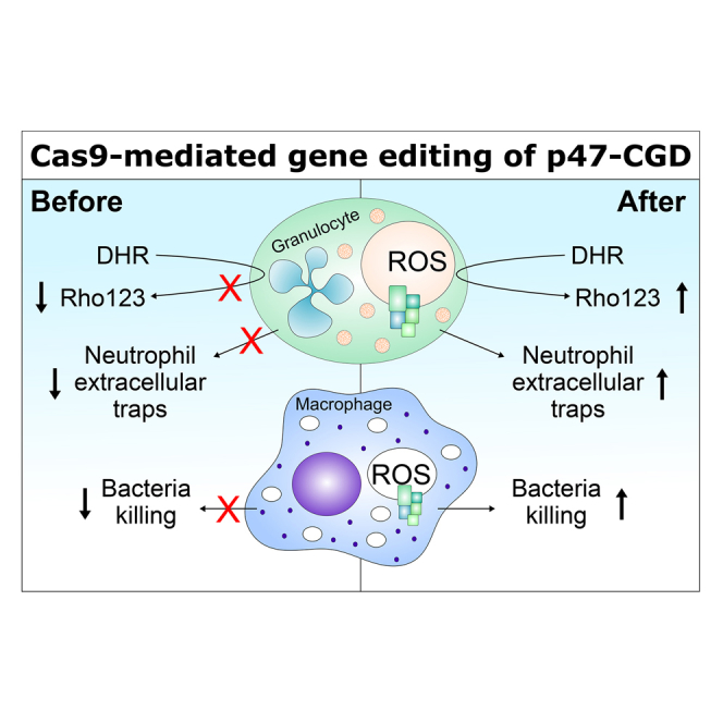
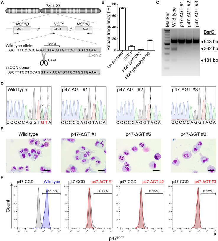
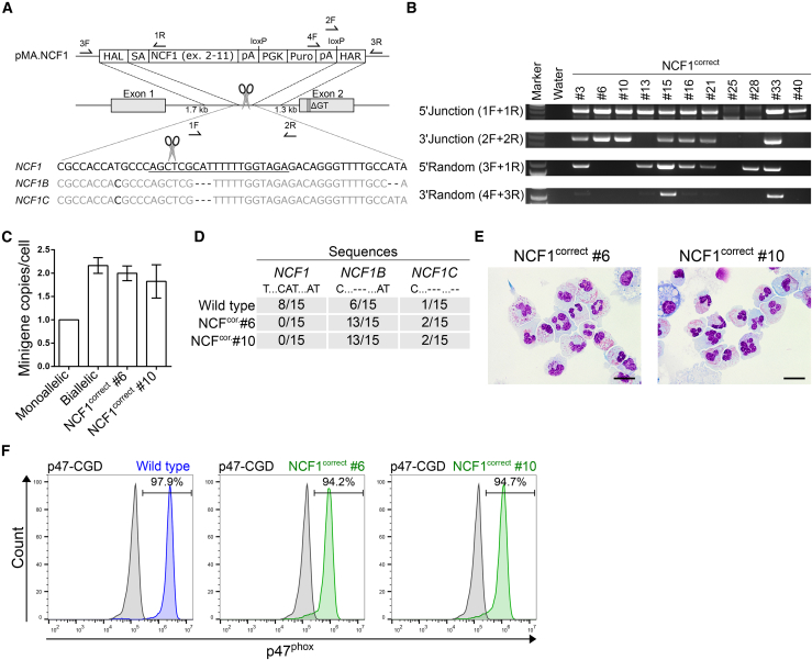
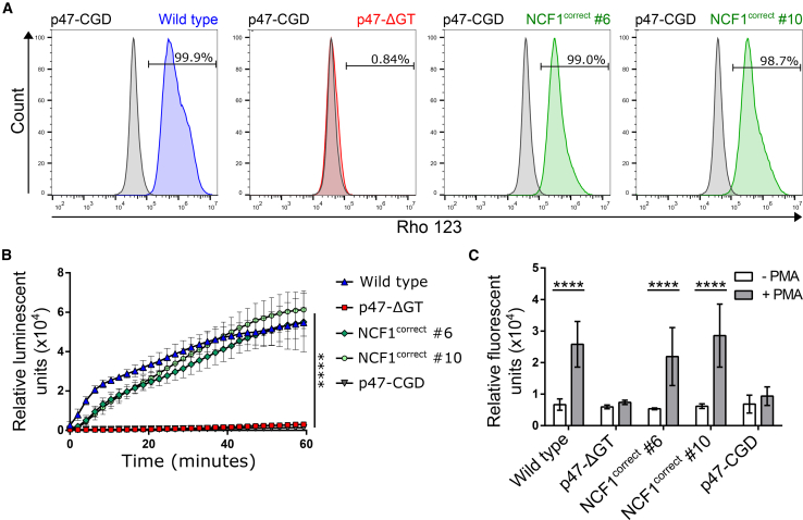
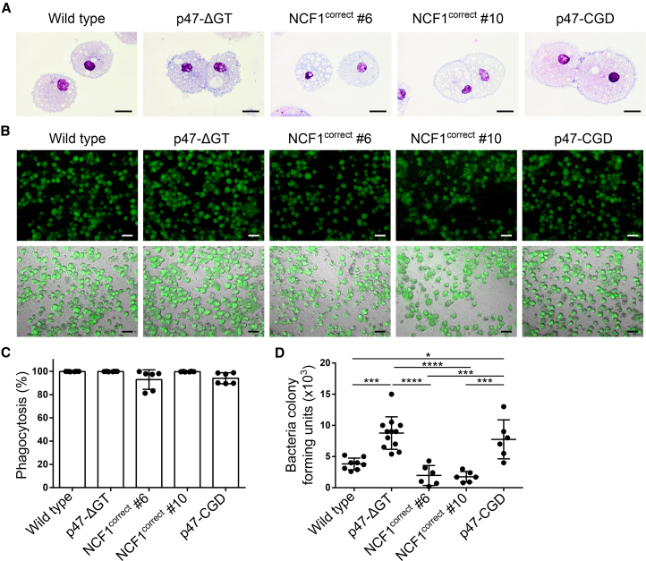
Mutations in the NADPH oxidase, which is crucial for the respiratory burst in phagocytes, result in chronic granulomatous disease (CGD). The only curative treatment option for CGD patients, who suffer from severe infections, is allogeneic bone marrow transplantation. Over 90% of patients with mutations in the p47phox subunit of the oxidase complex carry the deletion c.75_76delGT (ΔGT). This frequent mutation most likely originates via gene conversion from one of the two pseudogenes NCF1B or NCF1C, which are highly homologous to NCF1 (encodes p47phox) but carry the ΔGT mutation. We applied CRISPR/Cas9 to generate patient-like p47-ΔGT iPSCs for disease modeling. To avoid unpredictable chromosomal rearrangements by CRISPR/Cas9-mediated cleavage in the pseudogenes, we developed a gene-correction approach to specifically target NCF1 but leave the pseudogenes intact. Functional assays revealed restored NADPH oxidase activity and killing of bacteria in corrected phagocytes as well as the specificity of this approach.

Keywords: CRISPR/Cas9; NADPH oxidase; NCF1; chronic granulomatous disease (CGD); gene editing; human induced pluripotent stem cells; p47phox; pseudogenes.


Figures

None
Graphical abstract
Figure 1
Figure 1
Generation of Patient-like p47-ΔGT iPSCs Using CRISPR/Cas9 (A) Schematic of NCF1 and its pseudogenes NCF1B/C located on chromosome 7. The sgRNA target sequence (underlined) is shown on the wild type allele in the 5′ region of exon 2 (gray box). The donor template (ssODN) carries the ΔGT mutation and a silent point mutation (bold). (B) Frequency of repaired alleles by non-homologous end-joining (NHEJ) or homology-directed repair (HDR) after Cas9-mediated cleavage. The frequency was determined in sorted bulk populations by next-generation sequencing. The name in parentheses indicates the donor template used (n = 3, mean ± SD). (C) PCR amplification and BsrGI digest to discriminate correctly modified iPSC clones (BsrGI site removed) from wild type (BsrGI site present). (D) Sanger sequencing results of wild type iPSCs and p47-ΔGT clones. The wild type sequence shows overlaid sequences of NCF1 and the pseudogenes due to co-amplification via PCR (asterisk). (E) Cell morphology of differentiated iPSC-derived granulocytes for wild type and p47-ΔGT cells after Pappenheim staining. Scale bars, 20 μm. (F) Intracellular staining followed by flow cytometry to detect p47phox expression in wild type and p47-ΔGT granulocytes (gated on p47-CGD granulocytes).
Figure 2
Figure 2
Genetic Correction of p47-ΔGT iPSCs by Targeted Insertion of a Minigene into Intron 1 of NCF1 (A) Schematic of gene editing and correction strategy. The sgRNA target sequence (underlined) differs from the pseudogene sequence by three additional nucleotides. Arrows indicate positions of primers used for genotyping. F, forward primer; R, reverse primer; HAL, homology arm left; SA, splice acceptor site; NCF1, cDNA encoding the NCF1 gene excluding exon 1; pA, polyadenylation signal; PGK, phosphoglycerate kinase promoter; Puro, puromycin resistance gene; HAR, homology arm right. (B) PCR-based genotyping. Applied primers were used as indicated, and their binding sites are depicted in (A). (C) Determination of the minigene copy number normalized to the PTBP2 gene via qPCR (n = 3, mean ± SD, technical replicates). (D) Sequencing analysis of the region adjacent to the p47.in1 target site in NCF1 and the pseudogenes. The PCR fragment was subcloned and single clones were analyzed by Sanger sequencing. Sequences were identified as NCF1, NCF1B, or NCF1C based on specific point mutations and deletions as indicated. (E) Cell morphology of corrected iPSC-derived granulocytes after Pappenheim staining. Scale bars, 20 μm. (F) Intracellular staining followed by flow cytometry to detect p47phox expression in wild type and corrected granulocytes (gated on p47-CGD granulocytes).
Figure 3
Figure 3
Functional Characterization of Corrected iPSC-Derived Granulocytes (A) DHR assay. After PMA stimulation, DHR is converted in the presence of ROS to green fluorescent Rho123 and assessed by flow cytometry (gated on stimulated p47-CGD granulocytes). (B) Chemiluminescent ROS assay. ROS production was measured in the presence of luminol over time in granulocytes after PMA stimulation (n = 4–7 pooled from three independent experiments; mean ± SD, one-way ANOVA; ∗∗∗∗p ≤ 0.0001). (C) NET formation assay. NET formation was quantified by Sytox green staining 2 h after PMA stimulation and compared with unstimulated granulocytes (n = 3–9 pooled from three independent experiments; mean ± SD, two-way ANOVA; ∗∗∗∗p ≤ 0.0001). Significance is only shown for stimulation.
Figure 4
Figure 4
Corrected iPSC-Derived Macrophages Kill Phagocytosed Bacteria (A) Cell morphology of iPSC-derived macrophages after Pappenheim staining. Scale bars, 20 μm. (B–D) E. coli killing assay. (B) Macrophages are infected with sfGFP-labeled E. coli at an MOI of 1. Shown are microscopic pictures of macrophages that phagocytosed bacteria. Top panel: sfGFP; bottom panel: bright-field + sfGFP (scale bars, 50 μm). (C) Phagocytosis rate of total macrophages measured 6 h after infection with sfGFP-labeled E. coli by flow cytometry (n = 6 pooled from three independent experiments; mean ± SD, one-way ANOVA—no significance). (D) Colony-forming units of E. coli bacteria isolated from cell lysates of infected macrophages 24 h after infection (n = 6–12 pooled from three independent experiments; mean ± SD, one-way ANOVA; p ≤ 0.05, ∗∗∗p ≤ 0.001, ∗∗∗∗p ≤ 0.0001).

References

    1. Ackermann M., Kempf H., Hetzel M., Hesse C., Hashtchin A.R., Brinkert K., Schott J.W., Haake K., Kühnel M.P., Glage S. Bioreactor-based mass production of human iPSC-derived macrophages enables immunotherapies against bacterial airway infections. Nat. Commun. 2018;9:5088–6001. - PMC - PubMed
    1. Ackermann M., Lachmann N., Hartung S., Eggenschwiler R., Pfaff N., Happle C., Mucci A., Göhring G., Niemann H., Hansen G. Promoter and lineage independent anti-silencing activity of the A2 ubiquitous chromatin opening element for optimized human pluripotent stem cell-based gene therapy. Biomaterials. 2014;35:1531–1542. - PubMed
    1. Connelly J.A., Marsh R., Parikh S., Talano J. Allogeneic hematopoietic cell transplantation for chronic granulomatous disease: controversies and state of the art. J. Pediatr. Infect. Dis. Soc. 2018;7:31–39. - PMC - PubMed
    1. Greve B., Hoffmann P., Vonthein R., Kun J., Lell B., Mycko M.P., Selmaj K.W., Berger K., Weissert R., Kremsner P.G. NCF1 gene and pseudogene pattern: association with parasitic infection and autoimmunity. Malar. J. 2008;7:251–257. - PMC - PubMed
    1. Güngör T., Teira P., Slatter M., Stussi G., Stepensky P., Moshous D., Vermont C., Ahmad I., Shaw P.J., Telles da Cunha J.M. Reduced-intensity conditioning and HLA-matched haemopoietic stem-cell transplantation in patients with chronic granulomatous disease: a prospective multicentre study. Lancet. 2014;383:436–448. - PubMed

Publication types

MeSH terms

Substances

LinkOut - more resources