. 2019 Nov 8;294(45):16684-16697.
doi: 10.1074/jbc.RA119.009133. Epub 2019 Sep 22.

The myotubularin MTMR4 regulates phagosomal phosphatidylinositol 3-phosphate turnover and phagocytosis

Affiliations

The myotubularin MTMR4 regulates phagosomal phosphatidylinositol 3-phosphate turnover and phagocytosis

David A Sheffield et al. J Biol Chem. .

Abstract

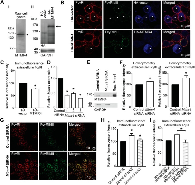
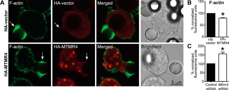
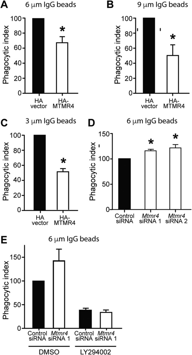
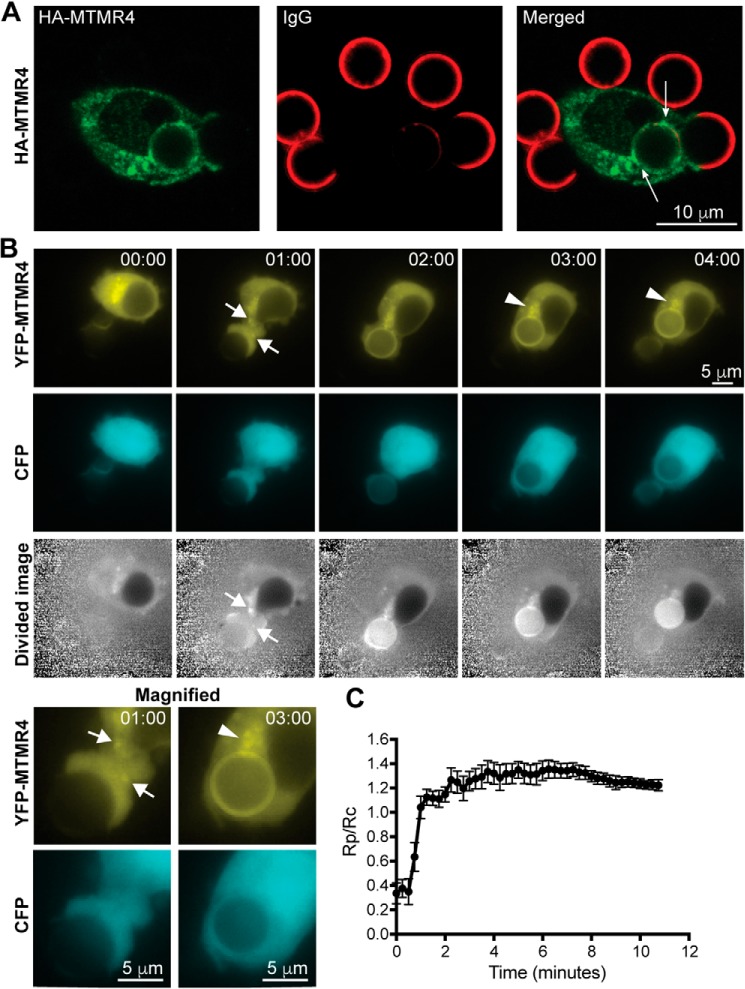
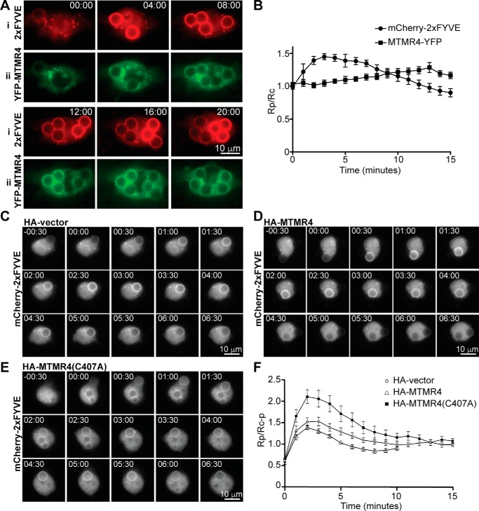
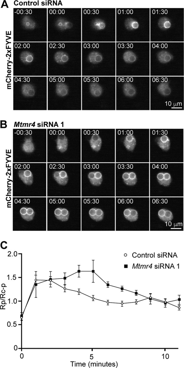
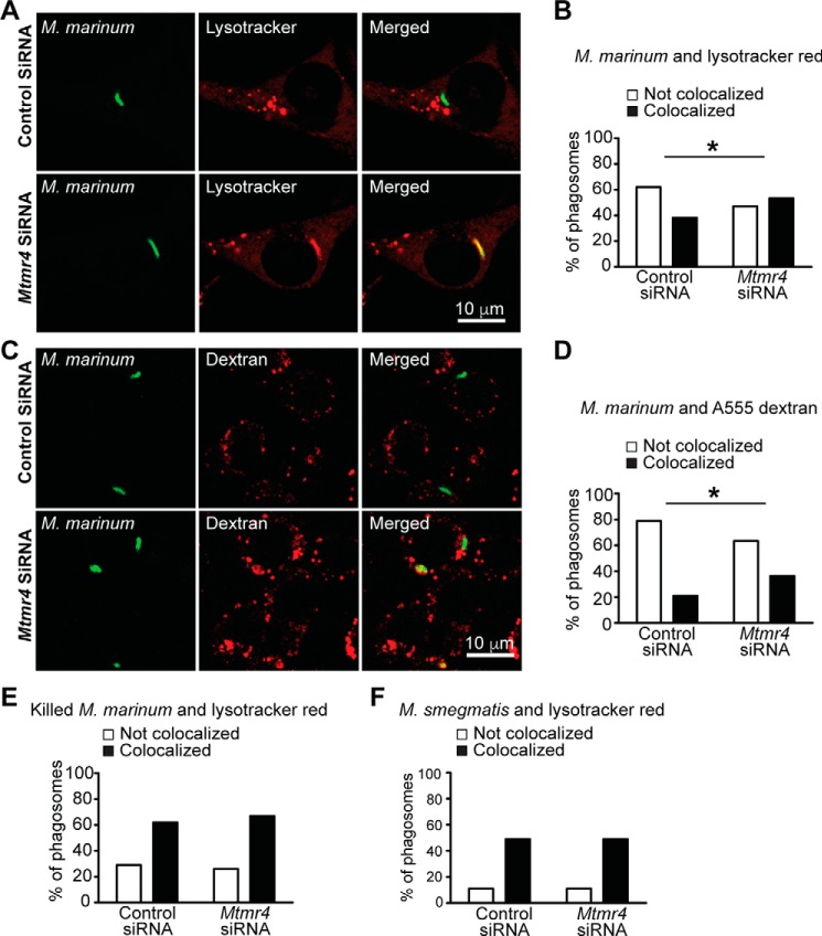
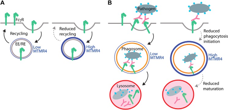
Macrophage phagocytosis is required for effective clearance of invading bacteria and other microbes. Coordinated phosphoinositide signaling is critical both for phagocytic particle engulfment and subsequent phagosomal maturation to a degradative organelle. Phosphatidylinositol 3-phosphate (PtdIns(3)P) is a phosphoinositide that is rapidly synthesized and degraded on phagosomal membranes, where it recruits FYVE domain- and PX motif-containing proteins that promote phagosomal maturation. However, the molecular mechanisms that regulate PtdIns(3)P removal from the phagosome have remained unclear. We report here that a myotubularin PtdIns(3)P 3-phosphatase, myotubularin-related protein-4 (MTMR4), regulates macrophage phagocytosis. MTMR4 overexpression reduced and siRNA-mediated Mtmr4 silencing increased levels of cell-surface immunoglobulin receptors (i.e. Fcγ receptors (FcγRs)) on RAW 264.7 macrophages, associated with altered pseudopodal F-actin. Furthermore, MTMR4 negatively regulated the phagocytosis of IgG-opsonized particles, indicating that MTMR4 inhibits FcγR-mediated phagocytosis, and was dynamically recruited to phagosomes of macrophages during phagocytosis. MTMR4 overexpression decreased and Mtmr4-specific siRNA expression increased the duration of PtdIns(3)P on phagosomal membranes. Macrophages treated with Mtmr4-specific siRNA were more resistant to Mycobacterium marinum-induced phagosome arrest, associated with increased maturation of mycobacterial phagosomes, indicating that extended PtdIns(3)P signaling on phagosomes in the Mtmr4-knockdown cells permitted trafficking of phagosomes to acidic late endosomal and lysosomal compartments. In conclusion, our findings indicate that MTMR4 regulates PtdIns(3)P degradation in macrophages and thereby controls phagocytosis and phagosomal maturation.

Keywords: Fc-gamma receptor; innate immunity; macrophage; mycobacteria; myotubularin-related protein-4 (MTMR4); phagocytosis; phagosome; phosphatidylinositol 3-phosphate (PtdIns(3)P); phosphatidylinositol phosphatase.


Conflict of interest statement

The authors declare that they have no conflicts of interest with the contents of this article

Figures

Figure 1.
Figure 1.
MTMR4 regulates surface levels of FcγR on RAW 264.7 macrophages. A, lysates of nontransfected (i) and HA-vector– or HA-MTMR4–transfected (ii) RAW 264.7 macrophages were subjected to SDS-PAGE and immunoblotted using a polyclonal antibody raised against recombinant MTMR4 or an anti-HA antibody for the detection of endogenous MTMR4 (arrowhead) and recombinant HA-MTMR4 (arrow), respectively. An anti-GAPDH antibody was used as loading control. B, cells were transfected with HA-vector or HA-MTMR4 as indicated and fixed. Unpermeabilized cells were stained with anti-FcγRI (red) and anti-FcγRII/III (green) antibodies and were thereafter permeabilized and stained again using an anti-HA antibody (blue) to detect transfected cells (marked with an asterisk). C, the FcγRI fluorescence signal in B was quantified in three independent experiments with 30 cells analyzed per condition. Within experiments, the fluorescence was normalized to that of the control condition, which was arbitrarily assigned a value of 100. D, RAW 264.7 cells were treated with negative control or Mtmr4 siRNA for 72 h (siRNA 1) or 48 h (siRNA 2 and siRNA 3), after which Mtmr4 mRNA levels were quantitated by RT-PCR analysis relative to Gapdh. mRNA levels were normalized to that of control siRNA cells, which was arbitrarily assigned a value of 1. E, siRNA-mediated knockdown of MTMR4 in lysates from control or Mtmr4 siRNA 1–treated cells was assessed by Western blotting using a polyclonal anti-MTMR4 antibody and anti-GAPDH antibody as loading control. F, cells were treated with control or Mtmr4 siRNA 3, and FcγRI and FcγRII/III signal fluorescence was quantified by flow cytometry in six independent experiments with >1000 cells analyzed. Fluorescence was normalized to that of control siRNA cells, which was arbitrarily assigned a value of 100. G, immunofluorescent micrographs of cells transfected with control siRNA or Mtmr4 siRNA 3, as indicated, and immunostained using anti-FcγRI and -FcγRII/III antibodies. H, the FcγRI signal fluorescence of cells transfected with control siRNA or Mtmr4 siRNA 2 or 3 was quantified in three independent experiments with 30 cells analyzed per condition. Within experiments, the fluorescence was normalized to that of the control condition, which was arbitrarily assigned a value of 100. I, the relative FcγRI fluorescence signal intensity of cells transfected with HA-vector or HA-MTMR4 and control or Mtmr4 siRNA 3 was quantified. Within experiments, the fluorescence was normalized to that of the control condition, which was arbitrarily assigned a value of 100. *, p < 0.05, two-tailed paired t test. Scale bars, 10 μm. Error bars, S.E. Images are representative of at least three independent experiments.
Figure 2.
Figure 2.
MTMR4 regulates pseudopodal F-actin during IgG-mediated phagocytosis. A, RAW 264.7 cells were transfected with HA-vector or HA-MTMR4, allowed to commence phagocytosis of bIgG-6μm, fixed, and stained with phalloidin (green) and anti-HA antibodies (red) to detect F-actin and transfected cells, respectively. Immunofluorescent micrographs of transfected cells as well as merged and bright-field images are shown. Arrows, phagocytic cups. Scale bars, 5 μm. B, phalloidin fluorescence was quantified on phagocytic cups in HA-vector– or HA-MTMR4–transfected cells undergoing phagocytosis of bIgG-6μm, after which they were fixed and stained as in A. 27–34 phagosomes were examined in three independent experiments, and fluorescence was normalized to the control condition, which was arbitrarily assigned a value of 100. C, quantification of pseudopodal phalloidin signal in cells treated with control or Mtrm4 siRNA 1 undergoing phagocytosis, fixed and stained as described in A. 176–200 phagosomes were examined in four independent experiments, and fluorescence was normalized to the control condition, which was arbitrarily assigned a value of 100. Error bars, S.E. *, p < 0.05, two-tailed paired t test. Images are representative of at least three independent experiments.
Figure 3.
Figure 3.
MTMR4 negatively regulates phagocytotic engulfment. 24 h post-transfection with HA-vector or HA-MTMR4, RAW 264.7 cells underwent synchronized phagocytosis of IgG-opsonized latex beads for 15 min. The phagocytic index was calculated as the number of beads fully internalized per 100 cells and normalized to the control condition, which was arbitrarily assigned a value of 100. A, phagocytic index upon phagocytosis of bIgG-6μm in n = 5 independent experiments; B, bIgG-9μm in n = 4 independent experiments; C, bIgG-3μm in n = 3 independent experiments. D, RAW 264.7 cells were transfected with control or Mtmr4 siRNA 1 or Mtmr4 siRNA 2, prior to phagocytosis of bIgG-6μm in n = 4 and 5 independent experiments, respectively. E, RAW 264.7 cells were transfected with control or Mtmr4 siRNA 1, incubated with vehicle (DMSO) or 100 μm LY294002 for 30 min, and then allowed to phagocytose bIgG-6μm in the presence of LY294002 for 15 min, and the phagocytic index was scored in n = 3 independent experiments. *, p < 0.05, two-tailed paired t test. Error bars, S.E.
Figure 4.
Figure 4.
MTMR4 is recruited to phagosomes in a time-dependent fashion. A, RAW 264.7 cells were transfected with HA-MTMR4 and allowed to phagocytose bIgG-6μm before fixation, staining (anti-HA in green and anti-IgG in red), and confocal imaging. HA-MTMR4 punctate signals were observed in a perinuclear distribution and on the phagosome (arrows) containing internalized IgG beads. Scale bars, 10 μm. B, RAW 264.7 cells were cotransfected with YFP-MTMR4 and CFP and imaged live during the phagocytosis of bIgG-6μm. Time = 00:00 was defined as the time of the first extension of pseudopods onto the bead. Shown are YFP, CFP, and divided (YFP/CFP) fluorescent images. Magnified images at 01:00 min and 03:00 min in the fluorescent channels are shown. Arrows, YFP-MTMR4 vesicles mobilizing toward the phagocytic cup. Arrowheads, YFP-MTMR4 on perinuclear vesicles. Scale bars, 5 μm. C, average fluorescence ratios of Rp (phagosomal YFP signal/phagosomal CFP signal)/Rc (cell YFP signal/cell CFP signal) analyzed for n = 5 cells (10 phagosomes). Measurements at the phagosome (Rp) and of the cell (Rc) were taken from the divided cell image. Error bars, S.E. Images are representative of at least three independent experiments.
Figure 5.
Figure 5.
MTMR4 negatively regulates the duration of PtdIns(3)P signaling on phagosomes. A, RAW 264.7 cells were cotransfected with MTMR4-YFP and 2xFYVE and imaged live during the phagocytosis of bIgG-6μm. Time = 00:00 was defined as the time of pseudopod closure around the first internalized bead. Scale bars, 10 μm. B, average mCherry-2xFYVE and MTMR4-YFP fluorescence ratios of Rp (phagosomal fluorescence signal)/Rc (cell fluorescence signal) of bIgG-6μm phagosomes in cotransfected RAW 264.7 cells. n = 5 beads from two experiments combined. C–E, cells were cotransfected with mCherry-2xFYVE and HA-vector (C), HA-MTMR4 (D), or HA-MTMR4(C407A) (E) at a 1:10 ratio, and time-lapse imaging was conducted during the phagocytosis of bIgG-6μm, with recording of mCherry fluorescence at 30-s intervals. Time = 00:00 was defined as the time of pseudopod closure around the first internalized bead. Scale bars, 10 μm. F, average mCherry fluorescence was quantitated and Rp (phagosomal mCherry fluorescence)/Rc − p (total cellular mCherry fluorescence-phagosomal mCherry fluorescence) was calculated at each time point with n = 15 beads (HA), 11 beads (HA-MTMR4), and 22 beads (HA-MTMR4(C407A)). Error bars, S.E. Images are representative of at least three independent experiments.
Figure 6.
Figure 6.
siRNA knockdown of Mtmr4 results in sustained phagosomal PtdIns(3)P signaling. A and B, RAW 264.7 cells were treated with control siRNA (A) or Mtmr4 siRNA 1 (B) for 48 h and then transfected with mCherry-2xFYVE and the following day imaged live during the phagocytosis of bIgG-6μm, with recording of mCherry fluorescence at 30-s intervals. Time = 00:00 was defined as the time of pseudopod closure around the first internalized bead. Scale bars, 10 μm. C, average mCherry fluorescence was quantitated, and Rp (phagosomal mCherry fluorescence)/Rc − p (total cellular mCherry fluorescence-phagosomal mCherry fluorescence) was calculated at each time point with n = 6–10 cells for each condition. Error bars, S.E. Images are representative of at least three independent experiments.
Figure 7.
Figure 7.
siRNA knockdown of Mtmr4 results in increased maturation of phagosomes containing pathogenic M. marinum. A, RAW 264.7 cells were treated with control siRNA or Mtmr4 siRNA 1 for 72 h. Cells underwent synchronized phagocytosis of GFP-M. marinum for 2 h, after which they were incubated with LysoTracker Red to stain acidic compartments, prior to fixation and confocal microscopy. B, quantification of mycobacterial colocalization with acidic compartments in cells treated with control or Mtmr4 siRNA 1 as shown by the percentage of GFP-positive phagosomes not colocalizing or colocalizing with LysoTracker Red–positive compartments. Results are pooled from three independent experiments with 120 phagosomes analyzed for each condition. C, RAW 264.7 cells were treated with control or Mtmr4 siRNA 1 for 72 h. Late endosomal and lysosomal compartments were labeled by incubation with Alexa Fluor 555–dextran for 2 h prior to mycobacterial exposure. Cells underwent synchronized phagocytosis of live GFP-M. marinum for 2 h, prior to fixation and visualization by confocal microscopy. D, quantification of colocalization of M. marinum with late endosomal and lysosomal compartments as shown by the percentage of GFP-positive phagosomes not colocalizing or colocalizing with Alexa Fluor 555–dextran–positive compartments. Results are pooled from two independent experiments with 100–107 phagosomes analyzed for each condition. E and F, RAW 264.7 cells were treated with control or Mtmr4 siRNA 1 for 72 h prior to synchronized mycobacterial phagocytosis and 2-h chase and incubated with LysoTracker Red to label acidic compartments prior to fixation. E, quantification of LysoTracker Red colocalization with paraformaldehyde-killed GFP-M. marinum. Results are pooled from three independent experiments with 91–93 phagosomes analyzed for each condition. F, quantification of LysoTracker Red colocalization with nonpathogenic M. smegmatis. Results are pooled from two independent experiments with 60 phagosomes analyzed for each condition. *, p < 0.05, Fisher's exact test. Scale bars, 10 μm. Images are representative of at least three independent experiments.
Figure 8.
Figure 8.
MTMR4 potentially regulates phagocytosis by two mechanisms. A, recycling of FcγRs under conditions of low and high levels of MTMR4 (shown in blue). MTMR4 associates with early and recycling endosomes (EE/RE) where it dephosphorylates PtdIns(3)P, potentially resulting in reduced recycling and therefore reduced FcγR surface levels. B, FcγR-mediated phagocytosis of a pathogen under conditions of low and high levels of MTMR4. High levels of MTMR4 reduce FcγR surface levels, resulting in reduced initiation of phagocytosis. MTMR4 also localizes to the phagosome, where PtdIns(3)P dephosphorylation potentially reduces phagosome maturation and fusion with lysosomes. PtdIns(3)P-dependent phagosomal arrest induced by pathogenic mycobacteria may thus be overcome by MTMR4 depletion, which prolongates PtdIns(3)P signaling on phagosomes.

Similar articles

Cited by

References

    1. Flannagan R. S., Cosío G., and Grinstein S. (2009) Antimicrobial mechanisms of phagocytes and bacterial evasion strategies. Nat. Rev. Microbiol. 7, 355–366 10.1038/nrmicro2128 - DOI - PubMed
    1. Poon I. K., Hulett M. D., and Parish C. R. (2010) Molecular mechanisms of late apoptotic/necrotic cell clearance. Cell Death Differ. 17, 381–397 10.1038/cdd.2009.195 - DOI - PubMed
    1. Yeung T., Ozdamar B., Paroutis P., and Grinstein S. (2006) Lipid metabolism and dynamics during phagocytosis. Curr. Opin. Cell Biol. 18, 429–437 10.1016/j.ceb.2006.06.006 - DOI - PubMed
    1. Ellson C. D., Anderson K. E., Morgan G., Chilvers E. R., Lipp P., Stephens L. R., and Hawkins P. T. (2001) Phosphatidylinositol 3-phosphate is generated in phagosomal membranes. Curr. Biol. 11, 1631–1635 10.1016/S0960-9822(01)00447-X - DOI - PubMed
    1. Vieira O. V., Botelho R. J., Rameh L., Brachmann S. M., Matsuo T., Davidson H. W., Schreiber A., Backer J. M., Cantley L. C., and Grinstein S. (2001) Distinct roles of class I and class III phosphatidylinositol 3-kinases in phagosome formation and maturation. J. Cell Biol. 155, 19–25 10.1083/jcb.200107069 - DOI - PMC - PubMed

Publication types

MeSH terms

LinkOut - more resources