. 2019 Sep 23;10(10):705.
doi: 10.1038/s41419-019-1942-1.

MicroRNA-29a represses osteoclast formation and protects against osteoporosis by regulating PCAF-mediated RANKL and CXCL12

Affiliations

MicroRNA-29a represses osteoclast formation and protects against osteoporosis by regulating PCAF-mediated RANKL and CXCL12

Wei-Shiung Lian et al. Cell Death Dis. .

Abstract

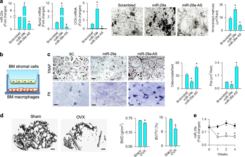
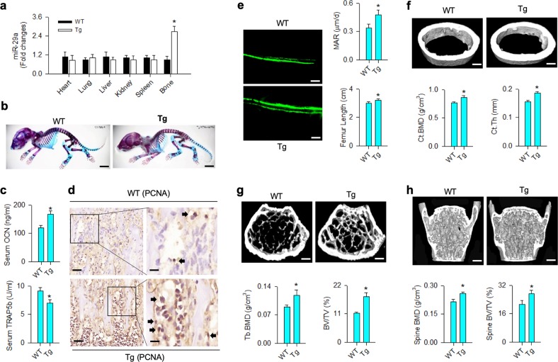
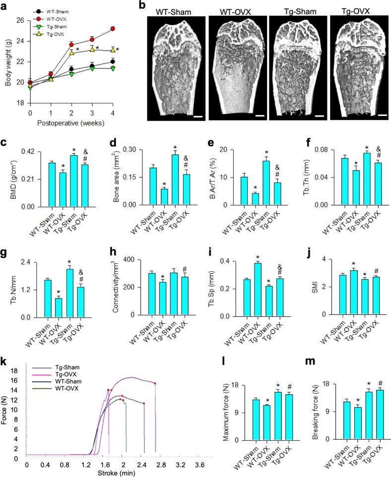
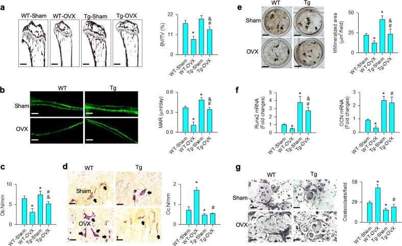
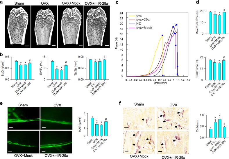
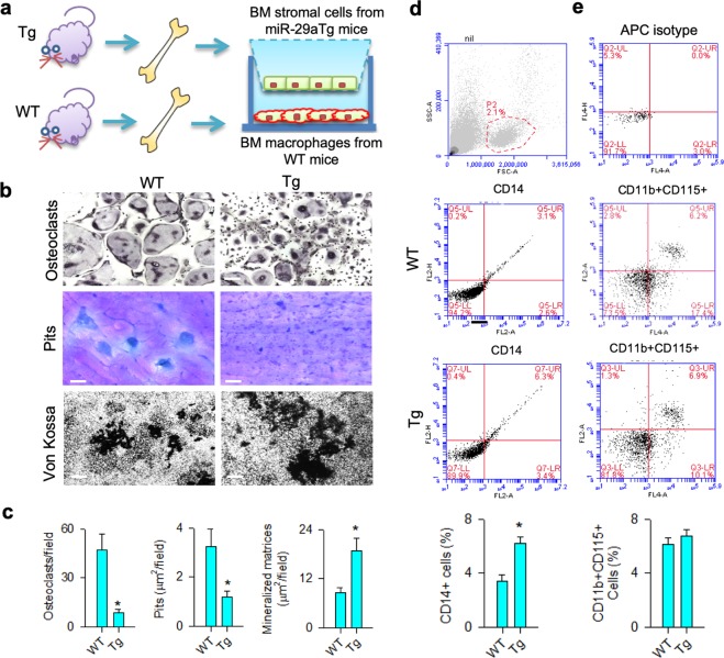
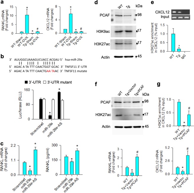
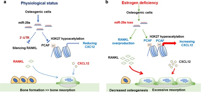
Osteoporosis deteriorates bone mass and biomechanical strength, becoming a life-threatening cause to the elderly. MicroRNA is known to regulate tissue remodeling; however, its role in the development of osteoporosis remains elusive. In this study, we uncovered that silencing miR-29a expression decreased mineralized matrix production in osteogenic cells, whereas osteoclast differentiation and pit formation were upregulated in bone marrow macrophages as co-incubated with the osteogenic cells in transwell plates. In vivo, decreased miR-29a expression occurred in ovariectomy-mediated osteoporotic skeletons. Mice overexpressing miR-29a in osteoblasts driven by osteocalcin promoter (miR-29aTg/OCN) displayed higher bone mineral density, trabecular volume and mineral acquisition than wild-type mice. The estrogen deficiency-induced loss of bone mass, trabecular morphometry, mechanical properties, mineral accretion and osteogenesis of bone marrow mesenchymal cells were compromised in miR-29aTg/OCN mice. miR-29a overexpression also attenuated the estrogen loss-mediated excessive osteoclast surface histopathology, osteoclast formation of bone marrow macrophages, receptor activator nuclear factor-κ ligand (RANKL) and C-X-C motif chemokine ligand 12 (CXCL12) expression. Treatment with miR-29a precursor improved the ovariectomy-mediated skeletal deterioration and biomechanical property loss. Mechanistically, miR-29a inhibited RANKL secretion in osteoblasts through binding to 3'-UTR of RANKL. It also suppressed the histone acetyltransferase PCAF-mediated acetylation of lysine 27 in histone 3 (H3K27ac) and decreased the H3K27ac enrichment in CXCL12 promoters. Taken together, miR-29a signaling in osteogenic cells protects bone tissue from osteoporosis through repressing osteoclast regulators RANKL and CXCL12 to reduce osteoclastogenic differentiation. Arrays of analyses shed new light on the miR-29a regulation of crosstalk between osteogenic and osteoclastogenic cells. We also highlight that increasing miR-29a function in osteoblasts is beneficial for bone anabolism to fend off estrogen deficiency-induced excessive osteoclastic resorption and osteoporosis.


Conflict of interest statement

The authors declare that they have no conflict of interest.

Figures

Fig. 1
Fig. 1. Analysis of miR-29a actions to osteogenic and osteoclastogenic differentiation.
Increased Runx2 and osteocalcin expression and von Kossa-stained mineralized matrix formation in miR-29a-transfected bone marrow mesenchymal cells (a); scale bar: 40 μm. Schematic drawing of bone marrow macrophages and mesenchymal cells co-incubated in transwell plates (b). Increasing miR-29a in osteoblasts decreased TRAP-stained osteoclast differentiation (scale bar: 8 μm) and pit formation (scale bar: 20 μm) of bone marrow macrophages (c). The data of in vitro model are expressed as mean ± SEM calculated from four experiments. Asterisks * indicate significant difference from the scrambled control. Sparse trabecular microstructure (scale bar: 5 mm) along with decreased BMD, BV/TV (d), and miR-29a expression in bone tissue (e) occurred in ovariectomized mice. Investigations are expressed mean ± SEM calculated from six mice. Asterisks * indicate significant difference from sham controls
Fig. 2
Fig. 2. Skeletal phenotypes and bone microstructure of miR-29aTg/OCN mice.
Increased miR-29a expression solely occurred in bone tissue in miR-29aTg/OCN mice (a). Macroscopic morphology of 7-day-old miR-29aTg/OCN mice was similar to wild-type mice (b); scale bar: 20 mm. Increased serum osteocalcin and reduced TRAP5b levels (c) and strong PCNA immunostaining (d) in osteoblasts in miR-29aTg/OCN mice; scale bar: 30 μm (low magnification); 10 μm (high magnification). Increased fluorescent calcein accumulation and MAR along with femur length in 8-week-old miR-29aTg/OCN mice (e); scale bar: 30 μm. Significant increases in cortical BMD and Ct.Th (f), trabecular BMD and BV/TV (g) together with spinal BMD and BV/TV (h) in miR-29aTg/OCN mice; scale bar: 5 mm. The data are expressed mean ± SEM calculated from six mice. Asterisks * indicate significant difference from wild-type mice
Fig. 3
Fig. 3. Analysis of bone mass and biomechanical property of skeletal tissue.
miR-29a overexpression reduced the ovariectomy-induced excessive body weight gain (a) and retained abundant trabecular microstructure (b); scale bar: 5 mm. The ovariectomy-induced loss of BMD (c), bone area (d), B.Ar/T.Ar (e), Tb.Th (f), Tb.N (g), and connectivity (h) were attenuated in miR-29aTg/OCN mice, whereas Tb.Sp (i) and SMI (j) were improved. Load-displacement curve (k) showed minor responses to ovariectomy-induced downregulation of maximum force (l) and breaking force (m) in miR-29aTg/OCN mice. The data are expressed mean ± SEM calculated from six mice. Asterisks * and hashtags # indicate significant difference from sham controls and OVX, respectively. Ampersands & indicate significant difference from Tg-Sham and Tg-OVX
Fig. 4
Fig. 4. Analysis of bone histology and ex vivo osteogenesis and osteoclastogenesis.
miR-29a overexpression repressed the ovariectomy-mediated loss of trabecular histology (scale bar: 120 μm), BV/TV (a), fluorescent calcein labeling (scale bar: 30 μm), MAR (b) and Ob.N (c), as well as compromised excessive TRAP-positive osteoclast distribution (scale bar: 8 μm) and Oc.N (d). miR-29aTg/OCN mice showed minor response to the ovariectomy-induced loss of mineralized matrix formation (scale bar: 7 mm) (e), Runx2 and osteocalcin expression (f) of primary bone marrow mesenchymal cells and downregulated osteoclast formation of primary bone marrow macrophages (g); scale bar: 8 μm. The data are expressed mean ± SEM calculated from six mice. Asterisks * and hashtags # indicate significant difference from sham controls and OVX, respectively. Ampersands & indicate significant difference from Tg-Sham and Tg-OVX
Fig. 5
Fig. 5. miR-29a precursor treatment for bone microstructure and histology.
miR-29a precursor attenuated the ovariectomy-induced loss of trabecular microarchitecture (a); scale bar: 5 mm; BMD, BV/TV, and Tb.Th (b), as well as improved load-displacement profile (c), maximum force and breaking force (d). The treatment retained fluorescent calcein accumulation (scale bar: 40 μm) and MAR (e) and compromised the ovariectomy upregulation of osteoclast distribution (scale bar: 8 μm) and Oc.N (f). The data are expressed mean ± SEM calculated from eight mice. Asterisks * and hashtags # indicate significant difference from sham controls and OVX, respectively
Fig. 6
Fig. 6. Osteoclast-formation capacity of bone marrow macrophages in miR-29aTg/OCN mice.
Schematic drawing of bone marrow macrophages co-incubated with bone marrow mesenchymal stem cells (a). Decreased osteoclast differentiation (scale bar: 8 μm) and pit formation (scale bar: 20 μm) of wild-type bone marrow macrophage and increased mineralized matrix formation (scale bar: 40 μm) of wild-type bone marrow cells co-incubated with miR-29aTg/OCN bone marrow mesenchymal cells (b, c). Investigations are expressed mean ± SEM calculated from six mice. High CD14 + macrophages (d) rather than CD11 + CD115 + monocytes (e) existed in bone marrow in miR-29aTg/OCN mice. Investigations are expressed mean ± SEM calculated from four mice. Asterisks * indicate significant difference from wild-type mice
Fig. 7
Fig. 7. Analysis of miR-29a inhibition of RANKL and CXCL12 expression in bone marrow mesenchymal cells.
miR-29a compromised the ovariectomy upregulation of RANKL and CXCL12 expression in bone marrow mesenchymal cells (a). The data are expressed mean ± SEM calculated from six mice. Asterisks * and hashtags # indicate significant difference from sham controls and OVX, respectively. Forced miR-29a expression decreased luciferase activity of 3′-UTR of RANKL mRNA (b) and downregulated RANKL mRNA expression and protein abundances, whereas miR-29 interference upregulated RANKL expression (c) of mesenchymal stem cell cultures. miR-29a overexpression reduced PCAF and H3K27ac abundances (d) and decreased H3K27ac enrichment in CXCL12 promoters (e). Forced PCAF expression repressed the miR-29a downregulation of H3K27ac levels and RANKL expression (f). It increased H3K27ac occupancy in CXCL12 promoters and CXCL12 expression (g) in bone marrow mesenchymal cells. Investigations are expressed mean ± SEM calculated from three experiments. Asterisks * and hashtags # indicate significant difference from WT and miR-29aTg/OCN, respectively
Fig. 8
Fig. 8. Schematic drawing of miR-29a signaling in osteoblasts repressed osteoclastic activity and osteoporosis.
miR-29a targets RANKL expression and downregulates PCAF signaling-mediated CXCL12 expression to reduced osteoclastic activity, sustaining bone mass (a). Estrogen loss decreases miR-29a loss, which augments RANKL and CXCL12 expression in osteogenic cells, accelerating osteoclast formation and osteoporosis development (b)

Similar articles

Cited by

References

    1. Black DM, Rosen CJ. Clinical Practice. Postmenopausal osteoporosis. N. Engl. J. Med. 2016;374:254–262. doi: 10.1056/NEJMcp1513724. - DOI - PubMed
    1. Khosla S, Hofbauer LC. Osteoporosis treatment: recent developments and ongoing challenges. Lancet Diabetes Endocrinol. 2017;5:898–907. doi: 10.1016/S2213-8587(17)30188-2. - DOI - PMC - PubMed
    1. Saag KG, et al. Romosozumab or alendronate for fracture prevention in women with osteoporosis. N. Engl. J. Med. 2017;377:1417–1427. doi: 10.1056/NEJMoa1708322. - DOI - PubMed
    1. Fan Y, et al. Parathyroid hormone directs bone marrow mesenchymal cell fate. Cell Metab. 2017;25:661–672. doi: 10.1016/j.cmet.2017.01.001. - DOI - PMC - PubMed
    1. Compston JE, McClung MR, Leslie WD. Osteoporosis. Lancet. 2019;393:364–376. doi: 10.1016/S0140-6736(18)32112-3. - DOI - PubMed

Publication types

MeSH terms