. 2019 Sep 20;92(3):413-422.
eCollection 2019 Sep.

Identifying Specific Subcellular Organelle Damage by Photosensitized Oxidations

Affiliations

Identifying Specific Subcellular Organelle Damage by Photosensitized Oxidations

Tayana Mazin Tsubone et al. Yale J Biol Med. .

Abstract

The search for conditions that maximize the outcome of Photodynamic Therapy (PDT) continues. Recent data indicate that PDT-induced cell death depends more on the specific intracellular location of the photosensitizer (PS) than on any other parameter. Indeed, knowledge of the PS intracellular location allows the establishment of clear relationships between the mechanism of cell death and the PDT efficacy. In order to determine the intracellular localization sites of a given PS, classical co-localization protocols, which are based in the comparison of the emissive profiles of organelle-specific probes to those of the PS, are usually performed. Since PSs are usually not efficient fluorophores, co-localization protocols require relatively high PS concentrations (micromolar range), distorting the whole proposal of the experiment, as high PS concentration means accumulation in many low-affinity sites. To overcome this difficulty, herein we describe a method that identifies PS intracellular localization by recognizing and quantifying the photodamage at intracellular organelles. We propose that irradiation protocols and characterization of major sites of photodamage results from many cycles of photosensitized oxidations, furnishing an integrated picture of the PS location. By comparing the results of protocols based in either method, we showed that the analysis of the damaged organelles can be conducted at optimal conditions (low PS concentrations), providing clear correlations with cell death mechanisms, which is not the case for the results obtained with co-localization protocols. Experiments using PSs that target either mitochondria or lysosomes were described and investigated in detail, showing that evaluating organelle damage is as simple as performing co-localization protocols.

Keywords: Photodamage; Photodynamic Therapy; intracellular localization; organelles; photosensitizer; specificity.


Figures

Figure 1
Figure 1
Mechanism of photosensitization. The photosensitizer (PS) absorbs light forming an excited singlet state (PS(S1)). The PS(S1) is converted to triplet excited state (PS(T1)) via the intersystem crossing. Then, PS(T1) can transfer energy to oxygen, generating singlet oxygen (1O2) or abstraction of electron or hydrogen-producing radicals such as O2•- and OH.
Figure 2
Figure 2
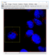
Representative image to show the selected cell in the yellow rectangle. Blue color represents the fluorescence of DAPI and red color the fluorescence of lysosomal probe.
Figure 3
Figure 3
Representative image of only the red channel evident to quantify the pixels. Yellow rectangle depicts the analyzed cell and red color the fluorescence of lysosomal probe.
Figure 4
Figure 4
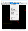
Steps to measure the red pixels.
Figure 5
Figure 5
Finally measuring it and getting the values.
Figure 6
Figure 6
Fluorescence images 3 hours after PDT-treatment of HeLa cells with 100 nM CisDiMPyP and 30 nM TPPS2a and non-treated cells (Control). First column: blue fluorescence of DAPI-stained nuclei; second column: red fluorescence of lysosomes; third column: both channels (blue and red) merged. Scale bar represents 20 µm. This figure was reproduced from Tsubone et al. [21], with permission from Springer Nature under the terms of the Creative Commons Attribution 4.0 International License.
Figure 7
Figure 7
Fluorescence images 3 hours after PDT-treatment of HeLa cells with 100 nM CisDiMPyP and 30 nM TPPS2a and non-treated cells (Control). First column: blue fluorescence of DAPI-stained nuclei; second column: red fluorescence of mitochondria; third column: both channels (blue and red) merged. Scale bar represents 20 µm. This figure was reproduced from Tsubone et al. [21], with permission from Springer Nature under the terms of the Creative Commons Attribution 4.0 International License.
Figure 8
Figure 8
Average of red fluorescence calculated by relative to the fluorescent pixels number in the treated cells compared to control (taken as 100%). (A) Lysosomal probe and (B) Mitochondrial probe. Each circle represents one cell, and lines indicate the mean ± SD. *p<0.05, **p<0.03 and ***p<0.001 are considered statistically significant.
Figure 9
Figure 9
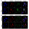
(A) Confocal fluorescence microscopy images of HeLa cells, showing blue fluorescence for DAPI-stained nuclei, red fluorescence related to photosensitizer (1 μM) and green fluorescence related to mitochondria (150 nM of the probe). The right column shows the overlay from three channels (blue, red and green). (B) Confocal fluorescence microscopy images of HeLa cells with blue fluorescence of nucleus (DAPI), red fluorescence of porphyrins (1 μM) and green fluorescence of lysosomes (150 nM of probe). The right column shows the overlay from three channels (blue, red and green). Scale bars correspond to 20 µm. This figure was reproduced from Tsubone et al. [21], with permission from Springer Nature under the terms of the Creative Commons Attribution 4.0 International License.

References

    1. Spring BQ, Rizvi I, Xu N, Hasan T. The role of photodynamic therapy in overcoming cancer drug resistance. Photochem Photobiol Sci. 2015;14(8):1476–91. - PMC - PubMed
    1. Allison RR, Sibata CH, Downie GH, Cuenca RE. A clinical review of PDT for cutaneous malignancies. Photodiagn Photodyn Ther. 2006;3(4):214–26. - PubMed
    1. Hasan T, Ortel B, Moor AC, Pogue BW. Photodynamic therapy of cancer. In: Radiation Oncology. 1988. p. 43–55.
    1. Usuda J, Kato H, Okunaka T, Furukawa K, Yamada K, Suga Y, et al. Photodynamic Therapy (PDT) for Lung Cancers. J Thorac Oncol. 2006;1(5):489–93. - PubMed
    1. Babilas P, Schreml S, Landthaler M, Szeimies RM. Photodynamic therapy in dermatology: state-of-the-art. Photodermatol Photoimmunol Photomed. 2010;26(3):118–32. - PubMed

Publication types

LinkOut - more resources