

The Golgin Protein Giantin Regulates Interconnections Between Golgi Stacks
- PMID: 31544102
- PMCID: PMC6732663
- DOI: 10.3389/fcell.2019.00160
The Golgin Protein Giantin Regulates Interconnections Between Golgi Stacks
Abstract
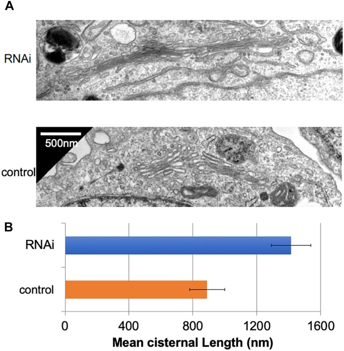
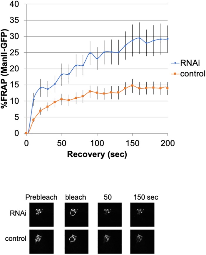
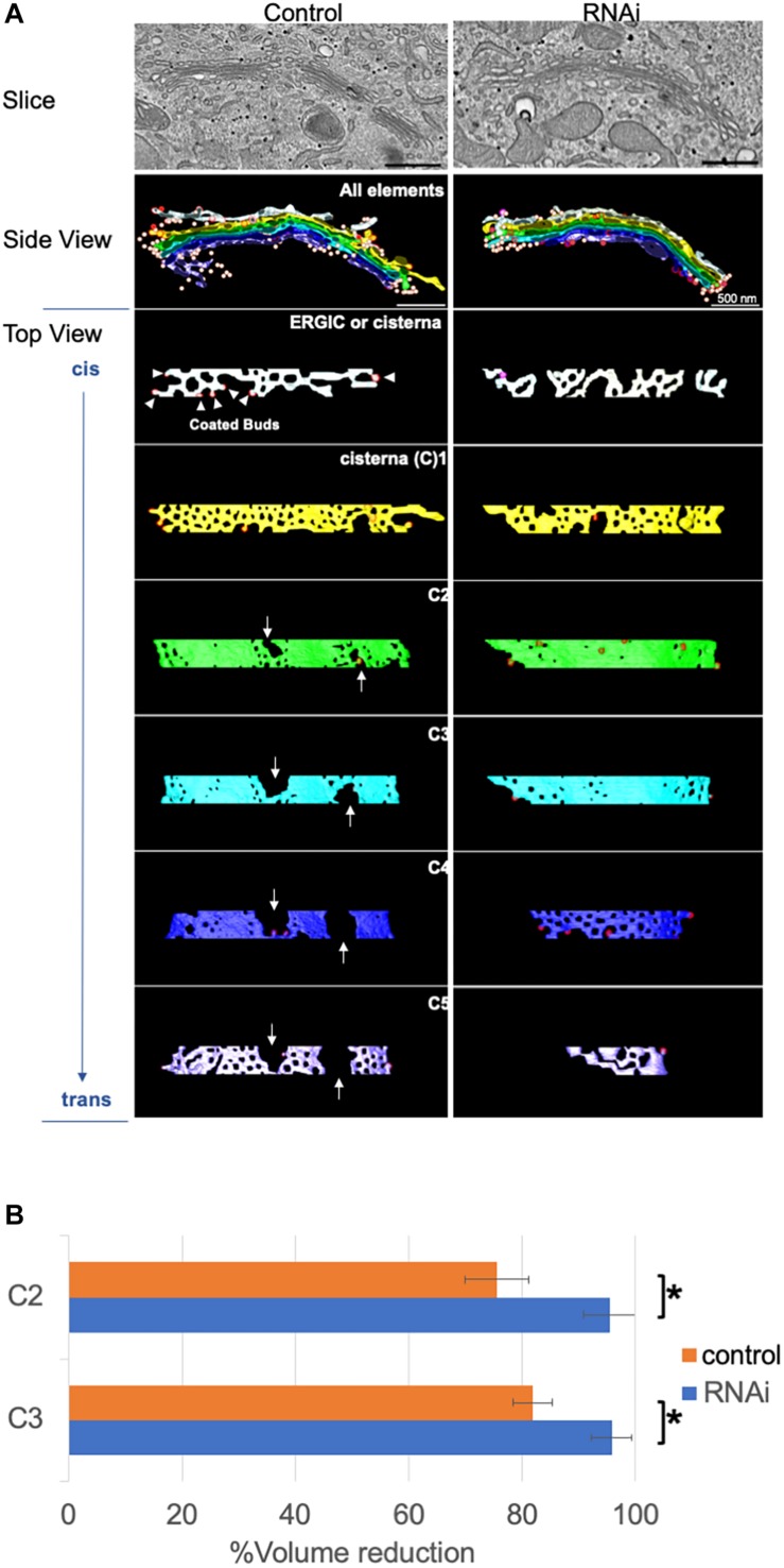
Golgins are a family of Golgi-localized long coiled-coil proteins. The major golgin function is thought to be the tethering of vesicles, membranes, and cytoskeletal elements to the Golgi. We previously showed that knockdown of one of the longest golgins, Giantin, altered the glycosylation patterns of cell surfaces and the kinetics of cargo transport, suggesting that Giantin maintains correct glycosylation through slowing down transport within the Golgi. Giantin knockdown also altered the sizes and numbers of mini Golgi stacks generated by microtubule de-polymerization, suggesting that it maintains the independence of individual Golgi stacks. Therefore, it is presumed that Golgi stacks lose their independence following Giantin knockdown, allowing easier and possibly increased transport among stacks and abnormal glycosylation. To gain structural insights into the independence of Golgi stacks, we herein performed electron tomography and 3D modeling of Golgi stacks in Giantin knockdown cells. Compared with control cells, Giantin-knockdown cells had fewer and smaller fenestrae within each cisterna. This was supported by data showing that the diffusion rate of Golgi membrane proteins is faster in Giantin-knockdown Golgi, indicating that Giantin knockdown structurally and functionally increases connectivity among Golgi cisternae and stacks. This increased connectivity suggests that contrary to the cis-golgin tether model, Giantin instead inhibits the tether and fusion of nearby Golgi cisternae and stacks, resulting in transport difficulties between stacks that may enable the correct glycosylation of proteins and lipids passing through the Golgi.
Keywords: Golgi; electron tomography; endoplasmic reticulum; glycosylation; golgins.
Figures
Similar articles
-
An Electron Tomographic Analysis of Giantin-Deficient Golgi Proposes a New Function of the Golgin Protein Family.Methods Mol Biol. 2023;2557:235-246. doi: 10.1007/978-1-0716-2639-9_15. Methods Mol Biol. 2023. PMID: 36512219
-
The golgin tether giantin regulates the secretory pathway by controlling stack organization within Golgi apparatus.PLoS One. 2013;8(3):e59821. doi: 10.1371/journal.pone.0059821. Epub 2013 Mar 21. PLoS One. 2013. PMID: 23555793 Free PMC article.
-
A novel Rab6-interacting domain defines a family of Golgi-targeted coiled-coil proteins.Curr Biol. 1999 Apr 8;9(7):381-4. doi: 10.1016/s0960-9822(99)80167-5. Curr Biol. 1999. PMID: 10209123
-
The Golgin Family of Coiled-Coil Tethering Proteins.Front Cell Dev Biol. 2016 Jan 11;3:86. doi: 10.3389/fcell.2015.00086. eCollection 2015. Front Cell Dev Biol. 2016. PMID: 26793708 Free PMC article. Review.
-
Finding the Golgi: Golgin Coiled-Coil Proteins Show the Way.Trends Cell Biol. 2016 Jun;26(6):399-408. doi: 10.1016/j.tcb.2016.02.005. Epub 2016 Mar 11. Trends Cell Biol. 2016. PMID: 26972448 Review.
Cited by
-
Getting Sugar Coating Right! The Role of the Golgi Trafficking Machinery in Glycosylation.Cells. 2021 Nov 23;10(12):3275. doi: 10.3390/cells10123275. Cells. 2021. PMID: 34943782 Free PMC article. Review.
-
The factory, the antenna and the scaffold: the three-way interplay between the Golgi, cilium and extracellular matrix underlying tissue function.Biol Open. 2023 Feb 15;12(2):bio059719. doi: 10.1242/bio.059719. Epub 2023 Feb 21. Biol Open. 2023. PMID: 36802341 Free PMC article. Review.
-
Giantin is required for intracellular N-terminal processing of type I procollagen.J Cell Biol. 2021 Jun 7;220(6):e202005166. doi: 10.1083/jcb.202005166. Epub 2021 May 4. J Cell Biol. 2021. PMID: 33944912 Free PMC article.
-
Post-ER Stress Biogenesis of Golgi Is Governed by Giantin.Cells. 2019 Dec 13;8(12):1631. doi: 10.3390/cells8121631. Cells. 2019. PMID: 31847122 Free PMC article.
-
Alcohol induces α2-6sialo mucin O-glycans that kill U937 macrophages mediated by sialic acid-binding immunoglobulin-like lectin 7 (Siglec 7).FEBS Open Bio. 2025 Jan;15(1):165-179. doi: 10.1002/2211-5463.13919. Epub 2024 Nov 26. FEBS Open Bio. 2025. PMID: 39592427 Free PMC article.
References
LinkOut - more resources
Full Text Sources