. 2019 Aug 30:7:180.
doi: 10.3389/fcell.2019.00180. eCollection 2019.

Zinc Maintains Embryonic Stem Cell Pluripotency and Multilineage Differentiation Potential via AKT Activation

Affiliations

Zinc Maintains Embryonic Stem Cell Pluripotency and Multilineage Differentiation Potential via AKT Activation

Hayk Mnatsakanyan et al. Front Cell Dev Biol. .

Abstract

Embryonic stem cells (ESCs) possess remarkable abilities, as they can differentiate into all cell types (pluripotency) and be self-renewing, giving rise to two identical cells. These characteristics make ESCs a powerful research tool in fundamental embryogenesis as well as candidates for use in regenerative medicine. Significant efforts have been devoted to developing protocols to control ESC fate, including soluble and complex cocktails of growth factors and small molecules seeking to activate/inhibit key signaling pathways for the maintenance of pluripotency states or activate differentiation. Here we describe a novel method for the effective maintenance of mouse ESCs, avoiding the supplementation of complex inhibitory cocktails or cytokines, e.g., LIF. We show that the addition of zinc to ESC cultures leads to a stable pluripotent state that shares biochemical, transcriptional and karyotypic features with the classical LIF treatment. We demonstrate for the first time that ESCs maintained in long-term cultures with added zinc, are capable of sustaining a stable ESCs pluripotent phenotype, as well as differentiating efficiently upon external stimulation. We show that zinc promotes long-term ESC self-renewal (>30 days) via activation of ZIP7 and AKT signaling pathways. Furthermore, the combination of zinc with LIF results in a synergistic effect that enhances LIF effects, increases AKT and STAT3 activity, promotes the expression of pluripotency regulators and avoids the expression of differentiation markers.

Keywords: AKT; ZIP7; embryonic stem cells (ESC); stemness maintenance; zinc.


Figures

FIGURE 1
FIGURE 1
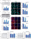
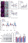
Role of zinc in ESC self-renewal. (A) Intracellular Zn2+ wave determined by FluoZin3-AM labeling (data points were taken every 40 s). Cells were kept at 37°C throughout the analysis (n = 6). (B) AP staining of ESCs after 5 days of culture in BM, medium supplemented with LIF or medium supplemented with different concentrations of Zn2+ (n = 3). (C) Immunofluorescence detection of pluripotency markers (POU5F1 and SOX2) and staining of AP in ESCs after 5 days of culture in BM, medium supplemented with LIF or medium supplemented with different concentrations of Zn2+ (n = 5). (D) Immunofluorescence detection of pluripotency (POU5F1 and SOX2) and differentiation markers (endoderm: SOX17) determined after 5 days of culture in accelerated differentiation conditions (medium supplemented with 5% KSR) (n = 5). (E) qPCR detection of pluripotency markers (Pou5f1) and differentiation markers (endoderm: Foxa2, mesoderm: Brachyury/T and ectoderm: Sox1) determined after 5 days of culture in accelerated differentiation conditions (medium supplemented with 5% KSR). Gapdh was used as housekeeping gene (n = 4). Significant differences were determined by ANOVA test; p < 0.05. Scale bar: 200 μm.
FIGURE 2
FIGURE 2
Role of ZIP7 and AKT in ESCs stemness maintenance. (A) Analysis of pAKT/AKT ratio by western blot after AKT inhibition with LY-294002 10 μM during 24 h in cells cultured in BM, medium supplemented with LIF or medium supplemented with 100 μM of Zn2+. GAPDH was used as loading control protein (n = 4). (B) Analysis of ZIP7 expression and pAKT/AKT ratio by western blot after ZIP7 silencing with RNAi after 3 days of culture. GAPDH was used as loading control protein (n = 4). Cells were cultured in BM and a medium supplemented with LIF or medium supplemented with 100 μM of Zn2+. (C,D) Immunofluorescence detection of pluripotency markers (POU5F1-green and alkaline phosphatase-red, AP) determined after 7 days of culture in BM, medium supplemented with LIF or medium supplemented with 100 μM of Zn2+, and treated 24 h with AKT inhibitor LY-294002, or ZIP7 silenced with RNAi during 3 days. Scale bar: 100 μm. (E) Analysis of pAKT/AKT (S473) and pGSK3β/GSK3β (S9) ratios by western blot in ESCs cultured in BM, medium supplemented with LIF or medium supplemented with 100 μM of Zn2+ for 4 or 24 h. GAPDH was used as loading control protein (n = 4). Significant differences were determined by ANOVA test; between groups: p < 0.05; within group: #p < 0.05.
FIGURE 3
FIGURE 3
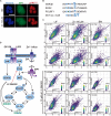
Relationship between nuclear pAKT and the core transcriptional network for ESC self-renewal. (A) Immunostaining of Zn2+ transporter ZIP7 and pAKT (S473) in ESCs cultured for 24 h in BM only or BM supplemented with 100 μM of Zn2+. Scale bar: 50 μm. (B) Alignments of aminoacid sequences of mouse GSK3β, POU5F1, and SOX2 showing the target motifs for AKT-mediated phosphorilation. NetPhos 3.1 Server was used to identify these sequences, which then were aligned with the minimal recognition motif of AKT. (C) Immunofluorescence detection and quantification of pluripotency markers POU5F1, SOX2, and pAKT (S473) in ESCs cultured for 3 days in normal medium and medium supplemented with 10 μM of PI3K inhibitor LY-294002. Obtained data were represented and Pearson’s correlation r-values were obtained using R. (D) Proposed mechanism illustrating the effects of Zn2+ in AKT phosphorylation and ESCs self-renewal. Addition of extracellular Zn2+ in the culture medium, activate ZIP7 and AKT downstream pathways. Note that these results have been determined experimentally in this work and the illustrated steps activated by AKT to maintain ESCs pluripotency have been previously reported in the literature (Saxe et al., 2009; Jeong et al., 2010; Lin et al., 2012).
FIGURE 4
FIGURE 4
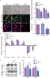
Role of zinc in ESCs pluripotency maintenance after 30 days of culture. (A) Immunofluorescence detection of pluripotency markers: POU5F1-green, SOX2-red and alkaline phosphatase-red, determined after 30 days of ESCs culture, in BM and a medium supplemented with LIF or medium supplemented with 100 μM of Zn2+ (n = 5). Scale bar: 200 μm. (B) qPCR detection of pluripotency markers (Pou5f1, Nanog, Klf4) and differentiation markers (endoderm: Foxa2; mesoderm: Brachyury/T and ectoderm: Sox1). Gapdh was used as housekeeping gene (n = 4). (C) Western blot detection of AKT and pAKT (S473) on ESCs cultured in BM and a medium supplemented with LIF or medium supplemented with 100 μM of Zn2+after 1, 5, and 30 days of culture (n = 4). Significant differences were determined by ANOVA test; p < 0.05.
FIGURE 5
FIGURE 5
Role of zinc in spontaneous embryoid body differentiation after 30 days of culture. (A) Embryoid body formation of ESCs cultured for 30 days in BM and a medium supplemented with LIF or 100 μM Zn2+. New EBs formed from low passage ESCs (not previously cultured for 30 days under different conditions) were included as a control of embryoid body formation (1d-LIF). Measurements of EB diameters (n = 9). Scale bar: 200 μm. (B) Evaluation of spontaneous differentiation of EBs formed after culture of ESCs for 30 days in BM and a medium supplemented with LIF or 100 μM Zn2+. qPCR detection of primary germ layer markers: endoderm (Foxa2), mesoderm (Brachyury/T) and ectoderm (Sox1). Gapdh was used as housekeeping gene (n = 4). (C) Hematoxylin/eosin staining of histological sections of EBs after 15 days of culture. EBs were formed after culture of ESCs for 30 days in BM, medium supplemented with LIF or 100 μM Zn2+. Scale bar: 100 μm. Significant differences were determined by ANOVA test: p < 0.05.
FIGURE 6
FIGURE 6
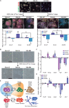
Zinc maintains naïve ESC state after long-term cultures. (A) ESCs previously cultured for 30 days in BM (30d-BM) and a medium supplemented with LIF (30d-LIF) or 100 μM Zn2+ (30d-Zn) were treated with LIF or BM for 3 more days. Pluripotency markers (POU5F1-green and SOX2-red) were determined by immunofluorescence (n = 5). Scale bar: 200 μm. (B) Bright field pictures of 30d-ESC cultured in BM (30d-BM) and a medium supplemented with LIF (30d-LIF) or 100 μM Zn2+ (30d-Zn) after 3 days of culture in the presence of LIF. Scale bar: 200 μm. (C) qPCR evaluation of stemness-related markers of pluripotency (Pou5f1, Nanog) indicative of naïve state, and differentiation markers (Gata6, Brachyury/T, Fgf5) indicative of primed state, of ESCs previously cultured for 30 days in BM (30d-BM), medium supplemented with LIF (30d-LIF) or 100 μM Zn2+(30d-Zn). Gapdh was used as housekeeping gene (n = 4). (D) qPCR evaluation of stemness related markers of pluripotency (Pou5f1, Nanog) indicative of naïve state, and differentiation markers (Gata6, Brachyury/T, Fgf5) indicative of primed state, of ESCs previously cultured for 30 days in BM (30d-BM), medium supplemented with LIF (30d-LIF) or 100 μM Zn2+ (30d-Zn) and treated with LIF for 3 more days in order to reverse primed to naïve state. Gapdh was used as housekeeping gene (n = 4). (E) Scheme of ESC pluripotency dynamics, indicating the state of the ESC population cultured with LIF (blue) and Zn (orange) after 30 days of culture. Significant differences were determined by ANOVA test; p < 0.05.
FIGURE 7
FIGURE 7
The combination of LIF with Zn2+ sustains ESC pluripotency with greater efficiency. (A) ESCs were cultured for 15 days, passaging cells each 3 days, in presence of either 1,000 U/ml of LIF (LIF), 1,000 U/ml LIF + 100 μM Zn2+ (LIF-Zn), 250 U/ml of LIF (1/4 LIF), and 250 U/ml LIF + 100 μM Zn2+ (1/4LIF-Zn). After each passage, ESCs were fixed and stained for alkaline phosphatase (AP) (n = 4) and immunostained for SOX2 and SOX17 (n = 5). Scale bar: 200 μm. (B) qPCR evaluation of stemness-related markers of pluripotency (Nanog) indicative of naïve state, and differentiation markers (Gata6, Brachyury/T, Fgf5) indicative of primed state, of ESCs previously cultured for 15 days. Gapdh was used as housekeeping gene (n = 4). (C) Western blot detection of AKT, pAKT (S473), pSTAT3 (T705), and SOX2 on ESCs cultured for 15 days. GAPDH was used as a housekeeping gene (n = 4). Significant differences were determined by ANOVA test; p < 0.05.

Similar articles

Cited by

References

    1. Anzellotti A. I., Farrell N. P. (2008). Zinc metalloproteins as medicinal targets. Chem. Soc. Rev. 37 1629–1651. 10.1039/b617121b - DOI - PubMed
    1. Armstrong L., Hughes O., Yung S., Hyslop L., Stewart R., Wappler I., et al. (2006). The role of PI3K/AKT, MAPK/ERK and NFκβ signalling in the maintenance of human embryonic stem cell pluripotency and viability highlighted by transcriptional profiling and functional analysis. Hum. Mol. Genet. 15 1894–1913. 10.1093/hmg/ddl112 - DOI - PubMed
    1. Bechard M., Dalton S. (2009). Subcellular localization of glycogen synthase kinase 3 controls embryonic stem cell self-renewal. Mol. Cell. Biol. 29 2092–2104. 10.1128/MCB.01405-08 - DOI - PMC - PubMed
    1. Berge D. T., Kurek D., Blauwkamp T., Koole W., Maas A., Eroglu E., et al. (2011). Embryonic stem cells require Wnt proteins to prevent differentiation to epiblast stem cells. Nat. Cell Biol. 13 1070–1077. 10.1038/ncb2314 - DOI - PMC - PubMed
    1. Cardone M. H. (1998). Regulation of cell death protease caspase-9 by phosphorylation. Science 282 1318–1321. 10.1126/science.282.5392.1318 - DOI - PubMed