. 2018 Jul;1(3):1800032.
doi: 10.1002/adtp.201800032. Epub 2018 May 28.

Decellularized Extracellular Matrix Hydrogels as a Delivery Platform for MicroRNA and Extracellular Vesicle Therapeutics

Affiliations

Decellularized Extracellular Matrix Hydrogels as a Delivery Platform for MicroRNA and Extracellular Vesicle Therapeutics

Melissa J Hernandez et al. Adv Ther (Weinh). 2018 Jul.

Abstract

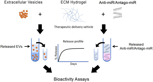
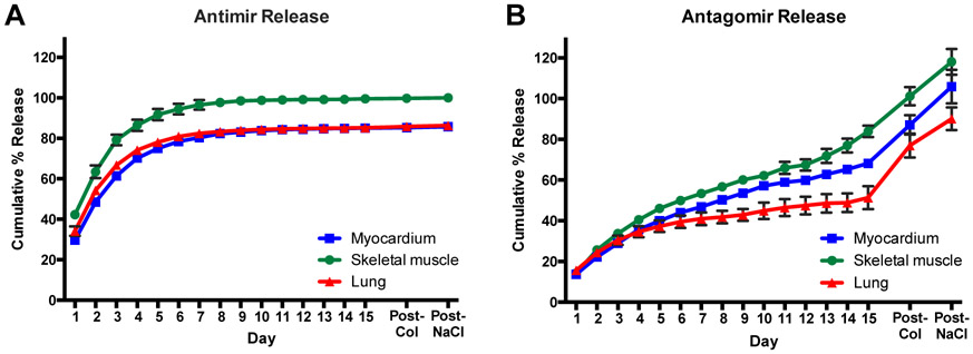
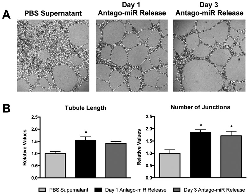
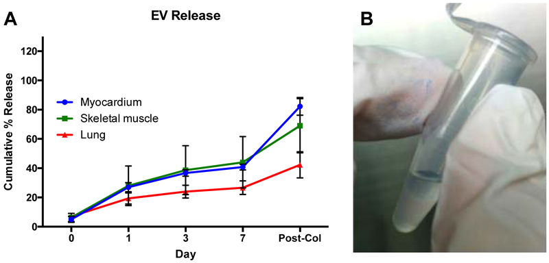
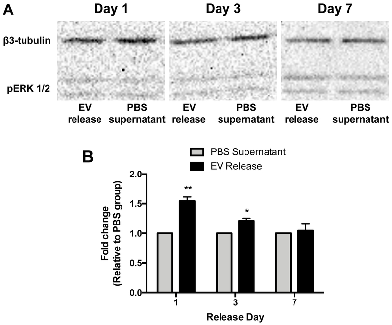
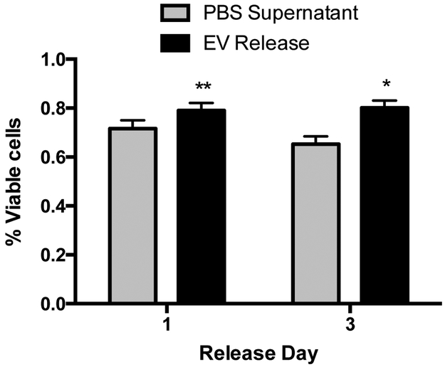
In the last decade, the use of microRNA (miRNA) and extracellular vesicle (EV) therapies has emerged as an alternative approach to mitigate the negative effects of several disease pathologies ranging from cancer to tissue and organ regeneration; however, delivery approaches towards target tissues have not been optimized. To alleviate these challenges, including rapid diffusion upon injection and susceptibility to degradation, porcine-derived decellularized extracellular matrix (ECM) hydrogels are examined as a potential delivery platform for miRNA and EV therapeutics. The incorporation of EVs and miRNA antagonists, including anti-miR and antago-miR, in ECM hydrogels results in a prolonged release as compared to the biologic agents alone. In addition, individual in vitro assessments confirm the bioactivity of the therapeutics upon release from the ECM hydrogels. This work demonstrates the feasibility of encapsulating miRNA and EV therapeutics in ECM hydrogels to enhance delivery and potentially efficacy in later in vivo applications.

Keywords: extracellular matrix; extracellular vesicles; hydrogels; microRNAs.


Conflict of interest statement

Conflict of Interest K.L.C. is co-founder, consultant, board member, and holds equity interest in Ventrix, Inc.

Figures

Figure 1.
Figure 1.
Schematic of the workflow for assessing decellularized ECM hydrogels as a delivery platform for miRNA and EV therapeutics. Anti-miRs, antago-miRs, and EVs were encapsulated in ECM hydrogels, and the release profiles were first generated. Antago-miR and EV release samples were then further analyzed to ensure the biologics remained bioactive.
Figure 2.
Figure 2.
Release profiles for miRNA inhibitors of miR-214, an anti-miR and antago-miR. Values were obtained from fluorescence measurements using the Cy3 dye molecule conjugated to each miRNA, and values exceeding 100% likely resulted from errors due to the linear fit of the generated standard curves. The anti-miR (A) yielded a more rapid release rate, likely due to the absence of a cholesterol group, which is present on the antago-miR (B). The cholesterol group introduces hydrophobic interactions, which appear to affect the release rate. Some of the error bars are too small to be visualized at each time point. n = 3/gel type
Figure 3.
Figure 3.
Bioactivity of released antago-miRs in a Matrigel tube formation assay. (A) Representative images are shown for the tube formation of HCAECs on Matrigel. Since ECM soluble factors are present in the PBS supernatant group, some tube formation is seen but with a large degree of cell clustering. However, released samples from days 1 and 3 produce more organized tubes that yield relative increases in (B) tubule length and the number of junctions over the PBS control (n = 3/group). *p < 0.05 compared to the PBS supernatant control using a one-way ANOVA with a Dunnett’s post hoc test.
Figure 4.
Figure 4.
Cumulative release of hCPC-derived EVs from porcine ECM hydrogels. (A) Conditioned PBS was collected at days 0, 1, 3 and 7, and the concentration of detected EVs is shown as a percentage of the mean fluorescent intensity of 4 μg untreated EVs. Fluorescent intensities were determined with magnetic bead capture flow cytometry. Myocardial ECM hydrogels (n = 4), skeletal muscle ECM hydrogels (n = 3), and lung ECM hydrogels (n = 3) were examined. (B) PKH26 labeled EVs confer a pink color to the gels, which is still visible after 7 days and indicates the presence of the encapsulated EVs.
Figure 5.
Figure 5.
The effect of CPC-derived EVs released from cardiac ECM hydrogels on pERK 1/2 levels in HCAECs. Cells were incubated with conditioned PBS collected at days 1 (n = 4/group), 3 (n = 3/group), and 7 (n = 4/group). The expression of pERK 1/2 was determined with western blot analysis, normalized to β3-tubulin and relative to conditioned PBS from empty ECM hydrogels. *p < 0.05 and **p < 0.01 compared to PBS supernatant using a Student’s t-test.
Figure 6.
Figure 6.
The protective effect of CPC-derived EVs released from cardiac ECM hydrogels on H2O2-induced apoptosis of hCPCs. Cells were incubated with conditioned PBS, collected at days 1 (n = 6/group) and 3 (n = 7/group) in combination with 25 μM H2O2. The survival rate was determined with an alamarBlue cell viability assay and normalized to alamarBlue baseline values. *p < 0.05 and **p < 0.01 compared to PBS supernatant using Student’s t-test.

References

    1. Segers VF, Lee RT, Stem-cell therapy for cardiac disease, Nature 2008, 451, 937. - PubMed
    1. Hastings CL, Roche ET, Ruiz-Hernandez E, Schenke-Layland K, Walsh CJ, Duffy GP, Drug and cell delivery for cardiac regeneration, Adv Drug Deliv Rev 2015, 84, 85. - PubMed
    1. Woodburn JR, The epidermal growth factor receptor and its inhibition in cancer therapy, Pharmacol Ther 1999, 82, 241. - PubMed
    1. Tugues S, Koch S, Gualandi L, Li X, Claesson-Welsh L, Vascular endothelial growth factors and receptors: anti-angiogenic therapy in the treatment of cancer, Mol Aspects Med 2011, 32, 88. - PubMed
    1. Robbins PD, Morelli AE, Regulation of immune responses by extracellular vesicles, Nat Rev Immunol 2014, 14, 195. - PMC - PubMed