Review
. 2020 Apr 1;41(2):273-319.
doi: 10.1210/endrev/bnz005.

The Role of Sex and Sex Hormones in Neurodegenerative Diseases

Affiliations
Review

The Role of Sex and Sex Hormones in Neurodegenerative Diseases

Elisabetta Vegeto et al. Endocr Rev. .

Abstract

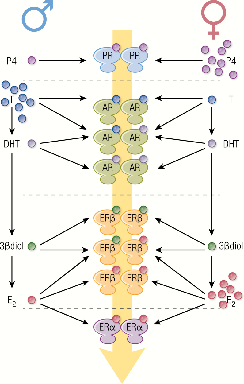
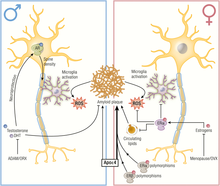
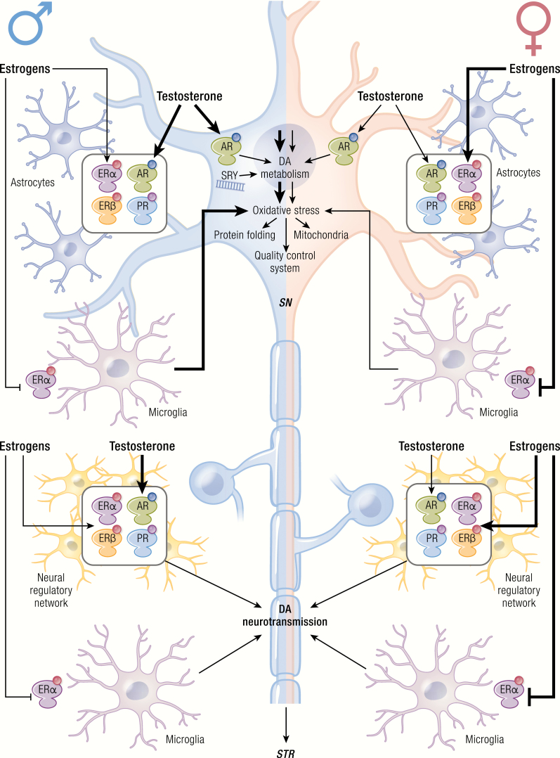
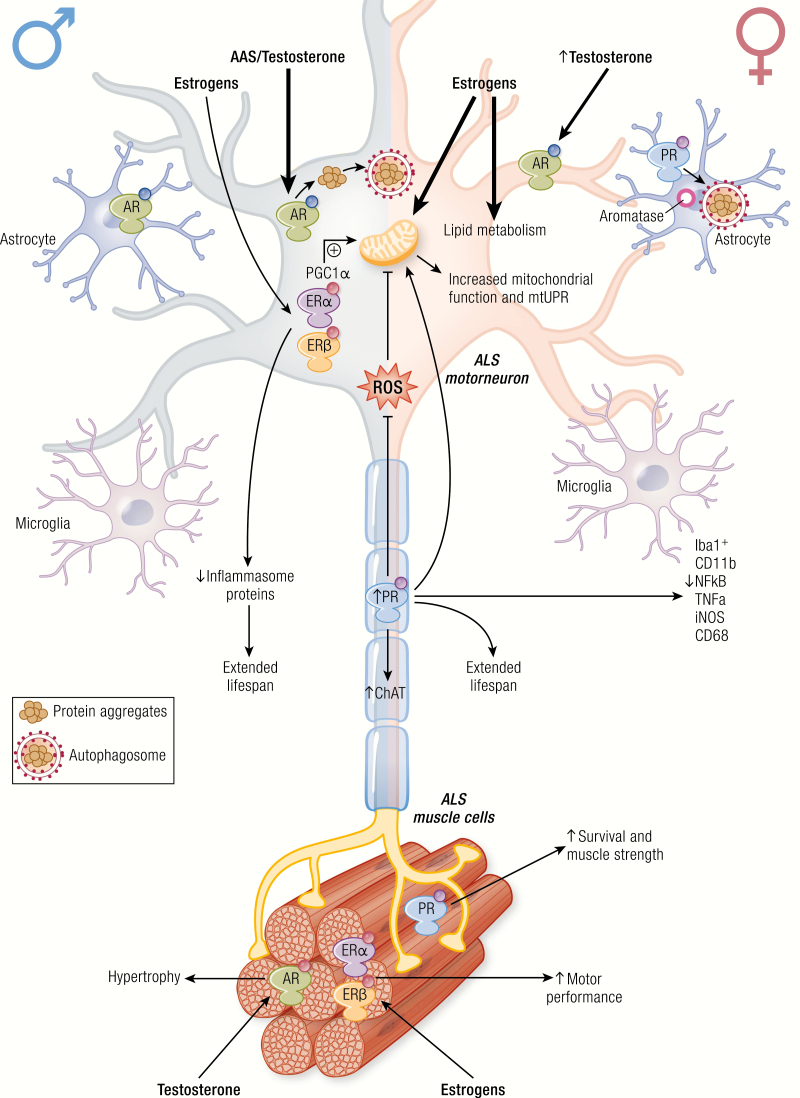
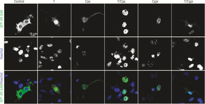
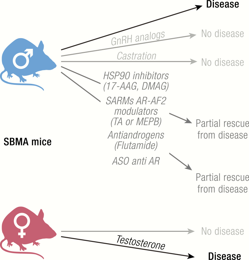
Neurodegenerative diseases (NDs) are a wide class of disorders of the central nervous system (CNS) with unknown etiology. Several factors were hypothesized to be involved in the pathogenesis of these diseases, including genetic and environmental factors. Many of these diseases show a sex prevalence and sex steroids were shown to have a role in the progression of specific forms of neurodegeneration. Estrogens were reported to be neuroprotective through their action on cognate nuclear and membrane receptors, while adverse effects of male hormones have been described on neuronal cells, although some data also suggest neuroprotective activities. The response of the CNS to sex steroids is a complex and integrated process that depends on (i) the type and amount of the cognate steroid receptor and (ii) the target cell type-either neurons, glia, or microglia. Moreover, the levels of sex steroids in the CNS fluctuate due to gonadal activities and to local metabolism and synthesis. Importantly, biochemical processes involved in the pathogenesis of NDs are increasingly being recognized as different between the two sexes and as influenced by sex steroids. The aim of this review is to present current state-of-the-art understanding on the potential role of sex steroids and their receptors on the onset and progression of major neurodegenerative disorders, namely, Alzheimer's disease, Parkinson's diseases, amyotrophic lateral sclerosis, and the peculiar motoneuron disease spinal and bulbar muscular atrophy, in which hormonal therapy is potentially useful as disease modifier.

Keywords: Alzheimer’s disease; Parkinson’s disease; amyotrophic lateral sclerosis; sex hormones; spinal and bulbar muscular atrophy.


Figures

Graphical Abstract
Graphical Abstract
Figure 1.
Figure 1.
Biosynthetic pathways of neurosteroid formation. HMG-CoA is the precursor for cholesterol synthesis. HMG-CoA reductase catalyzes the production of mevalonic acid from HMG-CoA, with a reaction that is rate-limiting for cholesterol synthesis. Mevalonic acid is then converted via the the mevalonate pathway to lanosterol, which is, in turn, converted to cholesterol via either the Bloch pathway or the Kandutsch–Russell pathway. CYP11A1 (P450 side-chain cleavage, named P450scc in rodents), more expressed in females than in males, catalyzes the conversion of cholesterol to pregnenolone through the first rate-limiting step of de novo steroidogenesis. Further steroid conversions are catalyzed by 3β-HSD, CYP17 (P450c17), 5α-R, and CYP19 (aromatase). Full-line arrows indicate active steroidogenic biosynthetic pathways in the brain. Dashed line arrows represent undetermined steroidogenic biosynthetic pathways. Steroidogenic enzymes are represented by dashed line boxes. The color codes indicate their distribution in brain cells; cyan: neurons; green: microglia; orange: astrocytes.
Figure 2.
Figure 2.
The complexity of sex hormone-receptor interactions in the nervous system. In neurons, 5α-R converts testosterone into DHT, which can be converted to 3β-diol by 3β-HSD, while aromatase converts testosterone to E2, giving rise to a variety of androgen metabolites, particularly in males. Blood-derived or locally produced sex hormones interact with specific homo or heterodimeric receptors, giving rise to a range of ligand-receptor complexes. Each hormone-receptor complex binds to gene promoters with specific preference and affinity, resulting in a combinatorial mechanism of transcriptional regulation by sex hormones within a cell. Furthermore, sex steroid receptors are expressed in neural and microglial cells in which they activate cell-specific genetic and metabolic programs. All these molecular and cellular mechanisms make up the response to the initial hormonal signal and participate in the sexual dimorphism of neurological functions.
Figure 3.
Figure 3.
Main sexual differences and activity of sex steroid hormones on amyloid plaque deposition, neuroinflammation, and neuroprotection. The accumulation of aggregates and deposits of misfolded proteins, like amyloid beta peptides, represent the main disease-specific histopathological change in the brain of AD patients. Additional hallmarks include a diffused neuroinflammation during the advanced stages of AD. There is a strong association between APOε4 genotype, the most established genetic risk factor, with sporadic AD onset. In particular, woman carrying both the homo- and heterozygous Apoε4 isoform have a higher rate of amyloid plaque deposition, while Apoε4 variant in men has marginal effects in both homo- and heterozygous subjects. An interaction between the APOε4 variant and SNPs of the ESR1 (rs9340799, rs2234693, rs2228480) and ESR2 (rs4986938) genes is possibly involved. Estrogens, by affecting the secretase pathway in neurons and the lipid metabolism in the periphery, decrease the production of beta-amyloid peptides, thus reducing Aβ accumulation in AD. Moreover, estrogens exert anti-inflammatory effects on microglia, limiting the Aβ-induced production of ROS. The beneficial effects of estrogens are lost following menopause, or after OVX. Progesterone exerts beneficial effects on neurons, but it is still unclear if this is due to an anti-inflammatory action of progesterone per se or if the effect is mediated by the progesterone metabolite allopregnanolone. In males, AD is associated with a decrease in circulating testosterone, which, in turn, could dysregulate Aβ deposition and hinder the testosterone-mediated neuroprotective actions including the regulation of spine density. The beneficial effects of testosterone and DHT are lost in androgen deficiency in the aging male (ADAM), or after ORX.
Figure 4.
Figure 4.
Sexual differences in the activity of sex steroid hormones on dopaminergic neurons of the NSDA system. Dopamine metabolism in the NSDA system, consisting of dopaminergic neurons that from the SN innervate the striatum (STR), triggers oxidative stress that may lead to dysfunctions in protein folding, mitochondrial activity, and quality control systems, which are pathogenic mechanisms of Parkinson’s disease. Astrocytes and microglia residing in the microenvironment of the SN and STR sustain DA neurotransmission, thus increasing oxidative reactions. Among sex steroid hormone receptors, only AR is expressed by DA neurons, both in males and females. In males (left side), SRY expression has been involved in DA metabolism, while high levels of testosterone sustain DA neurotransmission by targeting DA neurons and astrocytes, while only its conversion to estrogens activates microglia. Thus, male sex and hormones correlate with the potentiation DA metabolism. Higher levels of estrogens in females (right side) induce DA neurotransmission only through astrocytes and microglia. ERα-mediated microglia responses to estrogens provide anti-inflammatory and antioxidant effects.
Figure 5.
Figure 5.
Effects of sex steroid hormones on motoneurons, glial and muscle cells of ALS models. Estrogens, progestagens, and androgens through their receptors (respectively, ERα, ERβ, PR, and AR) modulate different pathways in motoneurons and neighbor cells. Estrogens and progestagens extend the life span of ALS animal models through a reduced production of inflammasome proteins, directly on motoneuron or through microglial cells. Estrogens also act on motoneuron lipid metabolism, and mitochondrial functions. Furthermore, progestagens block oxidative reactions allowing a better mitochondrial activity. Androgens and AAS may lead to dysfunctions in protein folding. Skeletal muscle cells express sex steroid receptors, but while ER and PR activation in female triggers a better motor performance, in male an excessive activation of AR leads to muscle fiber hypertrophy and diminished survival of the ALS animal model.
Figure 6.
Figure 6.
Effects of testosterone and anti-androgens on ARpolyQ aggregation in SBMA cell model. Confocal fluorescence microscopy analysis on immortalized motoneuronal (NSC34) cells transfected with plasmid coding for a chimera of green fluorescent protein (GFP) and the AR containing an elongated polyglutamine tract (GFP-AR.Q48). Cells have been treated with ethanol (as vehicle control), 10 nM testosterone, 100 nM Casodex (Cas), or 100 nM Cyproterone acetate (Cypr) for 48 hours. Nuclei were stained with Hoechst (blue) (63X magnification). Scale bar = 10 µm. Aggregation of GFP-AR.Q48 is induced by testosterone and cyproterone acetate, but not by Casodex. Casodex, but not cyproterone acetate reverts the testosterone induced aggregation of GFP-AR.Q48.
Figure 7.
Figure 7.
Ligand-dependent toxicity of mutant ARpolyQ in SBMA. Neurotoxicity of the mutant ARpolyQ associated with SBMA is triggered by the endogenous AR ligand testosterone. Pharmacological treatments (GnRH analogs) or surgical castration, which abolish testosterone production from the male gonads, completely rescue from the aberrant motor behavior phenotype and extend survival of all tg SBMA mouse models tested so far. Antiandrogens (eg, flutamide), SARMs inhibiting the AF-2 of AR, inhibitors of HSP90 also ameliorate motor behavior in the same tg SBMA mice. Similar results have been obtained using a genetic approach based on the administration of antisense oligonucleotides (ASO) against the AR mRNA (particularly active in muscle tissue). Conversely, female tg SBMA mice normally do not develop SBMA; testosterone treatment in female tg SBMA mice induces similar motor alteration to those described in male tg SBMA mice.

References

    1. Kettenmann H, Kirchhoff F, Verkhratsky A. Microglia: new roles for the synaptic stripper. Neuron. 2013;77(1):10–18. - PubMed
    1. Bartzokis G. Alzheimer’s disease as homeostatic responses to age-related myelin breakdown. Neurobiol Aging. 2011;32(8):1341–1371. - PMC - PubMed
    1. Villa A, Vegeto E, Poletti A, Maggi A. Estrogens, neuroinflammation, and neurodegeneration. Endocr Rev. 2016;37(4):372–402. - PMC - PubMed
    1. Paolicelli RC, Bolasco G, Pagani F, et al. Synaptic pruning by microglia is necessary for normal brain development. Science. 2011;333(6048):1456–1458. - PubMed
    1. Rodríguez-Arellano JJ, Parpura V, Zorec R, Verkhratsky A. Astrocytes in physiological aging and Alzheimer’s disease. Neuroscience. 2016;323:170–182. - PubMed

Publication types

Substances