

Review
doi: 10.1002/cpt.1643.
Epub 2019 Dec 9.
PharmVar GeneFocus: CYP2D6
Affiliations
- PMID: 31544239
- PMCID: PMC6925641
- DOI: 10.1002/cpt.1643
Item in Clipboard
Review
PharmVar GeneFocus: CYP2D6
Clin Pharmacol Ther.
2020 Jan.
Abstract
The Pharmacogene Variation Consortium (PharmVar) provides nomenclature for the highly polymorphic human CYP2D6 gene locus. CYP2D6 genetic variation impacts the metabolism of numerous drugs and, thus, can impact drug efficacy and safety. This GeneFocus provides a comprehensive overview and summary of CYP2D6 genetic variation and describes how the information provided by PharmVar is utilized by the Pharmacogenomics Knowledgebase (PharmGKB) and the Clinical Pharmacogenetics Implementation Consortium (CPIC).
© 2019 The Authors Clinical Pharmacology & Therapeutics © 2019 American Society for Clinical Pharmacology and Therapeutics.
Figures
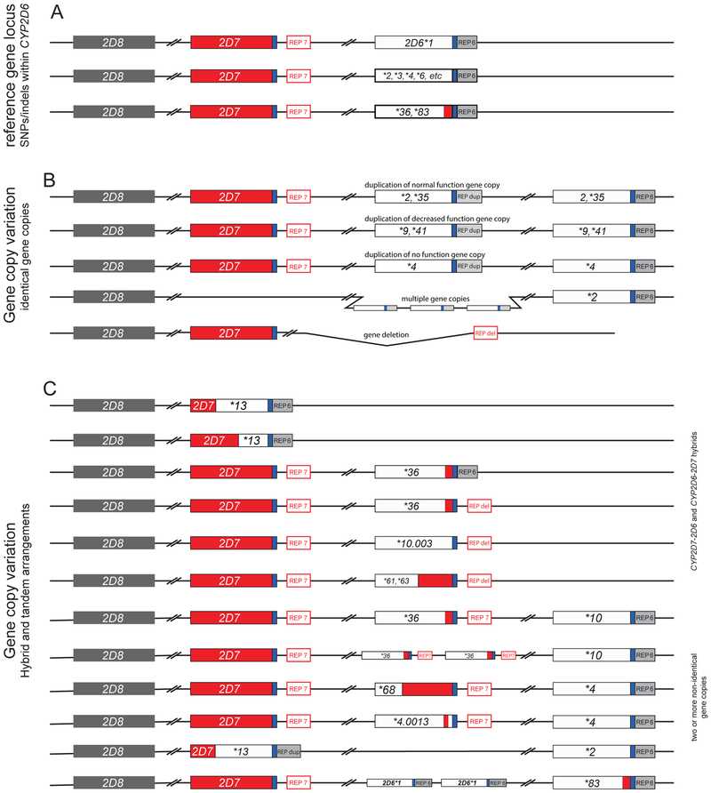
The top line in Panel A depicts the reference gene locus containing a single copy of the CYP2D6 gene. The 2nd line represents CYP2D6*2, *3, *4, *6, etc. Panel B exemplifies allelic variants carrying two or more (multiple) normal function (e.g. *2xN, *35xN), decreased function (e.g. *9xN, *41xN) or nonfunctional (e.g. *4xN) gene copies. The duplicated and multiplied gene copies shown in this panel are believed to be identical. The last line in B represents the CYP2D6*5 gene deletion allele. Of note, alleles with duplicated gene copies have a CYP2D6-like REP-dup sequence without the 1.6 kb long CYP2D7 spacer sequence. Panel C depicts the most complex structural variants. These harbor a singleton hybrid gene or carry two or more non-identical gene copies. The duplicated gene in such arrangements often, but not always, has a CYP2D7-like downstream region including the 1.6 kb long spacer sequence. CYP2D6 and CYP2D7-derived sequences are shown in white/gray and red, respectively. A common downstream element is shown in blue and repetitive elements (REP6, REP7, REPdup, and REPdel) are gray or red based on whether they resemble CYP2D6 (without the spacer) or CYP2D7 (with the spacer).
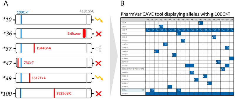
Many platforms test for a panel of the more commonly observed SNVs, but not all known SNVs or all alleles defined by PharmVar. As a consequence, allele assignments are made by ‘default’ as exemplified on those that are defaulted to CYP2D6*10. It is imperative to know which SNVs are tested in order to garner a full understanding of how phenotype is derived as well as to fully understand a test’s limitations. Panel A depicts a selection of allelic variants all carrying g.100C>T. An unequivocal CYP2D6*10 call can only be made after ruling out the presence of numerous other SNVs, e.g. those defining *36, *37, *47, *49, *100 and others. These alleles can be nonfunctional, have decreased or uncertain function as indicated by the function symbols, or await function assignment by CPIC in which case alleles are labeled as ‘awaits curation’ on the PharmVar CYP2D6 gene page. Panel B shows all currently defined alleles containing the g.100C>T core SNV (see Figure 4 and text for more information regarding core allele definitions). The depicted graph was generated with the PharmVar’s CAVE tool.
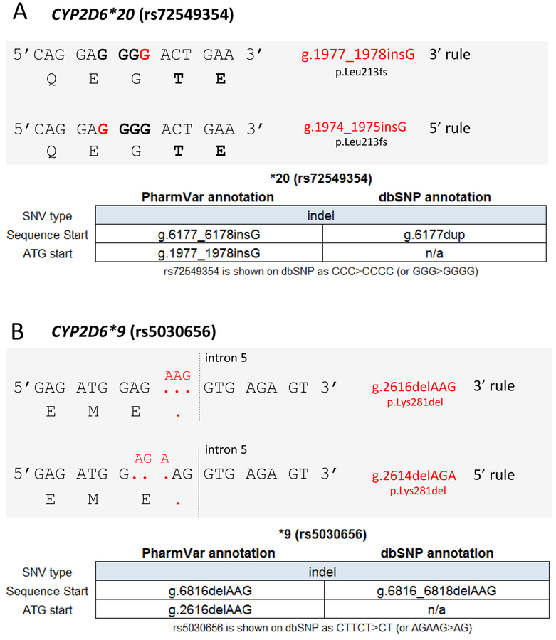
Panel A shows CYP2D6*20 which is characterized by a g.1977_1978insG (rs72549354) (red), which causes a frameshift at amino acid position 213 that obliterates function. This SNV is embedded within three ‘G’ (bold) that are flanked on each side by one ‘A’ (5’ AGGGA 3’). Because of the nature of the surrounding sequence, the actual insertion site is unclear. PharmVar displays the position of this variant as g.1977_1978 (per the 3’ rule) as opposed to g.1974_1975 (5’ rule). Of note, dbSNP uses the same position (g.6177 counting from sequence start) for this variant for the RefSeq annotation, but describes the ‘insertion’ of the ‘G’ as ‘duplication’. Panel B shows CYP2D6*9 which is characterized by a 3-base pair deletion (red) commonly described as g.2616delAAG (rs5030656) that results in the loss of a lysine (K281del) causing decreased function. Because of the nature of the surrounding sequence it remains unclear, however, which three bases are deleted. PharmVar displays the position of this in-frame deletion as g.2616delAAG (per the 3’ rule) as opposed to g.2614delAGA (per the 5’ rule). Of note, dbSNP uses the same position (g.6816 counting from the sequence start) for this variant for the RefSeq annotation.
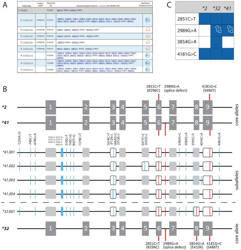
Panel A shows the CYP2D6*2 core allele definition (gray bar). Core SNVs and the allele’s PharmVar ID (PVID) are as shown. A selection of suballeles is provided under the core allele definition (as of July 2019, there were 20 CYP2D6*2 suballeles). Legacy allele designations are cross-referenced (e.g. *2.001 corresponds to *2A). Two of the suballeles depicted, *2.002 and *2.003 have been defined by exon sequencing only and therefore are assigned a ‘Lim’ evidence level. Panel B is a graphical representation of the CYP2D6*2, *32 and *41 core alleles and their core SNVs. g.2851C>T (R296C) and g.4181G>C (S486T) are present on all three, while g.2989G>A (splice defect) is present on CYP2D6*32 and *41, and g.3854G>A (E410K) is only found on *32. Gray boxes represent the nine exons. The middle section shows four of the five CYP2D6*41 suballeles defined to date. Core SNVs (causing an amino acid change or aberrant splicing) are shown in red, all other SNVs are highlighted in blue; core SNVs are found on all suballeles. Panel C depicts the CAVE output visualizing the core SNVs shared among CYP2D6*2, *32 and *41. Blue boxes indicate the presence of a SNV and the function symbol ( ) indicates that g.2989G>A alters function. It remains unknown, however, whether 3854G>A (E410K) exerts a functional impact.
) indicates that g.2989G>A alters function. It remains unknown, however, whether 3854G>A (E410K) exerts a functional impact.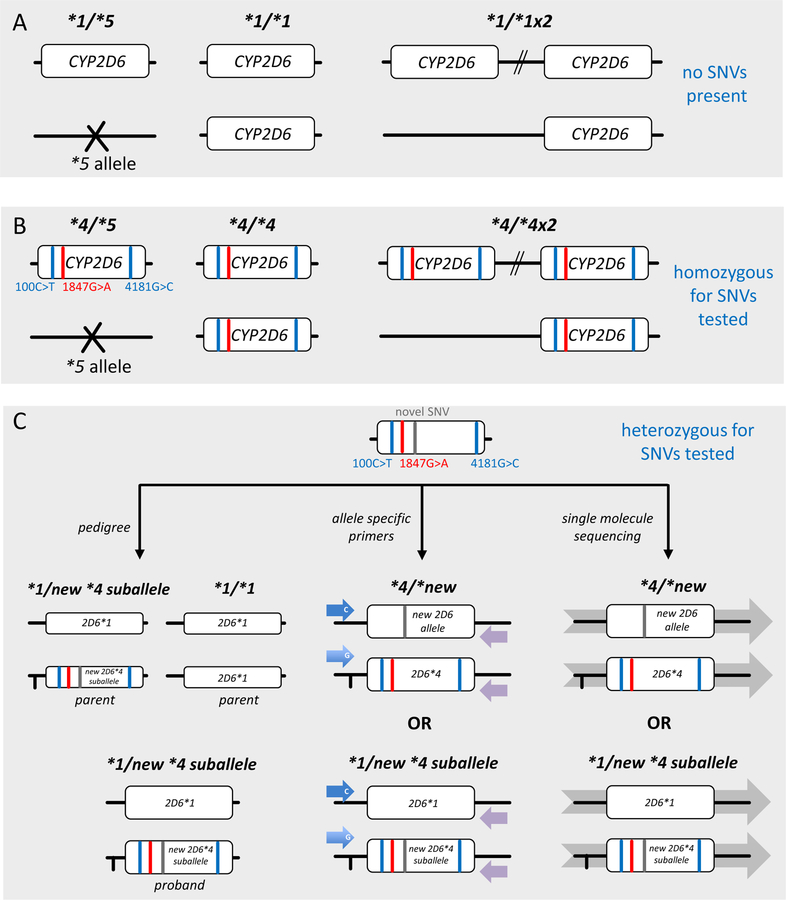
Panel A shows the CYP2D6 reference gene locus, i.e. no SNVs are present. This subject has a CYP2D6*1/*1 genotype in the absence of CNVs. If CNV testing yields 1 and 3 copies, genotypes will be assigned as CYP2D6*1/*5 and *1×2/*1, respectively. Panel B shows an example which is homozygous for three SNVs (depicted as red or blue lines), i.e. each SNV is present on each gene copy. This subject has a CYP2D6*4/*4 genotype in the absence of CNVs. If CNV testing yields 1 and 3 copies, genotypes will be assigned as CYP2D6*4/*5 and *4×2/*4 (or alternatively *4×3/*5, not shown). Panel C shows a sample that is heterozygous for three SNVs (same as in B) and a novel SNV. It is impossible, however, to know whether the novel SNV is in cis (on the same allele as other three SNVs signifying CYP2D6*4) or in trans (by itself on the opposite allele). The bottom panel visualizes different approaches of how haplotype can be inferred, e.g. inheritance (left-hand graph) or experimentally determined, e.g. allele-specific long-range PCR followed by sequencing (center graph) or single molecule sequencing (right-hand graph).
References
-
- Mahgoub A, Idle JR, Dring LG, Lancaster R & Smith RL Polymorphic hydroxylation of Debrisoquine in man. Lancet 2, 584–6 (1977). - PubMed
-
- Eichelbaum M, Spannbrucker N, Steincke B & Dengler HJ Defective N-oxidation of sparteine in man: a new pharmacogenetic defect. Eur J Clin Pharmacol 16, 183–7 (1979). - PubMed
-
- Brosen K & Gram LF Clinical significance of the sparteine/debrisoquine oxidation polymorphism. Eur J Clin Pharmacol 36, 537–47 (1989). - PubMed
Publication types
MeSH terms
Substances
Grants and funding
LinkOut - more resources
Full Text Sources
Other Literature Sources
