Review
. 2019 Jan 11;8(1):12.
doi: 10.3390/antib8010012.

Targeted Nanobody-Based Molecular Tracers for Nuclear Imaging and Image-Guided Surgery

Affiliations
Review

Targeted Nanobody-Based Molecular Tracers for Nuclear Imaging and Image-Guided Surgery

Pieterjan Debie et al. Antibodies (Basel). .

Abstract

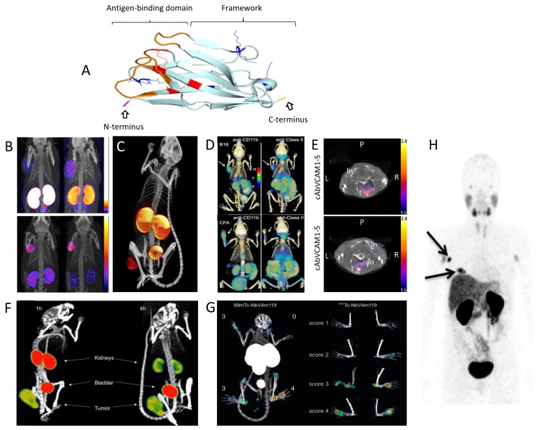
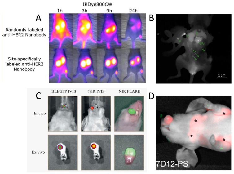
Molecular imaging is paving the way towards noninvasive detection, staging, and treatment follow-up of diseases such as cancer and inflammation-related conditions. Monoclonal antibodies have long been one of the staples of molecular imaging tracer design, although their long blood circulation and high nonspecific background limits their applicability. Nanobodies, unique antibody-binding fragments derived from camelid heavy-chain antibodies, have excellent properties for molecular imaging as they are able to specifically find their target early after injection, with little to no nonspecific background. Nanobody-based tracers using either nuclear or fluorescent labels have been heavily investigated preclinically and are currently making their way into the clinic. In this review, we will discuss different important factors in nanobody-tracer design, as well as the current state of the art regarding their application for nuclear and fluorescent imaging purposes. Furthermore, we will discuss how nanobodies can also be exploited for molecular therapy applications such as targeted radionuclide therapy and photodynamic therapy.

Keywords: intraoperative imaging; molecular imaging; molecular therapy; nuclear imaging; single-domain antibody fragments; targeted fluorescence imaging.


Conflict of interest statement

N.D. is a consultant of Camel-IDS and holds ownership interest (including patents) in camelid single-domain diagnostics and therapeutics. S.H. holds a patent in camelid single-domain diagnostics and therapeutics.

Figures

Figure 1
Figure 1
Schematic representation of the structure of a nanobody and illustrative positron emission tomography (PET) and single photon emission computed tomography (SPECT) preclinical and clinical images obtained using nanobodies that are labeled with distinct radionuclides in diverse medical applications, from oncology, immunology, atherosclerosis, and arthritis to the theranostic imaging of a radiotherapeutic probe. (A) Ribbon diagram of the nanobody 2Rs15d. The complementarity-determining regions (CDRs) are shown in orange, lysines (used for random conjugation methods) in blue, and cysteines and cysteine bridges in red. The C-terminus (in yellow) can be easily genetically modified for site-specific conjugation methods. (B) SPECT/Computed Tomography (CT) images of the biodistribution of 111In-labeled JV7 nanobodies at 3 h post-injection in PSMA+ tumor-bearing mice (on the left shoulder). Effect on renal retention by the removal of tags (top panels: Myc–Cys-tagged nanobody, bottom: Cys-tagged nanobody) and coinjection of positively charged amino acids and gelofusin (left panels: no injection, right panels: with coinjection) is shown. Adapted from [48]. (C) SPECT/CT imaging of an EGFR+ tumor-bearing mouse 1 h after injection of 99mTc-labeled 7C12 nanobody. Adapted from [64]. (D) PET/CT immune cell imaging 90 min after injection of 18F-labeled nanobodies against murine CD11b and major histocompatibility complex (MHC) class II. Top: C57Bl/6 mice inoculated with B16 tumor cells on the left shoulder; bottom: animals injected with complete Freund’s adjuvant on the left paw. Adapted from [72]. (E) SPECT/CT coronal image taken at 2–3 h post-injection of 99mTc-labeled cAbVCAM1-5 nanobody, showing uptake in atherosclerotic lesions (ao) of ApoE-/- mice (bottom) and absence of signals in the aortic arch of C57Bl/6J mice (top) [43]. (F) SPECT/CT images of the biodistribution of the 131I-labeled 2Rs15d therapeutic nanobody in a mouse model with subcutaneous HER2+ xenograft at 1 and 4 h post-injection. Adapted from [54]. (G) SPECT/CT imaging of arthritis in a mouse model with a VSIG4/CRIg-specific 99mTc-labeled nanobody. Adapted from [73]. (H) PET/CT image of the biodistribution of 68Ga-labeled anti-HER2 nanobody in a breast cancer patient 90 min post-injection showing uptake in breast tumor lesions. Adapted from [23].
Figure 2
Figure 2
Examples of in vivo fluorescent molecular imaging with nanobody tracers in mouse tumor models. (A) Comparison of the biodistribution and tumor-targeting potential of the anti-HER2 nanobody 2Rs15d conjugated with IRDye800CW either randomly (top) or site-specifically (bottom). Adapted with permission from [111]. Copyright 2017 American Chemical Society. (B) Fluorescence image acquired during the surgical resection of intraperitoneally disseminated HER2+ tumor lesions. Site-specifically IRDye800CW-labeled 2Rs15d nanobody was injected 90 min before surgery. Fluorescent signal in tumor lesions (indicated by green arrows) is clearly discernible from background signal. Adapted with permission from [123]. (C) Real-time fluorescence imaging of orthotopic tongue tumor 24 h post-injection of an EGFR-specific randomly IRDye800CW-conjugated nanobody. Colocalization with bioluminescence imaging (BLI) and green fluorescent protein (GFP) signals is shown. Adapted with permission from [109]. (D) Fluorescent imaging of an orthotopically inoculated tongue tumor at 1 h post-injection of an EGFR-specific randomly IRDye700DX-conjugated nanobody for photodynamic therapy (PDT). Stars denote the presence of fluorescent tracer uptake in invaded lymph nodes. Adapted with permission from [125].

References

    1. Mankoff D.A. A definition of molecular imaging. J. Nucl. Med. 2007;48:18N–21N. - PubMed
    1. Weber J., Haberkorn U., Mier W. Cancer stratification by molecular imaging. Int. J. Mol. Sci. 2015;16:4918–4946. doi: 10.3390/ijms16034918. - DOI - PMC - PubMed
    1. Vahrmeijer A.L., Hutteman M., van der Vorst J.R., van de Velde C.J.H., Frangioni J.V. Image-guided cancer surgery using near-infrared fluorescence. Nat. Rev. Clin. Oncol. 2013;10:507–518. doi: 10.1038/nrclinonc.2013.123. - DOI - PMC - PubMed
    1. Hingorani D.V., Whitney M.A., Friedman B., Kwon J.-K., Crisp J.L., Xiong Q., Gross L., Kane C.J., Tsien R.Y., Nguyen Q.T. Nerve-targeted probes for fluorescence-guided intraoperative imaging. Theranostics. 2018;8:4226–4237. doi: 10.7150/thno.23084. - DOI - PMC - PubMed
    1. Cha J., Nani R.R., Luciano M.P., Broch A., Kim K., Namgoong J.M., Kulkarni R.A., Meier J.L., Kim P., Schnermann M.J. A chemically stable fluorescent marker of the ureter. Bioorg. Med. Chem. Lett. 2018;28:2741–2745. doi: 10.1016/j.bmcl.2018.02.040. - DOI - PMC - PubMed

LinkOut - more resources