Review
. 2019 Apr 11;8(2):29.
doi: 10.3390/antib8020029.

IMGT® and 30 Years of Immunoinformatics Insight in Antibody V and C Domain Structure and Function

Affiliations
Review

IMGT® and 30 Years of Immunoinformatics Insight in Antibody V and C Domain Structure and Function

Marie-Paule Lefranc et al. Antibodies (Basel). .

Abstract

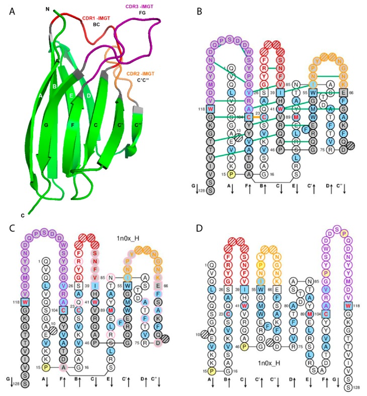
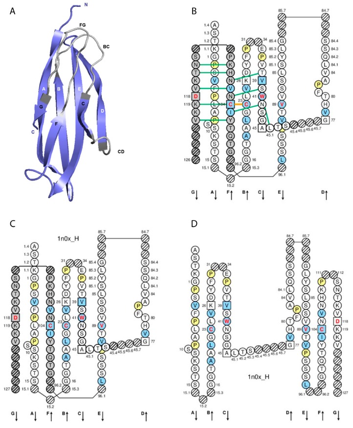
At the 10th Human Genome Mapping (HGM10) Workshop, in New Haven, for the first time, immunoglobulin (IG) or antibody and T cell receptor (TR) variable (V), diversity (D), joining (J), and constant (C) genes were officially recognized as 'genes', as were the conventional genes. Under these HGM auspices, IMGT®, the international ImMunoGeneTics information system®, was created in June 1989 at Montpellier (University of Montpellier and CNRS). The creation of IMGT® marked the birth of immunoinformatics, a new science, at the interface between immunogenetics and bioinformatics. The accuracy and the consistency between genes and alleles, sequences, and three-dimensional (3D) structures are based on the IMGT Scientific chart rules generated from the IMGT-ONTOLOGY axioms and concepts: IMGT standardized keywords (IDENTIFICATION), IMGT gene and allele nomenclature (CLASSIFICATION), IMGT standardized labels (DESCRIPTION), IMGT unique numbering and IMGT Collier de Perles (NUMEROTATION). These concepts provide IMGT® immunoinformatics insights for antibody V and C domain structure and function, used for the standardized description in IMGT® web resources, databases and tools, immune repertoires analysis, single cell and/or high-throughput sequencing (HTS, NGS), antibody humanization, and antibody engineering in relation with effector properties.

Keywords: IMGT; IMGT Collier de Perles; IMGT unique numbering; IMGT-ONTOLOGY; antibody; complementarity determining region; immunogenetics; immunoglobulin; immunoinformatics; paratope.


Conflict of interest statement

The authors declare no conflict of interest. The funders had no role in the design of the study; in the collection, analyses, or interpretation of data; in the writing of the manuscript, or in the decision to publish the results.

Figures

Figure 1
Figure 1
Variable (V) domain. An immunoglobulin (IG) variable heavy VH (V-DOMAIN) is shown as example. Reproduced with permission from IMGT®, the international ImMunoGeneTics information system®, http://www.imgt.org. (A) 3D structure ribbon representation with the IMGT strand and loop delimitations [6]. (B) IMGT Collier de Perles on two layers with hydrogen bonds. The IMGT Collier de Perles on two layers show, in the forefront, the GFCC′C′′ strands (forming the sheet located at the interface VH/VL of the IG) and, in the back, the ABED strands. The IMGT Collier de Perles with hydrogen bonds (green lines online, only shown here for the GFCC’C” sheet) is generated by the IMGT/Collier-de-Perles tool integrated in IMGT/3Dstructure-DB, from experimental 3D structure data [9,10,11]. (C) IMGT Collier de Perles on two layers generated from IMGT/DomainGapAlign [10,12,13]. Pink circles (online) indicate amino acid changes compared to the closest genes and alleles from the IMGT reference directory. (D) IMGT Collier de Perles on one layer. Amino acids are shown in the one-letter abbreviation. All proline (P) are shown online in yellow. IMGT anchors are in square. Hatched circles are IMGT gaps according to the IMGT unique numbering for V domain [6,14]. Positions with bold (online red) letters indicate the four conserved positions that are common to a V domain and to a C domain: 23 (1st-CYS), 41 (CONSERVED-TRP), 89 (hydrophobic), 104 (2nd-CYS) [4,5,6,7,14], and the fifth conserved position, 118 (J-TRP or J-PHE) which is specific to a V-DOMAIN and belongs to the motif F/W-G-X-G that characterizes the J-REGION [6,14] (Table 2). The hydrophobic amino acids (hydropathy index with positive value: I, V, L, F, C, M, A) and tryptophan (W) [15] found at a given position in more than 50% of sequences are shown (online with a blue background color). Arrows indicate the direction of the beta strands and their designations in 3D structures. IMGT color menu for the CDR-IMGT of a V-DOMAIN indicates the type of rearrangement, V-D-J (for a VH here, red, orange and purple) or V-J (for V-KAPPA or V-LAMBDA (not shown), blue, green and greenblue) [1]. The identifier of the chain to which the VH domain belongs is 1n0x_H (from the Homo sapiens b12 Fab) in IMGT/3Dstructure-DB (http://www.imgt.org). The CDR-IMGT lengths of this VH are [8.8.20] and the FR-IMGT are [25.17.38.11]. The 3D ribbon representation was obtained using PyMOL (http://www.pymol.org) and ‘IMGT numbering comparison’ of 1n0x_H (VH) from IMGT/3Dstructure-DB (http://www.imgt.org).
Figure 2
Figure 2
Constant (C) domain. An IG IGHG1 CH1 (C-DOMAIN) is shown as example. Reproduced with permission from IMGT®, the international ImMunoGeneTics information system®, http://www.imgt.org. (A) 3D structure ribbon representation with the IMGT strand and loop delimitations [7]. (B) IMGT Collier de Perles on two layers with hydrogen bonds. The IMGT Colliers de Perles on two layers show, in the forefront, the GFC strands and, in the back, the ABED strands (located at the interface CH1/CL of the IG), linked by the CD transversal strand. The IMGT Collier de Perles with hydrogen bonds (green lines online, only shown here for the GFC sheet) is generated by the IMGT/Collier-de-Perles tool integrated in IMGT/3Dstructure-DB, from experimental 3D structure data [9,10,11]. (C) IMGT Collier de Perles on two layers from IMGT/DomainGapAlign [10,12,13]. (D) IMGT Colliers de Perles on one layer. Amino acids are shown in the one-letter abbreviation. All proline (P) are shown online in yellow. IMGT anchors are in square. Hatched circles are IMGT gaps according to the IMGT unique numbering for C domain [7,14]. Positions with bold (online red) letters indicate the four conserved positions that are common to a V domain and to a C domain: 23 (1st-CYS), 41 (CONSERVED-TRP), 89 (hydrophobic), 104 (2nd-CYS) [4,5,6,7,14], and position 118 which is only conserved in V-DOMAIN. The identifier of the chain to which the CH1 domain belongs is 1n0x_H (from the Homo sapiens b12 Fab, in IMGT/3Dstructure-DB, http://www.imgt.org). The 3D ribbon representation was obtained using PyMOL and ‘IMGT numbering comparison’ of 1n0x_H (CH1) from IMGT/3Dstructure-DB (http://www.imgt.org).
Figure 3
Figure 3
V-DOMAIN Contact analysis results. Reproduced with permission from IMGT®, the international ImMunoGeneTics information system®, http://www.imgt.org. (A) IMGT/3Dstructure-DB domain pair contacts between the VH domain of motavizumab (3ixt_H) and the Fusion glycoprotein F1 (ligand) (3ixt_P). (B) IMGT/3Dstructure-DB Domain pair contacts between the V-KAPPA domain of motavizumab (3ixt_L) and the Fusion glycoprotein F1 (ligand) (3ixt_P). ‘Polar’, ‘Hydrogen bonds’, and ‘Nonpolar’ were selected prior to display, in ‘Atom contact types’. Amino acids belonging to the CDR1-IMGT, CDR2-IMGT and CDR3-IMGT are colored according to the IMGT color menu (red, orange, and purple, respectively, for VH; blue, light green and green, respectively, for V-KAPPA). In this 3D structure, all but one of the amino acids contacting the antigen belong to the CDR-IMGT. Clicking on R@P gives access to the IMGT Residue@Position cards [9,10,11].
Figure 3
Figure 3
V-DOMAIN Contact analysis results. Reproduced with permission from IMGT®, the international ImMunoGeneTics information system®, http://www.imgt.org. (A) IMGT/3Dstructure-DB domain pair contacts between the VH domain of motavizumab (3ixt_H) and the Fusion glycoprotein F1 (ligand) (3ixt_P). (B) IMGT/3Dstructure-DB Domain pair contacts between the V-KAPPA domain of motavizumab (3ixt_L) and the Fusion glycoprotein F1 (ligand) (3ixt_P). ‘Polar’, ‘Hydrogen bonds’, and ‘Nonpolar’ were selected prior to display, in ‘Atom contact types’. Amino acids belonging to the CDR1-IMGT, CDR2-IMGT and CDR3-IMGT are colored according to the IMGT color menu (red, orange, and purple, respectively, for VH; blue, light green and green, respectively, for V-KAPPA). In this 3D structure, all but one of the amino acids contacting the antigen belong to the CDR-IMGT. Clicking on R@P gives access to the IMGT Residue@Position cards [9,10,11].

References

    1. Lefranc M.-P., Lefranc G. The Immunoglobulin FactsBook. Academic Press; London, UK: 2001.
    1. Lefranc M.-P., Lefranc G. The T Cell Receptor FactsBook. Academic Press; London, UK: 2001.
    1. Lefranc M.-P. Immunoglobulin and T Cell Receptor Genes: IMGT® and the Birth and Rise of Immunoinformatics. Front. Immunol. 2014;5:22. doi: 10.3389/fimmu.2014.00022. - DOI - PMC - PubMed
    1. Lefranc M.-P. Unique database numberings system for immunogenetic analysis. Immunol. Today. 1997;18:509. doi: 10.1016/S0167-5699(97)01163-8. - DOI - PubMed
    1. Lefranc M.-P. The IMGT unique numbering for Immunoglobulins, T cell receptors and Ig-like domains. Immunologist. 1999;7:132–136.

LinkOut - more resources