Review
. 2019 Jul 3;8(3):41.
doi: 10.3390/antib8030041.

Bispecific T-Cell Redirection versus Chimeric Antigen Receptor (CAR)-T Cells as Approaches to Kill Cancer Cells

Affiliations
Review

Bispecific T-Cell Redirection versus Chimeric Antigen Receptor (CAR)-T Cells as Approaches to Kill Cancer Cells

William R Strohl et al. Antibodies (Basel). .

Abstract

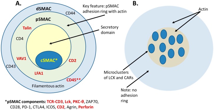
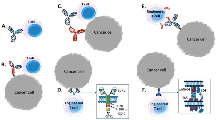
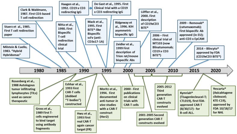
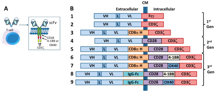
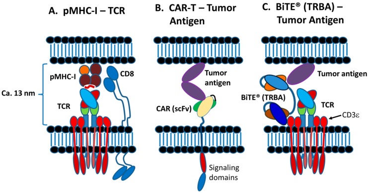
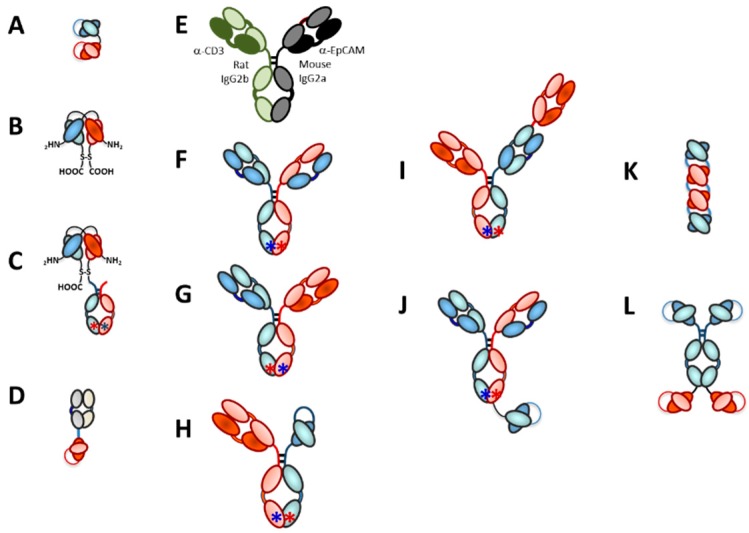
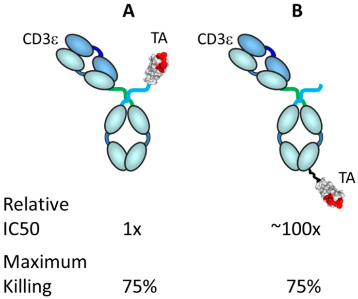
The concepts for T-cell redirecting bispecific antibodies (TRBAs) and chimeric antigen receptor (CAR)-T cells are both at least 30 years old but both platforms are just now coming into age. Two TRBAs and two CAR-T cell products have been approved by major regulatory agencies within the last ten years for the treatment of hematological cancers and an additional 53 TRBAs and 246 CAR cell constructs are in clinical trials today. Two major groups of TRBAs include small, short-half-life bispecific antibodies that include bispecific T-cell engagers (BiTE®s) which require continuous dosing and larger, mostly IgG-like bispecific antibodies with extended pharmacokinetics that can be dosed infrequently. Most CAR-T cells today are autologous, although significant strides are being made to develop off-the-shelf, allogeneic CAR-based products. CAR-Ts form a cytolytic synapse with target cells that is very different from the classical immune synapse both physically and mechanistically, whereas the TRBA-induced synapse is similar to the classic immune synapse. Both TRBAs and CAR-T cells are highly efficacious in clinical trials but both also present safety concerns, particularly with cytokine release syndrome and neurotoxicity. New formats and dosing paradigms for TRBAs and CAR-T cells are being developed in efforts to maximize efficacy and minimize toxicity, as well as to optimize use with both solid and hematologic tumors, both of which present significant challenges such as target heterogeneity and the immunosuppressive tumor microenvironment.

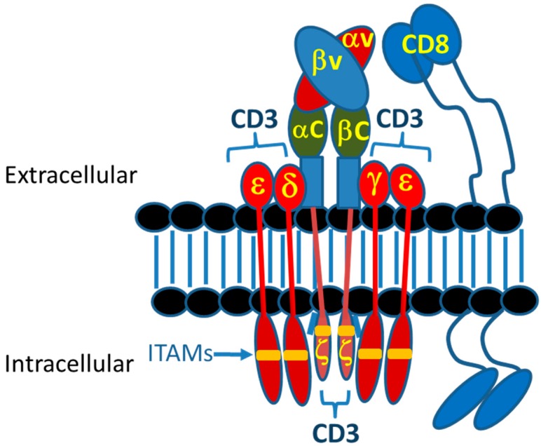
Keywords: CD3ε, T cells; NK cells; T-cell redirection; bispecific antibody; chimeric antigen receptor; immune synapse; tumor cell killing; tumor microenvironment.


Conflict of interest statement

The authors declare the following potential conflicts of interest: W.R.S. and M.N. both were recent employees at Janssen R&D, Johnson & Johnson and hold stock in that company. W.R.S. currently is a consultant for several small biotechnology companies and is a member of the Board of Directors of IGM Biosciences. M.N. currently is an employee of Century Therapeutics, Philadelphia, PA.

Figures

Figure 4
Figure 4
Classical immune synapse as compared with a bispecific T-cell engager (BiTE®)-induced synapse and a CAR-T synapse. (A) Diagrammatic representation of the immune synapse, adapted and modified from Huppa and Davis [110] and Watanabe et al. [111]. The classical immune synapse forms as a “bullseye” with the center central supramolecular activation cluster (cSMAC) surrounded by the peripheral SMAC (pSMAC) adhesion ring and the distal dSMAC ring. CD3, PKC-θ, perforin, CD28, CTLA4 and Agrin are found in the cSMAC. Additionally, Lck initially accumulates in the cSMAC and then distributes more broadly [110]. A key feature of the immune synapse is exclusion of CD45 from the cSMAC (noted by **). The pSMAC ring includes Talin, LFA1, VAV1 and CD4. LFA-1 is a key synapse stabilizing force in the pSMAC. The dSMAC markers are CD43, CD44, CD45 and filamentous actin. Offne×r et al. [13] compared the synapses formed by an anti-EpCAM × CD3 BiTE® TRBA to those formed by MHC-Her2-peptide/TCR. The markers denoted in red were positioned similarly in both the normal peptide-loaded major histocompatibility complex (pMHC)/TCR synapse and the BiTE-induced synapse [13]. CD45 was found to be excluded from both the BiTE®-induced synapse and the control pMHC/TCR synapse [13]. (B) A diagrammatic representation of the synapse formed by CAR-T cells, adopted and modified from Davenport et al. [112]. They described the CAR-T/target cell synapse as disorganized, with multifocal clusters containing LCK, no apparent LFA-1 stabilization and the absence of the adhesion ring that helps to define the classical immune synapse [112,113].
Figure 1
Figure 1
Examples of T-cell based therapeutics in clinical development. (A) Inhibition of checkpoint receptors such as PD-1 and CTLA-4 to improve T-cell activity [18]; (B) T-cell redirection with bispecific antibodies (TRBAs) in which one binding arm recognizes a tumor antigen and the other binding arm recognizes CD3ε on T-cells [17,19,20]; (C) Autologous T-cells activated ex vivo, combined with bispecific antibody conjugates recognizing tumor antigen with one mAb and CD3ε on T cells with the other mAb, followed by re-administration to the patient to kill tumors [21]; (D) Genetically engineered autologous chimeric antigen receptor (CAR)-T cells in which an antibody, typically a single chain variable fragment (scFv), fused to intracellular T-cell activation domains such as CD28, 4-1BB, OX40 and CD3ζ, replace the function of the T-cell receptor (TCR), making the T-cells killers of specific antigen-bearing cells [22,23,24]; (E) Autologous or allogeneic T-cells or NK cells genetically engineered with FcγRIIIa (CD16a), which, when administered with an anti-tumor monoclonal antibody (mAb) such as the anti-CD20 mAb, rituximab, binds to the Fc of the antibodies and functionally redirects the T-or NK-cells to the tumor to kill the cancer cells [25]; (F). Autologous T cells with engineered TCRs.
Figure 2
Figure 2
Key milestones in the history of T-cell redirected bispecific antibodies (TRBAs; top) and chimeric antigen receptor (CAR)-T cell (bottom) therapeutics. Specific references cited for TRBAs are: Milstein and Cuello, 1983 [38], Staerz et al., 1985 [41], Clark and Waldmann, 1987 [48], Nitta et al., 1990 [49], Haagen et al., 1992 [50], De Gast et al., 1995 [51], Mack et al., 1995 [45], Ridgeway et al., 1996 [52], Zeidler et al., 1999 [53] and Löffler et al., 2000 [54]. Specific references for CAR-T development cited are: Rosenberg et al., 1988 [55], Gross et al., 1989 [35], Eshhar et al., 1993 [37], Hwu et al., 1993 [56] and Moritz et al., 1994 [57].
Figure 3
Figure 3
Generations of CAR-T cell therapeutics. Generations of CAR-T cell therapeutics as described in the text. (A) Generalized drawing of a CAR-T showing the fusion of the scFv to the transmembrane domain and intracellular activation domains. (B) Drawing depicting examples of first generation (1–3), second generation (4, 5) and third generation (6–9) CAR-T constructs as described in the text.
Figure 5
Figure 5
Diagram of the T-cell receptor (TCR) complex. Normal TCR-pMHC interactions are in the range of 1–100 µM, with the inherent avidity of clustered TCRs providing the required attraction [11]. Affinities of the MHC to the presented peptide have a profound influence on the ability of natural T cells to kill and eradicate tumors. When the peptide-MHC affinity was found to be <10 nM as determined in in vitro assays, it was shown that T cells recognizing those pMHC complexes were able to cause tumor rejection [116]. On the other hand, when the peptide-MHC affinity was >100 nM, the tumors relapsed, indicating that the T cells were not capable of killing those tumors cells [116]. Both CAR-T cells and TRBAs function independently of this parameter.
Figure 6
Figure 6
Diagram of the T-cell receptor complex. Diagram of the molecules driving synapse formation in pMHC-1/TCR T-cell/APC interactions, in CAR-T/target cell interactions and in TRBA-induced T-cell/target cell interactions. (A) Classic TCR/pMHC-1 type of interaction with a membrane to membrane spacing in the range of 13 nm [119]. (B) scFv-based CAR-T cell binding to tumor antigen on target cell. (C) BiTE® binding to CD3ε on T-cell and to tumor antigen on target cell to bring the cells into close proximity to form the cytolytic synapse.
Figure 7
Figure 7
Examples of bispecific antibody platforms used to make clinical stage TRBAs. (A) Bispecific T-cell engager (BiTE®) [45]; (B) dual affinity retargeting (DART®) antibody [153]; (C) DART®-Fc for elongated half-life in vivo [154]; (D) TCR fused to scFv called immune-mobilizing monoclonal TCRs against cancer (ImmTAC) [155]; (E) mouse/rat hybrid IgG [53]; (F) Asymmetric IgG with common LC [59]; (G) Asymmetric IgG with different light chains [156]; (H) Asymmetric IgG-like molecule with a Fab arm and an scFv arm to eliminate light chain resorting [157,158]; (I) Asymmetric IgG using cross-mab technology for LC fidelity and extra Fab arm to make a 2:1 (target cell antigen:CD3ε) antibody call “TCBs,” for “T-cell Bispecifics” [159,160]; (J) Chugai’s asymmetric IgG using “Asymmetric Re-engineering Technology–Immunoglobulin” (ART-Ig®) platform [161,162] technology, with an scFv fused to one HC to make an ART-Ig®-scFv 2:1 (target cell antigen:CDε) antibody; (K) tetravalent, bispecific tandem diabody (TandAb) [163]; (L) tetravalent, bispecific ADAPTIR™ platform with two different scFvs fused to each Fc [164].
Figure 8
Figure 8
Effect of geometry on in vitro activity of an example TRBA. Cartoon of two antibody-centyrin fusions targeting CD3ε with the Fab arm and a solid tumor antigen with the centyrin (~12 kDa engineered FN3 domain). (A). Centyrin fused to the hinge, making it close to the anti-CD3 Fab arm; (B). Centyrin fused to the C-terminus of the heavy chain, making it distal from the Fab arm. All components, molecule sizes and in vitro killing assay conditions (E:T 5:1, 24 h assay) were identical. Thus, only the architecture and geometry of the two TRBAs are different, resulting in a ca. 100-fold difference in potency. This simple example demonstrates how important the geometry of the binding arms can be in the design of future TRBAs. Data presented were derived from experiments provided Steve Jacobs, Janssen R&D. These data were previously presented at PEGS Boston, April 2018 with permission.
Figure 9
Figure 9
Drawing depicting the construction of an example allogeneic CAR-T cell. The CAR contains, from N-to-C terminus, anti-BCMA scFv, 136 amino acid residue RQR peptide, CD8α hinge and transmembrane (TM) domain, 4-1BB costimulatory domain and CDζ signaling domain in a cell in which TCR- α and CD52 have been knocked out using gene editing technology [301].
Figure 10
Figure 10
Schematic representation of various CAR formats. Left, natural organization of a CD8+ TCR expressed on cytotoxic T cells driving MHC-restricted CD8+ T cell effects. TCR-CARs are formed by fusing the TCR α/β variable domains to a second-generation CAR scaffold. The Eureka Therapeutics Artemis platform fuses antibody variable domains to TCR α/β constant regions. TCR2 platform fuses scFvs to CD3ε. Triumvira platform fusing two scFv chains to CD8 stalk, hinge and intracellular domains-one scFv binds to CD3ε engaging the rest of the TCR complex, while the second is free to interact with tumor antigens.

References

    1. Coley W.B. Contribution to the knowledge of sarcoma. Ann. Surg. 1891;14:199–220. doi: 10.1097/00000658-189112000-00015. - DOI - PMC - PubMed
    1. Hoption Cann S.A., Van Netten J.J., Van Netten C. Dr William Coley and tumor regression: A place in history or in the future. Postgrad. Med. 2003;79:672–680. - PMC - PubMed
    1. Vernon L.F. William Bradley Coley, MD and the phenomenon of spontaneous regression. Immunotargets Ther. 2018;7:29–34. doi: 10.2147/ITT.S163924. - DOI - PMC - PubMed
    1. Poltorak A., He X., Smirnova I., Liu M.-Y., Van Huffel C., Du X., Birdwell D., Alejos E., Silva M., Galanos C., et al. Defective LPS signaling in C3H/HeJ and C57BL/10ScCr mice: Mutations in Tlr4 gene. Science. 1998;282:2085–2088. doi: 10.1126/science.282.5396.2085. - DOI - PubMed
    1. Reisser D., Pance A., Jeannin J.F. Mechanisms of the antitumoral effect of lipid A. Bioessays. 2002;24:284–289. doi: 10.1002/bies.10053. - DOI - PubMed

LinkOut - more resources