. 2018 Dec 13;7(4):42.
doi: 10.3390/antib7040042.

Enhancers Improve the AID-Induced Hypermutation in Episomal Vector for Antibody Affinity Maturation in Mammalian Cell Display

Affiliations

Enhancers Improve the AID-Induced Hypermutation in Episomal Vector for Antibody Affinity Maturation in Mammalian Cell Display

Chuan Chen et al. Antibodies (Basel). .

Abstract

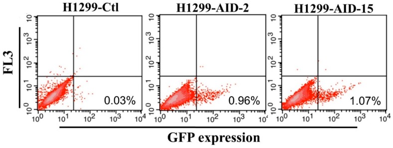
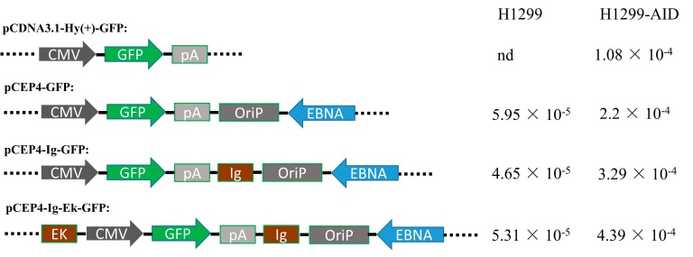
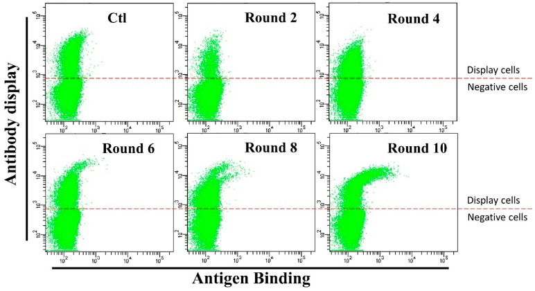
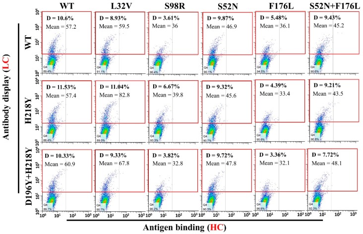
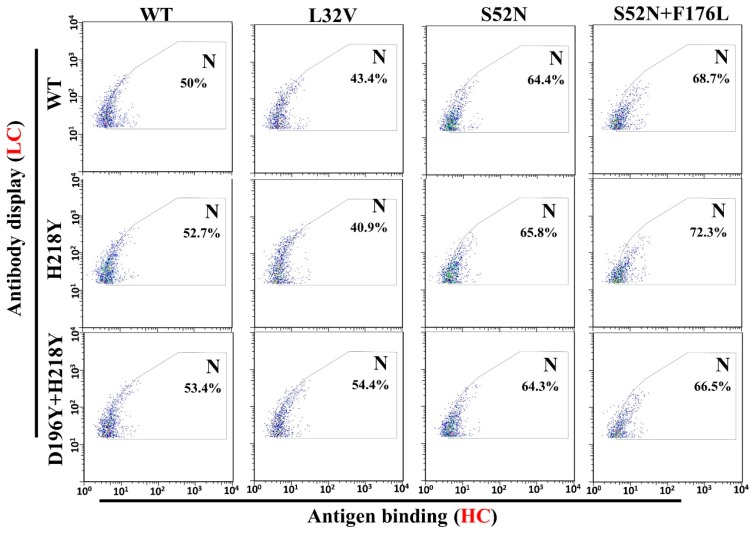
The induction of somatic hypermutation (SHM) in various cell lines by activation-induced cytidine deaminase (AID) has been used in protein-directed selection, especially in antibody affinity maturation. Several antibody affinity maturation systems based on mammalian cells have been developed in recent years, i.e., 293T, H1299, Raji and CHO cells. However, the efficiency of in vitro AID-induced hypermutation is low, restricting the application of such systems. In this study, we examined the role of Ig and Ek enhancers in enhancing SHM in the episomal vector pCEP4 that expresses an anti-high mobility group box 1 (HMGB1) full-length antibody. The plasmid containing the two enhancers exhibited two-fold improvement of mutation rate over pCEP4 in an AID expression H1299 cell line (H1299-AID). With the engineered episomal vector, we improved the affinity of this antibody in H1299-AID cells by 20-fold.

Keywords: activation-induced cytidine deaminase (AID); enhancer; episomal vector; somatic hypermutation (SHM).


Conflict of interest statement

The authors declare no conflict of interest.

Figures

Figure 1
Figure 1
H1299-AID cell clones reverse mutation rate (Clone 2 and Clone 15). H1299-AID clones were transfected with pCEP4-GFPm. 2 days after transfection, the GFP fluorescence (reverse mutation) was detected by flow cytometer. Ctl: negative control. AID: Activation-induced cytidine deaminase expression clones, FL3: FACSAria II FL-3 channel signal results.
Figure 2
Figure 2
Structure of the plasmids with or without enhancers and mutation rate (mutation rate/15 days) in H1299 and H1299-AID cells. CMV: Human cytomegalovirus immediate-early promoter/enhancer. GFP: green fluorescent protein. pA: SV40 polyadenylation signal. OriP: EBV origin of replication. EBNA: Epstein-Barr nuclear antigen. EK: Ek enhancer sequence. lg: Ig enhancer sequence.
Figure 3
Figure 3
Flow cytometry cell sorting results during affinity maturation. Each round of maturation, the 3B1 antibody on the cells were stained with anti-human-IgG-APC (1:20 dilution) and HMGB1-GFP (0.5 µg/mL). Cells with better HMGB1-GFP binding (0.05–0.1% of the cells) were sorted out and expanded for another round of sorting. Anti-human-IgG-APC: Allophycocyanin conjugated Mouse Anti-Human IgG antibody. HMGB1-GFP: High mobility group box 1 and GFP fusion protein.
Figure 4
Figure 4
Antibody mutants display on cell surface and binding with HMGB1-GFP. Various antibody light chain and heavy chain mutants with trans-membrane domain were co-transfected into H1299 cells. Two days after transfection, cells were stained with anti-human-IgG-APC and HMGB1-GFP. Antibody display was evaluated by flow cytometry (Gate D: Positive Cells). The heavy chain S98R and F176L mutant combinations groups have much fewer cells display the antibody on the cell surface (Gate D, S98R: 3.6–6.6%, F176L: 3.35–5.45%) than other heavy chain mutant combinations (Gate D, 7.7–11.5%). WT: Original wild type antibody.
Figure 5
Figure 5
Statistics of HMGB1-GFP binding with mutant combinations (well displayed combinations). Gate N was chosen according to the center of the WT/WT cells, and half of the cell were in the gate. Compared with the WT/WT, combinations with better antigen binding will have higher percentage of cells in gate N. According to the results, light chain H218Y, D196Y + H218Y, and heavy chain L32V mutants have no significant contribution to antigen binding (Lower or similar percentage of cells in gate N compared with WT/WT). While the heavy chain S52N and S52N + F176L mutants significantly improved the antigen binding (higher percentage of cell in gate N).

Similar articles

Cited by

References

    1. Leickt L., Grubb A., Ohlson S. Affinity screening for weak monoclonal antibodies. J. Immunol. Methods. 1998;220:19–24. doi: 10.1016/S0022-1759(98)00163-X. - DOI - PubMed
    1. Chappey O.N., Sandouk P., Scherrmann J.M.G. Monoclonal-antibodies in hapten immunoassays. Pharmaceut. Res. 1992;9:1375–1379. doi: 10.1023/A:1015890208714. - DOI - PubMed
    1. Yuan B., Schulz P., Liu R., Sierks M.R. Improved affinity selection using phage display technology and off-rate based selection. Electron. J. Biotechn. 2006;9:171–175. doi: 10.2225/vol9-issue2-fulltext-6. - DOI
    1. Lowman H.B., Wells J.A. Affinity maturation of human growth-hormone by monovalent phage display. J. Mol. Biol. 1993;234:564–578. doi: 10.1006/jmbi.1993.1612. - DOI - PubMed
    1. Sun S., Yang X., Wang H.F., Zhao Y., Lin Y., Ye C., Fang X.D., Hang H.Y. Antibody affinity maturation through combining display of two-chain paired antibody and precision flow cytometric sorting. Appl. Microbiol. Biot. 2016;100:5977–5988. doi: 10.1007/s00253-016-7472-1. - DOI - PubMed