. 2019 Nov 4;47(19):10426-10438.
doi: 10.1093/nar/gkz819.

The internal interaction in RBBP5 regulates assembly and activity of MLL1 methyltransferase complex

Affiliations

The internal interaction in RBBP5 regulates assembly and activity of MLL1 methyltransferase complex

Jianming Han et al. Nucleic Acids Res. .

Abstract

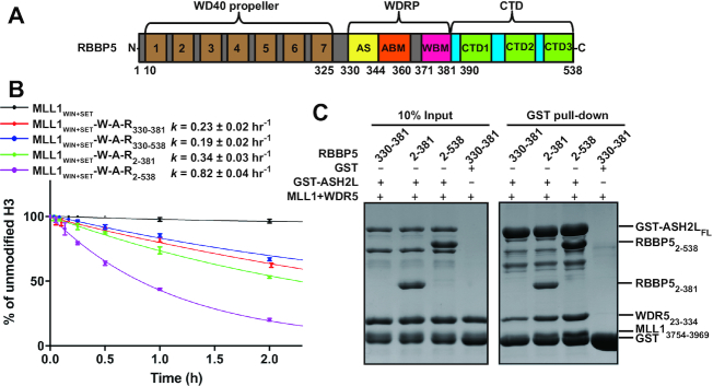
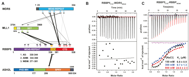
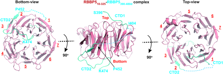
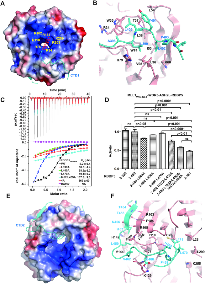
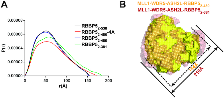
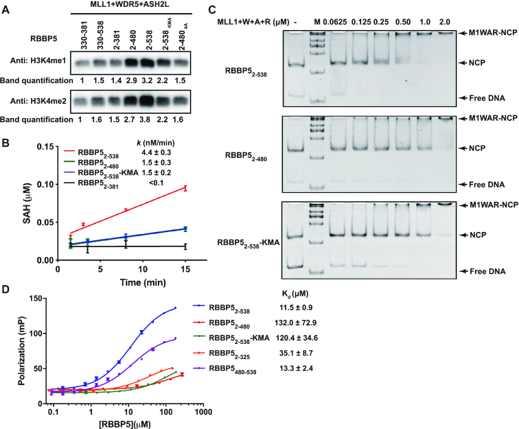
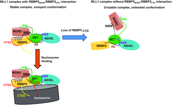
The Mixed Lineage Leukemia protein 1 (MLL1) plays an essential role in the maintenance of the histone H3 lysine 4 (H3K4) methylation status for gene expression during differentiation and development. The methyltransferase activity of MLL1 is regulated by three conserved core subunits, WDR5, RBBP5 and ASH2L. Here, we determined the structure of human RBBP5 and demonstrated its role in the assembly and regulation of the MLL1 complex. We identified an internal interaction between the WD40 propeller and the C-terminal distal region in RBBP5, which assisted the maintenance of the compact conformation of the MLL1 complex. We also discovered a vertebrate-specific motif in the C-terminal distal region of RBBP5 that contributed to nucleosome recognition and methylation of nucleosomes by the MLL1 complex. Our results provide new insights into functional conservation and evolutionary plasticity of the scaffold protein RBBP5 in the regulation of KMT2-family methyltransferase complexes.


Figures

Figure 1.
Figure 1.
The WD40 and CTD regions of RBBP5 are important for activity regulation of the MLL1 complex. (A) Domain organization of human RBBP5. The seven repeats of the WD40 propeller are indicated. WDRP, WD40 Repeat Proximity domain; AS, Activation Segment; ABM, ASH2L-Binding Motif; WBM, WDR5-Binding Motif; CTD, C-Terminal Distal domain. (B) Comparison of the overall rates of the methylation reactions on H3 peptides catalyzed by the MLL1 complex assembled with different RBBP5 fragments. Each data point was shown as mean ± s.d. from triplicate measurements. The reported rate constants and fitting errors were derived by exponential fitting of the decrease in the relative intensities of the unmodified H3 peptide peaks in MALDI–TOF mass spectra. (C) GST pull-down assays revealed the stabilities of the MLL1 complexes. GST-ASH2LFL was incubated with MLL13754–3969, WDR523–334, and different RBBP5 constructs. Bound proteins were eluted and separated by SDS-PAGE. MLL13754–3969 ran at a similar position as GST as indicated.
Figure 2.
Figure 2.
The internal interactions in RBBP5. (A) Schematic representation of inter-protein crosslinks detected by CX-MS analysis of the MLL1 complex. RBBP5 intra-protein crosslinks are also labeled. The width of the linking line is correlated to the peptide amount of one specific crosslink appeared in the MS. (B) WDR523–334 has no interaction with RBBP5CTD (residues 381–538) as shown by a representative ITC binding curve. The assay buffer is 150 mM NaCl, 25 mM Tris–HCl, pH8.0. (C) RBBP5WD40 (residues 2–333) has a direct interaction with RBBP5CTD (residues 381–538) as shown by representative ITC binding curves at three different salt concentrations (150, 300 and 800 mM NaCl in the presence of 25 mM Tris–HCl, pH 8.0 buffer). The binding affinity slightly decreased with the increase of salt concentration. The purple curve is the buffer titrated with RBBP5CTD. The dissociation constants (Kd) and the reported fitting errors were determined from the representative ITC curves by data fitting using one-site binding model.
Figure 3.
Figure 3.
Crystal structure of the RBBP5WD40-RBBP5CTDM complex. Three views of the RBBP5WD40–RBBP5CTDM complex are shown. RBBP5WD40 is colored in red and RBBP5CTDM in cyan. The two segments of RBBP5CTDM, CTD1 and CTD2, are indicated. The surface where CTD1 binds is defined as the top of the WD40 propeller.
Figure 4.
Figure 4.
The interaction interface between RBBP5WD40 and RBBP5CTDM. (A) The RBBP5CTD1-binding pocket on RBBP5WD40. RBBP5WD40 surface is colored according to its electrostatic potential (positive potential, blue; negative potential, red). RBBP5CTD1 is shown in cyan. One possible path of the absent N-terminal acidic extension of RBBP5CTD1 is indicated. (B) Details of hydrophobic interactions between RBBP5WD40 and RBBP5CTD1. The critical residues are presented as ball-and-stick models. RBBP5WD40 residues are colored in red and RBBP5CTD1 residues in cyan. Hydrogen bonding interactions are shown as dashed magenta lines. (C) Effects of mutations in the RBBP5CTDM on the interaction between RBBP52–333 and RBBP5CTDM (RBBP5390–480d) analyzed by ITC assays. ITC buffer is 25mM Tris–HCl, 300 mM NaCl, pH 8.0. The RBBP5–4A mutation is L399A/L400A/I457A/L459A mutation. The dissociation constants (Kd) and the reported fitting errors for each mutant were determined from one representative ITC curve by data fitting using one-site binding model. (D) HKMT activities of the MLL1 complexes (MLL13754–3969–WDR523–334–ASH2FL–RBBP5) containing different RBBP5 mutants determined by the MALDI-TOF-based methyltransferase assays. The HKMT activities are normalized to the activity of RBBP52–480-containing MLL1 complex. Mean ± s.d. (n = 3) are shown. The ANOVA test was used to determine the statistical difference of HKMT activities between groups. The label ‘ns’ stands for ‘not significant’ (P> 0.05). (E) The RBBP5CTD2-binding surface on RBBP5WD40. RBBP5WD40 surface is colored according to its electrostatic potential (positive potential, blue; negative potential, red). RBBP5CTD2 is shown in cyan. (F) Details of hydrophobic interactions between RBBP5WD40 and RBBP5CTD2. The critical hydrophobic residues are presented as ball-and-stick models. Salt bridge and hydrogen bonding interactions are shown as dashed magenta lines.
Figure 5.
Figure 5.
SAXS analyses of MLL1 complex. (A) Overlay of P(r) distributions from the MLL1 complexes assembled from different RBBP5 constructs. RBBP52–480-4A is RBBP52–480 with the L399A/L400A/I457A/L459A mutation. (B) Superimposition of the molecular envelops of the RBBP52–480-containing MLL1 complex (yellow) and the RBBP52–381-containing MLL1 complex (red). The sizes of the longest dimension of different complexes are indicated.
Figure 6.
Figure 6.
RBBP5CTD3 contributes to methylation on nucleosomes. (A) Methyltransferase activity assays were carried out using 167-bp nucleosomes as substrates. MLL1 complex (1 μM), nucleosome (1 μM), and SAM (20 μM) were incubated at 25°C for 1 h. At this condition, no H3K4me3 signal was detected. The band densities were quantified by setting the band density from the sample of MLL13754–3969–WDR523–334–ASH2LFL–RBBP5330–381 as one. RBBP5 2–538KMA is a mutation with 10 lysine residues in CTD3 mutated into alanine. RBBP5 2–4804A contains the RBBP52–480 L399A/L400A/I457A/L459A mutation. (B) Time-course monitoring of methyltransferase activities of different MLL1 complexes by MTase-Glo™ Methyltransferases Assay Kit. The assay system contains 50 nM MLL1 complex, 1 μM nuclesome, and 20 μM SAM. Each data point was presented as mean ± s.d. from triplicate measurements. The reaction rate and reported fitting errors were derived by linear fitting of the averaged dataset. (C) EMSA showed that MLL1 complexes bound to 167-bp nucleosomes. 0.1 μM nucleosome was incubated with increasing amounts of MLL1 complexes with different RBBP5 constructs as indicated. (D) The dissociation constants of the RBBP5-DNA interactions were determined by fluorescence polarization assays. 0.1 μM FAM-labelled 167-bp dsDNA was used. Each data point was shown as mean ± s.d. from triplicate measurements. Some error bars were not shown because the error bar was shorter than the size of the symbol. The dissociation constants (Kd) and the reported fitting errors were determined from the averaged data points by fitting with the sigmoidal dose-response model.
Figure 7.
Figure 7.
The model for the role of RBBP5CTD in regulating MLL1 complex. The interaction between RBBP5WD40 and RBBP5CTD facilitates the formation of a compact MLL1 complex with correct coordination of the structural elements required for methylation. The loss of RBBP5CTD generates an extended complex with low catalytic efficiency. A vertebrate-specific RBBP5CTD3 can bind to nucleosome DNA and increases the association of the MLL1 complex with nucleosomes, thereby promoting methylation of nucleosomal H3.

References

    1. Ruthenburg A.J., Allis C.D., Wysocka J.. Methylation of lysine 4 on histone H3: intricacy of writing and reading a single epigenetic mark. Mol. Cell. 2007; 25:15–30. - PubMed
    1. Shilatifard A. Molecular implementation and physiological roles for histone H3 lysine 4 (H3K4) methylation. Curr. Opin. Cell Biol. 2008; 20:341–348. - PMC - PubMed
    1. Barski A., Cuddapah S., Cui K., Roh T.Y., Schones D.E., Wang Z., Wei G., Chepelev I., Zhao K.. High-resolution profiling of histone methylations in the human genome. Cell. 2007; 129:823–837. - PubMed
    1. Heintzman N.D., Stuart R.K., Hon G., Fu Y., Ching C.W., Hawkins R.D., Barrera L.O., Van Calcar S., Qu C., Ching K.A. et al.. Distinct and predictive chromatin signatures of transcriptional promoters and enhancers in the human genome. Nat. Genet. 2007; 39:311–318. - PubMed
    1. Rao R.C., Dou Y.. Hijacked in cancer: the KMT2 (MLL) family of methyltransferases. Nat. Rev. Cancer. 2015; 15:334–346. - PMC - PubMed

Publication types

MeSH terms