. 2019 Nov 18;47(20):10744-10753.
doi: 10.1093/nar/gkz803.

Quantifying the impact of small molecule ligands on G-quadruplex stability against Bloom helicase

Affiliations

Quantifying the impact of small molecule ligands on G-quadruplex stability against Bloom helicase

Parastoo Maleki et al. Nucleic Acids Res. .

Abstract

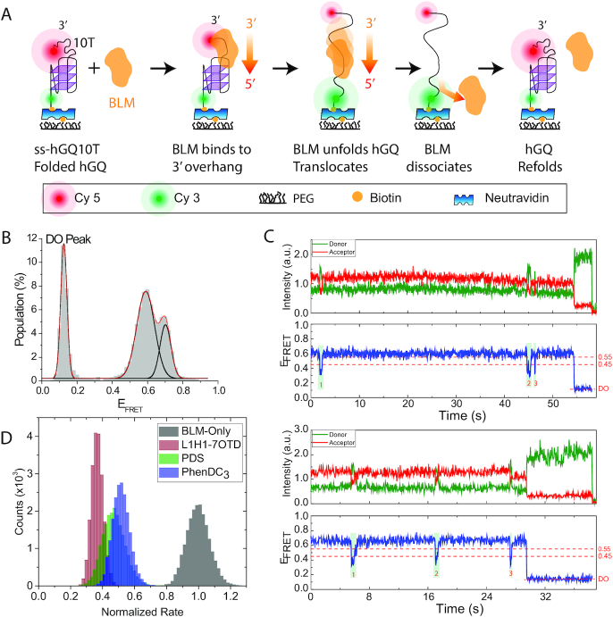
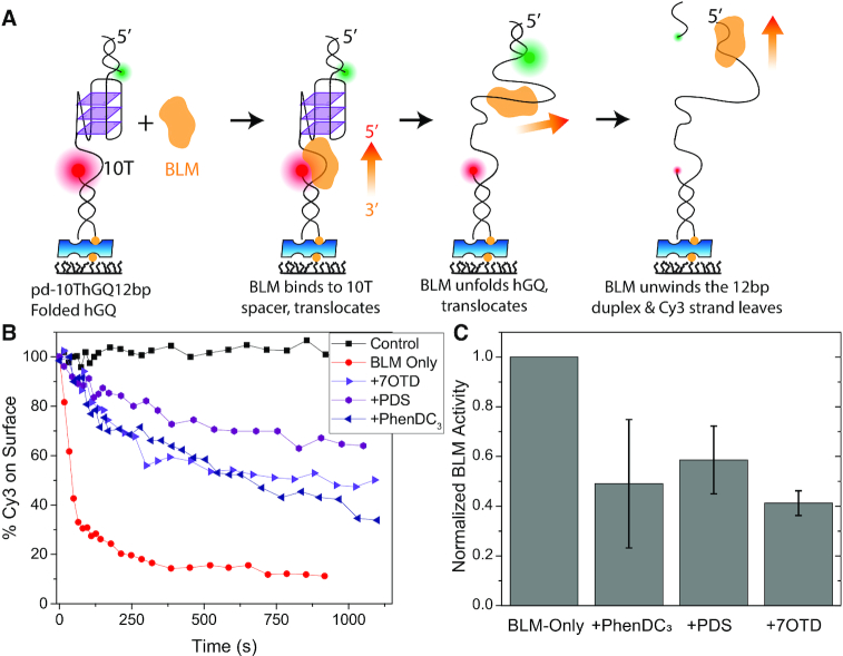
G-quadruplex (GQ) stabilizing small molecule (SM) ligands have been used to stabilize human telomeric GQ (hGQ) to inhibit telomerase activity, or non-telomeric GQs to manipulate gene expression at transcription or translation level. GQs are known to inhibit DNA replication unless destabilized by helicases, such as Bloom helicase (BLM). Even though the impact of SM ligands on thermal stability of GQs is commonly used to characterize their efficacy, how these ligands influence helicase-mediated GQ unfolding is not well understood. Three prominent SM ligands (an oxazole telomestatin derivative, pyridostatin, and PhenDC3), which thermally stabilize hGQ at different levels, were utilized in this study. How these ligands influence BLM-mediated hGQ unfolding was investigated using two independent single-molecule approaches. While the frequency of dynamic hGQ unfolding events was used as the metric in the first approach, the second approach was based on quantifying the cumulative unfolding activity as a function of time. All three SM ligands inhibited BLM activity at similar levels, 2-3 fold, in both approaches. Our observations suggest that the impact of SM ligands on GQ thermal stability is not an ideal predictor for their inhibition of helicase-mediated unfolding, which is physiologically more relevant.


Figures

Figure 1.
Figure 1.
Quantifying the impact of SM ligands on BLM-mediated hGQ unfolding based on analysis of dynamic interactions. (A) A schematic of different steps of the BLM-mediated hGQ unfolding are illustrated. Donor (green) and acceptor (red) fluorophores are placed across hGQ. BLM binds to the 3′-overhang, translocates towards 5′-end, and unfolds the hGQ before dissociating from the DNA, allowing hGQ to refold. (B) The smFRET histogram showing folded state peaks at EFRET = 0.70±0.03 and 0.59±0.05 at 150 mM KCl and 2 mM MgCl2. The peak at EFRET = 0.12±0.02 represents the DO state, resulting from hGQ molecules without Cy5. The solid black curves are individual Gaussian fit peaks and the red curve is cumulative of three Gaussian peaks. (C) Two smFRET traces showing unfolding events starting from different folded FRET levels. The unfolding events are marked with green boxes and numbered. EFRET = 0.45 and 0.55 (red dashed lines) represent the threshold FRET levels required for hGQ unfolding and refolding, respectively. An unfolding and refolding event has a signature that a high FRET state is followed by a dip to EFRET≤0.45 and a rise to EFRET≥0.55. (D) BLM-mediated hGQ unfolding rate without SM and with 1 μM SM ligand in the environment. The rates are normalized such that the peak of BLM-only rate is set to 1.0 and other rates are scaled accordingly. The distribution of rates is obtained by bootstrapping of rates obtained from single molecule traces. The BLM and ATP concentrations were 50 nM and 10 μM, respectively.
Figure 2.
Figure 2.
Quantification of cumulative BLM-mediated hGQ unfolding at 150 mM KCl and 2 mM MgCl2. (A) A schematic that summarizes the assay. BLM binds to the 10T spacer and proceeds to unfold the hGQ, followed by unwinding the 12-bp duplex. As a result, the Cy3-labeled strand leaves the surface while the Cy5-labeled strand remains bound via a biotin-neutravidin linker. The fraction of Cy3 molecules that leave the surface correlates with the number of hGQ molecules that have been unfolded by BLM at least once. (B) The fraction of Cy3 molecules that remains on the surface as a function of time. Control measurements (black squares) were performed in the absence of ATP, and represent the level of photobleaching in the assay, which is negligible. BLM-only (red circles) measurements do not contain any SM ligand and serve as reference to those with ligands. The concentration of SM is 1 μM for PhenDC3 (navy triangles), L1H1-7OTD (violet triangles), and PDS (purple hexagons). The lines between the data points are visual guides. BLM and ATP concentrations were 300 nM and 6 mM, respectively. (C) Fraction of Cy3 molecules that is removed from the surface 600 s after introducing the relevant agents. The data are presented in such a way that BLM-only activity is set to 1.0 (reference) and the activities in the presence of SM ligands are normalized accordingly. The results show that the BLM activity is suppressed by approximately 2-fold in the presence of SM ligands.

References

    1. McLuckie K.I., Di Antonio M., Zecchini H., Xian J., Caldas C., Krippendorff B.F., Tannahill D., Lowe C., Balasubramanian S.. G-quadruplex DNA as a molecular target for induced synthetic lethality in cancer cells. J. Am. Chem. Soc. 2013; 135:9640–9643. - PMC - PubMed
    1. Merle P., Gueugneau M., Teulade-Fichou M.P., Muller-Barthelemy M., Amiard S., Chautard E., Guetta C., Dedieu V., Communal Y., Mergny J.L. et al. .. Highly efficient radiosensitization of human glioblastoma and lung cancer cells by a G-quadruplex DNA binding compound. Sci. Rep. 2015; 5:16255. - PMC - PubMed
    1. Balasubramanian S., Hurley L.H., Neidle S.. Targeting G-quadruplexes in gene promoters: a novel anticancer strategy. Nat. Rev. Drug Discov. 2011; 10:261–275. - PMC - PubMed
    1. Fernando H., Reszka A.P., Huppert J., Ladame S., Rankin S., Venkitaraman A.R., Neidle S., Balasubramanian S.. A conserved quadruplex motif located in a transcription activation site of the human c-kit oncogene. Biochemistry. 2006; 45:7854–7860. - PMC - PubMed
    1. Fernando H., Sewitz S., Darot J., Tavare S., Huppert J.L., Balasubramanian S.. Genome-wide analysis of a G-quadruplex-specific single-chain antibody that regulates gene expression. Nucleic Acids Res. 2009; 37:6716–6722. - PMC - PubMed

Publication types