Multicenter Study
. 2020 Jan 1;77(1):82-93.
doi: 10.1001/jamaneurol.2019.2940.

Serial Anti-Myelin Oligodendrocyte Glycoprotein Antibody Analyses and Outcomes in Children With Demyelinating Syndromes

Affiliations
Multicenter Study

Serial Anti-Myelin Oligodendrocyte Glycoprotein Antibody Analyses and Outcomes in Children With Demyelinating Syndromes

Patrick Waters et al. JAMA Neurol. .

Abstract

Importance: Identifying the course of demyelinating disease associated with myelin oligodendrocyte glycoprotein (MOG) autoantibodies is critical to guide appropriate treatment choices.

Objective: To characterize serial anti-MOG antibody serologies and clinical and imaging features at presentation and during follow-up in an inception cohort of prospectively monitored children with acquired demyelination.

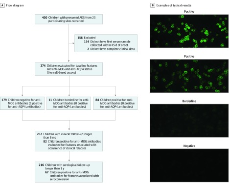
Design, setting, and participants: In this prospective cohort study, study participants were recruited from July 2004 to February 2017 through the multicenter Canadian Pediatric Demyelinating Disease Study. Inclusion criteria included (1) incident central nervous system demyelination, (2) at least 1 serum sample obtained within 45 days from onset, and (3) complete clinical information. Of 430 participants with acquired demyelinating syndrome recruited, 274 were included in analyses. Of 156 excluded participants, 154 were excluded owing to missing baseline samples and 2 owing to incomplete clinical information. Data were analyzed from May to October 2018.

Main outcomes and measures: Presence of anti-MOG antibodies was blindly assessed in serial samples collected over a median of 4 years. Clinical, magnetic resonance imaging, and cerebrospinal fluid features were characterized at presentation, and subsequent disease course was assessed by development of new brain magnetic resonance imaging lesions, total lesion volume at last evaluation, annualized relapse rates, Expanded Disability Status Scale score and visual functional score at 4 years, and any disease-modifying treatment exposure.

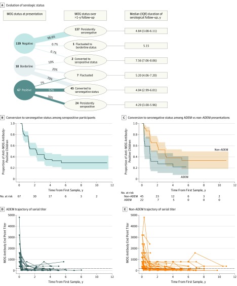
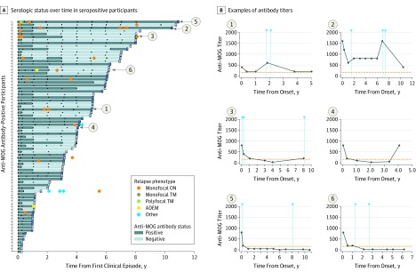
Results: Of the 274 included participants, 140 (51.1%) were female, and the median (interquartile range) age of all participants was 10.8 (6.2-13.9) years. One-third of children were positive for anti-MOG antibodies at the time of incident demyelination. Clinical presentations included a combination of optic neuritis, transverse myelitis, and acute disseminated encephalomyelitis for 81 of 84 anti-MOG antibody-positive children (96%). Brain lesions were present in 51 of 76 anti-MOG antibody-positive participants (67%), but magnetic resonance imaging characteristics differed with age at presentation. Complete resolution of baseline lesions was observed in 26 of 49 anti-MOG antibody-positive participants (53%). On serial serum analysis, 38 of 67 participants (57%) who were seropositive at onset became seronegative (median time to conversion, 1 year). Among all participants who were positive for anti-MOG antibodies at presentation, clinical relapses occurred in 9 of 24 children (38%) who remained persistently seropositive and in 5 of 38 children (13%) who converted to seronegative status.

Conclusions and relevance: Myelin oligodendrocyte glycoprotein antibodies are common in children with acquired demyelinating syndrome and are transient in approximatively half of cases. Even when persistently positive, most anti-MOG antibody-positive children experience a monophasic disease. The presence of anti-MOG antibodies at the time of incident demyelination should not immediately prompt the initiation of long-term immunomodulatory therapy.


Conflict of interest statement

Conflict of Interest Disclosures: Dr Waters has received grants from Euroimmun and personal fees from Euroimmun, Mereo BioPharma, Retrogenix, and the University of British Columbia; holds patent LGI1/CASPR2, which has been issued and licensed with royalties paid to Euroimmun, as well as patent GABAAR, which has been issued; and is codirector of the Oxford Autoimmune Neurology Diagnostic Laboratory, which provides myelin oligodendrocyte glycoprotein assays for patient diagnosis. Dr Fadda has received a clinical research training scholarship in multiple sclerosis from the American Academy of Neurology. Dr Brown has received personal fees from Biogen, NeuroRx Research, and the Population Health Council. Dr Irani has received grants and nonfinancial support from UCB as well as grants from Ono Pharmaceutical and holds patent LGI1/CASPR2, which has been issued and licensed with royalties paid to Euroimmun. Dr Yeh has received grants from the Multiple Sclerosis Scientific Foundation, Multiple Sclerosis Society of Canada, and Biogen. Dr Marrie has received grants from the Multiple Sclerosis Scientific Research Foundation. Dr Arnold has received grants from the Multiple Sclerosis Society of Canada. Dr Banwell has received personal fees from Novartis. Dr Ban-Or has received grants from the National Institutes of Health, National Multiple Sclerosis Society, Canadian Institutes of Health Research, Multiple Sclerosis Society of Canada, Multiple Sclerosis Scientific Foundation, and Melissa and Paul Anderson Chair in Neuroinflammation; has participated as a speaker in meetings sponsored by and received consulting fees from Janssen Pharmaceuticals/Actelion, Atara Biotherapeutics, Biogen, Celgene/Receptos, Roche/Genentech, Mapi Pharma, MedImmune, Merck & Co/EMD Serono, Novartis, and Sanofi Genzyme; and has received grant support from Biogen, Roche/Genentech, and Novartis. No other disclosures were reported.

Figures

Figure 1.
Figure 1.. Study Design
A, A total of 2022 serial samples from 274 participants with acquired demyelinating syndrome (ADS) with complete clinical data were tested for anti–myelin oligodendrocyte glycoprotein (MOG) IgG1 and anti–aquaporin 4 (AQP4) antibodies. Seven participants were recruited after July 2014, at which time the protocol entered a new phase, requiring participants to meet McDonald 2010 diagnostic criteria. None of these 7 patients were positive for anti-MOG antibodies. B, Example images of typical positive, borderline, and negative results for MOG IgG1 antibody testing.
Figure 2.
Figure 2.. Myelin Oligodendrocyte Glycoprotein (MOG) Results on Serial Samples
A, Evolution of serologic status in participants with follow-up greater than 1 year. Only 2 of 139 participants who were negative for anti-MOG antibodies at presentation changed serological status in subsequent examinations: 1 participant became seropositive at 3 months and was persistently positive for the subsequent 8 years and 1 had an isolated finding of a borderline result at 2 years from onset. Of the 10 participants with borderline results at onset, 7 became seronegative, 2 fluctuated between negative and borderline status and 1 became seropositive in the last follow-up sample (7 years from onset; titer, 1:200). B, Kaplan-Meier curve for the time to conversion to seronegative status in all participants who were positive for anti-MOG antibodies at time of presentation. The shaded areas indicate 95% CIs. C, Kaplan-Meier curves in participants with acute disseminated encephalomyelitis (ADEM) vs non-ADEM presentations. Participants with ADEM had a considerably shorter time to seroconversion than participants without ADEM. The shaded areas indicate 95% CIs. D and E, Trajectory of serial MOG titers of anti-MOG antibody–positive participants with ADEM at onset (D) and without ADEM at onset (E). The data points indicate the titers measured at each serological evaluation.
Figure 3.
Figure 3.. Evolution of Serologic Status and Clinical Relapses in Participants Who Were Positive for Anti–Myelin Oligodendrocyte Glycoprotein (MOG) Antibodies at Time of Initial Presentation
A, Serologic status over time in 77 anti-MOG antibody–positive participants with serial samples. Each bar represents an individual participant, and the square at the end of each bar indicates the serological status in the last follow-up sample. Dark blue bars indicate seropositive status, and light blue bars indicate seronegative status. Black dots represent the time of sampling. Colored circles indicate clinical relapses. B, Representative examples of antibody titers in participants during follow-up. Graphs 1 and 2 show 2 cases in which transient antibody titer increases were associated with clinical relapses (arrowheads). Graphs 3 and 4 show patients with reappearance of MOG antibodies after initial seroconversion from seropositive to seronegative status, in one case associated with a clinical attack. Graphs 5 and 6 show examples of patients who experienced clinical relapses after conversion to seronegative status.

References

    1. Reindl M, Waters P. Myelin oligodendrocyte glycoprotein antibodies in neurological disease. Nat Rev Neurol. 2019;15(2):82-93. doi:10.1038/s41582-018-0112-x - DOI - PubMed
    1. Fernandez-Carbonell C, Vargas-Lowy D, Musallam A, et al. . Clinical and MRI phenotype of children with MOG antibodies. Mult Scler. 2016;22(2):174-184. doi:10.1177/1352458515587751 - DOI - PMC - PubMed
    1. Hennes EM, Baumann M, Schanda K, et al. ; BIOMARKER Study Group . Prognostic relevance of MOG antibodies in children with an acquired demyelinating syndrome. Neurology. 2017;89(9):900-908. doi:10.1212/WNL.0000000000004312 - DOI - PubMed
    1. Höftberger R, Sepulveda M, Armangue T, et al. . Antibodies to MOG and AQP4 in adults with neuromyelitis optica and suspected limited forms of the disease. Mult Scler. 2015;21(7):866-874. doi:10.1177/1352458514555785 - DOI - PMC - PubMed
    1. Sepúlveda M, Armangue T, Martinez-Hernandez E, et al. . Clinical spectrum associated with MOG autoimmunity in adults: significance of sharing rodent MOG epitopes. J Neurol. 2016;263(7):1349-1360. doi:10.1007/s00415-016-8147-7 - DOI - PMC - PubMed

Publication types