. 2020 Jan 15;201(2):224-239.
doi: 10.1164/rccm.201903-0591OC.

Immunoglobulin-driven Complement Activation Regulates Proinflammatory Remodeling in Pulmonary Hypertension

Affiliations

Immunoglobulin-driven Complement Activation Regulates Proinflammatory Remodeling in Pulmonary Hypertension

Maria G Frid et al. Am J Respir Crit Care Med. .

Abstract

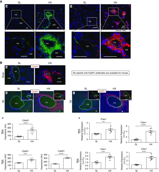
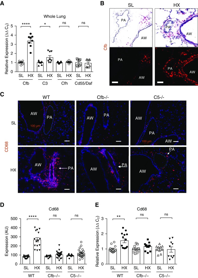
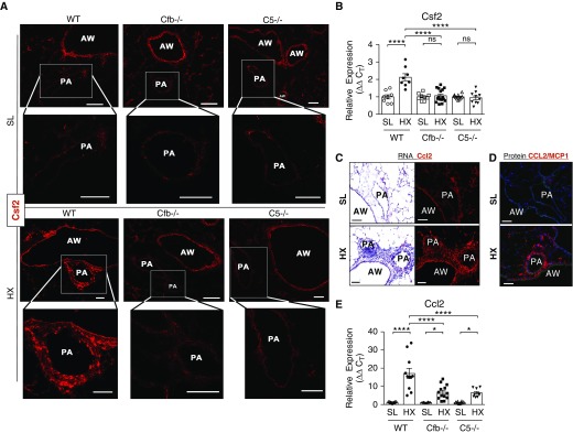
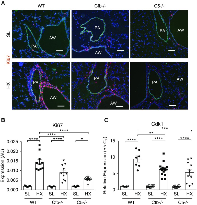
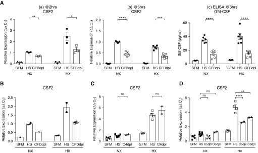
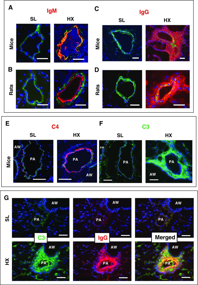
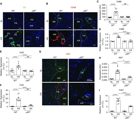
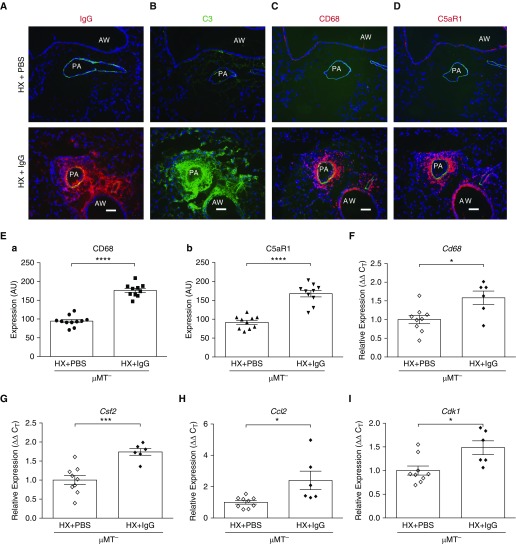
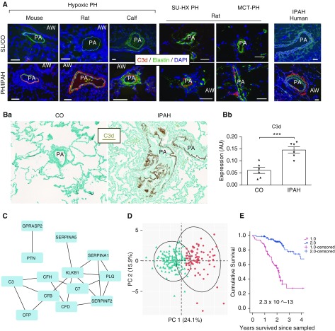
Rationale: Pulmonary hypertension (PH) is a life-threatening cardiopulmonary disorder in which inflammation and immunity have emerged as critical early pathogenic elements. Although proinflammatory processes in PH and pulmonary arterial hypertension (PAH) are the focus of extensive investigation, the initiating mechanisms remain elusive.Objectives: We tested whether activation of the complement cascade is critical in regulating proinflammatory and pro-proliferative processes in the initiation of experimental hypoxic PH and can serve as a prognostic biomarker of outcome in human PAH.Methods: We used immunostaining of lung tissues from experimental PH models and patients with PAH, analyses of genetic murine models lacking specific complement components or circulating immunoglobulins, cultured human pulmonary adventitial fibroblasts, and network medicine analysis of a biomarker risk panel from plasma of patients with PAH.Measurements and Main Results: Pulmonary perivascular-specific activation of the complement cascade was identified as a consistent critical determinant of PH and PAH in experimental animal models and humans. In experimental hypoxic PH, proinflammatory and pro-proliferative responses were dependent on complement (alternative pathway and component 5), and immunoglobulins, particularly IgG, were critical for activation of the complement cascade. We identified Csf2/GM-CSF as a primary complement-dependent inflammatory mediator. Furthermore, using network medicine analysis of a biomarker risk panel from plasma of patients with PAH, we demonstrated that complement signaling can serve as a prognostic factor for clinical outcome in PAH.Conclusions: This study establishes immunoglobulin-driven dysregulated complement activation as a critical pathobiological mechanism regulating proinflammatory and pro-proliferative processes in the initiation of experimental hypoxic PH and demonstrates complement signaling as a critical determinant of clinical outcome in PAH.

Keywords: GM-CSF; biomarkers; hypoxia; inflammation; vascular remodeling.


Figures

Figure 1.
Figure 1.
Lungs of hypoxic rodents demonstrate prominent vascular-specific deposition of complement C3 (component 3) and robust accumulation of cells expressing complement anaphylatoxin receptors. (A) Hypoxia-induced deposition of complement C3 in mice (a, green fluorochrome) and rats (b, red fluorochrome) is prominently observed in perivascular areas and also encompasses medial and luminal areas. (Aa and Ab) Lung cryosections were labeled with species-specific anti–complement C3 antibodies conjugated with green (a, for mouse) or red (b, for rat) fluorochrome. Cell nuclei are labeled with DAPI (blue). Scale bars, 100 μm. (B) Expression of receptors for complement anaphylatoxins C5a and C3a (C5aR1 and C3aR1, respectively) is markedly upregulated in the lungs of animals with experimental pulmonary hypertension. In rodents exposed to hypoxia (HX) for three days (a, mice; b and c, rats), cells expressing C5aR1 and C3aR1 (red fluorochrome) are markedly increased in numbers and localized to perivascular areas, whereas only a few pulmonary adventitial cells express these receptors in the lungs of sea-level (SL) animals. Lung cryosections were labeled with species-specific monoclonal antibodies against C5aR1 (mice and rats) and C3aR1 (available for rats only). Autofluorescence of elastic lamellae (green) defines tunica media, and cell nuclei are labeled with DAPI (blue). Scale bars, 100 μm. (Bd) Quantification of red fluorescence was performed as described in the online supplement and is presented in arbitrary units (AU). (Be) qRT-PCR analysis demonstrates that expression levels of C5ar1 and C3ar1 mRNA in the whole lungs of mice and rats are significantly upregulated by 3-day exposure to HX compared with SL controls. Unpaired/two-tailed test was performed for comparing two groups. **P ≤ 0.01, ***P ≤ 0.001, and ****P ≤ 0.0001. AW = airway; PA = pulmonary artery.
Figure 2.
Figure 2.
The alternative and terminal C5 (component 5) complement pathways are essential in driving hypoxia-induced proinflammatory processes in pulmonary perivascular areas. (A and B) The alternative pathway of complement is activated by hypoxic exposure. (A) qRT-PCR analysis of whole lung extracts from wild-type (WT) mice exposed to hypoxia (HX) for 3 days demonstrates robust augmentation of Cfb (complement factor B) and modest upregulation of complement C3 mRNA expression levels compared with sea-level (SL) controls. Unpaired/two-tailed test was performed for comparing two groups. (B) Lungs of 3-day HX WT mice demonstrate robust augmentation of Cfb mRNA expression, as detected via RNAscope in situ hybridization, in cells localized to pulmonary artery (PA) perivascular areas and airways (AWs). Fast Red chromogen (red), used for message detection in in situ hybridization, can be visualized by both light (upper panels) and fluorescent (bottom panels) microscopy. Gill’s hematoxylin (blue) was used for nuclear counterstaining in light microscopy imaging (upper panels). (C–E) Attenuated accumulation of CD68+ macrophages is observed in the lungs of complement (Cfb and C5)-deficient HX mice compared with WT HX counterparts, as validated by CD68 immunostaining (C, red fluorochrome) and its quantification (D) performed as described in the online supplement and presented in arbitrary units (AU), as well as by qRT-PCR of whole lung tissues (E). (Unpaired/two-tailed test was performed for comparing two groups in each cohort). Scale bars, 100 μm. *P < 0.05, **P ≤ 0.01, and ****P ≤ 0.0001; ns = not significant. Cfh = complement factor H; CT = cycle threshold.
Figure 3.
Figure 3.
Hypoxia-induced Csf2 and Ccl2 mRNA expression is markedly augmented in the lungs of wild-type (WT) mice but is abrogated (for Csf2) or partially decreased (for Ccl2) in the lungs of complement (Cfb [complement factor B] and C5 [component 5])-deficient mice. (A) RNAscope in situ hybridization demonstrates that Csf2 expression is markedly augmented in the pulmonary arteries (PAs) of WT mice exposed to 3-day hypoxia (HX) but is abrogated in the PAs of complement (Cfb and C5)-deficient mice. Airways (AWs) of all mouse strains, both sea level (SL) and HX, maintain Csf2 expression without any visually significant change. (B) qRT-PCR analysis of whole murine lung tissues demonstrates that hypoxia-induced upregulation of Csf2 mRNA expression, detected in WT mice, is abrogated in complement (Cfb and C5)-deficient mice. One-way ANOVA with a Sidak multiple-comparison test with single pooled variance was performed for multiple group comparison. (CE) HX-induced Ccl2 expression is complement (alternative and C5 pathways) dependent: levels of Ccl2 mRNA expression (C and E) and CCL2 protein expression (D) are markedly augmented in the lungs of 3-day HX WT mice; in contrast, the lungs of complement (Cfb and C5)-deficient HX mice demonstrate significantly decreased mRNA expression levels (E). One-way ANOVA with a Sidak multiple-comparison test with a single pooled variance was performed for multiple-group comparison. Scale bars, 100 μm. *P ≤ 0.05 and ****P ≤ 0.0001; ns = not significant. CT = cycle threshold.
Figure 4.
Figure 4.
Hypoxia (HX)-induced perivascular cell proliferation is complement (alternative and C5 [component 5] pathways) dependent. (A) Profound increases in perivascular cell proliferative responses are observed in the lungs of wild-type (WT) mice exposed to 3-day HX, as compared with sea-level (SL) controls, but are significantly attenuated in Cfb (complement factor B)- and C5-deficient HX mice, as evaluated by immunostaining for nuclear proliferation-associated Ki67 antigen (red). Scale bars, 100 μm. (B) Quantification of red fluorescence was performed as described in the online supplement and is presented in arbitrary units (AU). (C) qRT-PCR analysis of whole lung tissues for cell cycle–progression marker Cdk1 (cyclin-dependent kinase 1) expression. One-way ANOVA with a Sidak multiple-comparison test with single pooled variance was performed for multiple-group comparison. *P < 0.05, **P ≤ 0.01, ***P ≤ 0.001, and ****P ≤ 0.0001. AW = airway; PA = pulmonary artery.
Figure 5.
Figure 5.
Human pulmonary adventitial fibroblasts regulate Csf2/GM-CSF expression in a complement (alternative and C5 [component 5] pathways)-dependent manner. Fibroblasts, isolated from pulmonary arteries of patients with idiopathic pulmonary arterial hypertension (PH-Fibs; n = 5; Table E2), were serum-starved in serum-free medium (SFM) for 72 hours and incubated with complement-sufficient normal human serum (HS) or HS depleted in specific complement components. (A and B) PH-Fibs substantially upregulate CSF2 mRNA (Aa, Ab, and B) and protein (Ac) expression in response to complement-sufficient HS, whereas their responses to serum with inhibited alternative pathway (CFBdpl [complement factor B–depleted], Aa and Ab; CFDdpl [complement factor D–depleted], B) are significantly attenuated. This pattern was observed under both normoxia (NX; 21% O2) and hypoxia (HX; 3% O2) conditions; however, at a 2-hour time point (three different cell populations shown, Aa), the levels of HX-induced CSF2 mRNA expression were higher than those in NX, which was in contrast to a 6-hour time point (n = 5 cell populations shown, Ab). The qRT-PCR findings were confirmed at a protein level via ELISA (Ac) using conditioned medium generated at a 6-hour time point. (C) Expression of CSF2 by PH-Fibs was observed to be independent of complement C4 (main component of the classical and lectin pathways). (D) Two-hour exposure of PH-Fibs to HS depleted in complement C5 (C5dpl) or C6 (C6dpl) resulted in substantial attenuation of CSF2 expression compared with HS under HX (3% O2) but not under NX (21% O2) conditions. *P < 0.05, **P ≤ 0.01, ***P ≤ 0.001, and ****P ≤ 0.0001; ns = not significant. C4dpl = component 4–depleted; CT = cycle threshold.
Figure 6.
Figure 6.
Hypoxia (HX)-induced vascular deposition patterns of IgM and IgG are highly compartmentalized and correlate with deposition patterns of complement components C4 and C3, respectively. (AD) Highly compartmentalized patterns of IgM versus IgG deposition and complement components C4 versus C3 are observed in experimental rodents (mice and rats) exposed to 3-day HX: IgM deposition (red) in mice (A) and rats (B) is observed in the luminal and medial areas, whereas deposition of IgG (red) in mice (C) and rats (D) is detected mainly in perivascular/adventitial areas and additionally encompasses luminal/medial areas. (EG) In 3-day HX mice, luminal/medial deposition of complement C4 (red, E) correlates with that of IgM (red, A and B), whereas strong perivascular (in addition to luminal/medial) deposition of complement C3 (green, F and G) clearly correlates with that of IgG (red, C, D, and G). No deposition of IgM, IgG, C4, or C3 was detected in the lungs of sea-level (SL) rodents. Autofluorescence of elastic lamellae (green in A, B, and EG) or α-smooth muscle actin (green in C and D) defines tunica media. Cell nuclei are labeled with DAPI (blue). Scale bars, 100 μm. AW = airway; PA = pulmonary artery.
Figure 7.
Figure 7.
μMT mice deficient in all circulating immunoglobulins exhibit a “hypoxia-protective” phenotype. (A) Exposure to 3-day hypoxia (HX) fails to activate complement C3 (component 3) cascade in the lungs of μMT mice. Although the lungs of 3-day HX wild-type (WT) mice demonstrate activation of the complement cascade, as defined by strong perivascular deposition of complement C3 (green, lower left), no C3 deposition is observed in the lungs of 3-day HX μMT mice (lower right). Control sea-level (SL) mice of both strains do not display any C3 deposition. Cell nuclei are labeled with DAPI (blue). (BI) Hypoxia-induced proinflammatory and proliferative responses are attenuated in the lungs of μMT mice. Accumulation of CD68+ macrophages (BD) and Csf2 and Ccl2 cytokine/chemokine expression (E and F) are strongly (for Cd68 and Csf2) or moderately (for Ccl2) attenuated in the lungs of 3-day HX μMT mice compared with HX WT counterparts. (B) Immunofluorescent staining for CD68 macrophage marker (red fluorescence) and α-smooth muscle actin (green fluorescence). DAPI staining (blue) defines cell nuclei. (C) Quantification of red fluorescence was performed as described in the online supplement and is presented in arbitrary units (AU). (DF) qRT-PCR analysis of whole lung tissues for mRNA expression of macrophage marker Cd68 (D), Csf2 (E), and Ccl2 (F). One-way ANOVA with a Sidak multiple-comparison test with single pooled variance was performed for multiple-group comparison. (GI) Hypoxia-induced perivascular cell proliferative responses, defined by immunostaining for nuclear proliferation-associated Ki67 antigen (red, G, quantified in H) and qRT-PCR analysis of whole lung tissues for cell cycle–progression marker Cdk1 (cyclin-dependent kinase 1) expression (I) are significantly attenuated in the lungs of HX μMT mice compared with HX WT counterparts. (G) Autofluorescence of pulmonary artery elastic lamellae (green) defines tunica media; cell nuclei are labeled with DAPI (blue). *P ≤ 0.05 and ****P ≤ 0.0001; ns = not significant. Scale bars, 100 μm. AW = airway; CT = cycle threshold; PA = pulmonary artery.
Figure 8.
Figure 8.
Reconstitution of circulating IgG in hypoxic μMT mice restores a proinflammatory phenotype. Five consecutive daily injections of normoxic μMT mice with normal mouse IgG (2 mg/mouse) (36), equivalent to the “loading/initiation” phase of IgG reconstitution in immune-deficient human individuals (37), was followed by 3 “blank” days and subsequently by exposure to 3-day hypobaric hypoxia (HX). (A) Robust deposition of IgG (red) is observed mainly in pulmonary arteries (PAs) in a perivascular-specific manner but also encompasses vascular luminal and medial compartments. Some periairway deposition is also detected. (B) Deposition of complement C3 (component 3; green) is prominently observed in PA perivascular areas, as well as in vascular medial and luminal compartments and partially in periairway areas. (C and D) Robust accumulation of CD68+and C5aR1-expressing macrophages (red) is observed in perivascular areas. Cell nuclei are labeled with DAPI (blue). Scale bars, 100 μm. (E) Quantification of increased accumulation of CD68+and C5aR1+cells (red fluorescence) was performed as described in the online supplement and is presented in arbitrary units (AU). Quantification of IgG and C3 deposition was not performed because the sea-level lung samples were completely negative for the staining. (FI) qRT-PCR analysis demonstrates augmented expression of Cd68 (macrophage marker), Csf2 and Ccl2 (cytokine and chemokine, respectively), and Cdk1 (cyclin-dependent kinase 1; cell cycle–progression marker) in the whole lungs of HX immunoglobulin-deficient μMT mice that were reconstituted with circulating IgG (HX + IgG) compared with PBS-injected HX μMT mice.*P < 0.05, ***P ≤ 0.001, and ****P ≤ 0.0001. AW = airway; C5aR1 = receptor for anaphylatoxin C5a; CT = cycle threshold.
Figure 9.
Figure 9.
The complement cascade is activated in the lungs of experimental animal models of pulmonary hypertension (PH) and patients with idiopathic pulmonary arterial hypertension (IPAH). (A and B) Activation of the complement cascade, as defined by deposition of C3d fragment (terminal activation product of complement component 3 [C3]) is most prominently observed in a perivascular-specific manner in the lungs of experimental animal models of hypoxic PH (mouse, rat, and calf), sugen-hypoxia (SU-HX) PH and monocrotaline‐PH (MCT‐PH) rat models, and humans with IPAH. OCT-embedded lung cryosections of experimental animal specimens (A) and formalin-fixed paraffin-embedded sections of human lung specimens (B) were labeled with a biotinylated C3d-specific monoclonal antibody (mAb; red in A, brown in B), developed by Thurman and colleagues (38). This mAb distinguishes tissue-bound C3d from the intact C3 or C3b, allowing assessment of tissue-specific activation of the complement cascade. (A) Exposure of experimental animals to sustained hypoxia was performed for 3 weeks for mice and rats (n = 5, each group) and 2 weeks for calves (n = 5, each group); control (CO) animals were maintained at sea-level (SL) or ambient (Denver, Colorado) altitude. An image of the SU-HX rat model is shown at 2 weeks of hypoxic exposure (the most proinflammatory time point), and an image of the MCT-PH rat lung sample is shown at a 2-week time point. Autofluorescence of pulmonary artery (PA) elastic lamellae (green) defines tunica media in hypoxic experimental animal models and in human specimens, whereas α-smooth muscle actin defines tunica media in MCT-PH and SU-HX lung specimens. Images of C3d immunofluorescent staining for all analyzed human specimens are shown in Figure E8A. IPAH and normal (rejected for lung transplant) donor cohorts are described in Table E3A. Cell nuclei are labeled with DAPI (blue). Scale bars, 100 μm. (Ba) Immunohistochemistry (IHC) staining demonstrates C3d deposition in a medium-sized PA of a patient with IPAH. The C3d IHC signal is largely restricted to perivascular areas. Cohorts of patients with IPAH and CO (rejected for lung transplant) donors (n = 6, each) are described in Table E3. Images of C3d IHC staining of all analyzed human specimens (n = 6, each cohort) are shown in Figure E8B. (Bb) Quantification of IHC staining was performed via MetaMorph software, in which the dynamic range for gray intensity levels ranges between 0 (white) and 256 (black). Background levels of gray intensity are thus determined largely by the counterstain for the IHC (methyl green) and potentially represent baseline of nonspecific IHC signal to the detection system. The quantification of the IHC signal (presented in arbitrary units [AU]) for C3d revealed a 2.382-fold increase in IPAH lungs over CO donor lungs. (CE) Plasma complement is a critical determinant of clinical outcomes in patients with PAH. (C) Previously published data (12) identifying circulating proteins (Table E4) with prognostic importance to patients with PAH (n = 218) were analyzed using a network medicine approach. Differentially expressed proteins were mapped to the consolidated human interactome resulting in a network that was enriched with complement pathway intermediaries, referred to in this article as the complement–PAH network (13 proteins and 18 protein–protein interactions). (D) Two distinct PAH patient clusters were identified based on biological information derived solely from the complement–PAH network. Oval represents the estimated cluster boundaries determined by the patient data in each cluster. (E) Previously published data (12) identifying circulating proteins (Table E4) with prognostic importance to patients with PAH (n = 218) were analyzed using a network medicine approach. A total of 37 differentially expressed proteins were mapped to the consolidated human interactome resulting in the complement–PAH network. Kaplan–Meier survival estimates in patients with PAH divided into two clusters (as shown in D) based on the plasma levels of proteins in the complement–PAH network are presented; thin vertical marks indicate where patients were censored during the time course. ***P ≤ 0.001. AW = airway; OCT = optimal cutting temperature compound; PC = principal component.

Comment in

References

    1. Humbert M, Guignabert C, Bonnet S, Dorfmüller P, Klinger JR, Nicolls MR, et al. Pathology and pathobiology of pulmonary hypertension: state of the art and research perspectives. Eur Respir J. 2019;53:1801887. - PMC - PubMed
    1. Nicolls MR, Taraseviciene-Stewart L, Rai PR, Badesch DB, Voelkel NF. Autoimmunity and pulmonary hypertension: a perspective. Eur Respir J. 2005;26:1110–1118. - PubMed
    1. Rabinovitch M, Guignabert C, Humbert M, Nicolls MR. Inflammation and immunity in the pathogenesis of pulmonary arterial hypertension. Circ Res. 2014;115:165–175. - PMC - PubMed
    1. Frid MG, Brunetti JA, Burke DL, Carpenter TC, Davie NJ, Reeves JT, et al. Hypoxia-induced pulmonary vascular remodeling requires recruitment of circulating mesenchymal precursors of a monocyte/macrophage lineage. Am J Pathol. 2006;168:659–669. - PMC - PubMed
    1. Li M, Riddle SR, Frid MG, El Kasmi KC, McKinsey TA, Sokol RJ, et al. Emergence of fibroblasts with a proinflammatory epigenetically altered phenotype in severe hypoxic pulmonary hypertension. J Immunol. 2011;187:2711–2722. - PMC - PubMed

Publication types

MeSH terms