. 2019 Dec 1;317(6):G793-G801.
doi: 10.1152/ajpgi.00199.2019. Epub 2019 Sep 23.

Enteric neuron density correlates with clinical features of severe gut dysmotility

Affiliations

Enteric neuron density correlates with clinical features of severe gut dysmotility

Elisa Boschetti et al. Am J Physiol Gastrointest Liver Physiol. .

Abstract

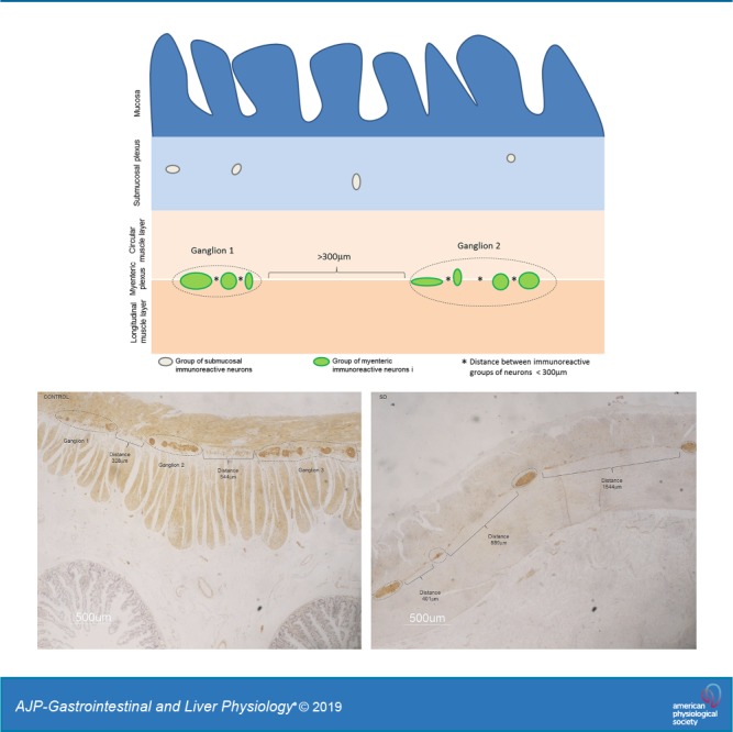
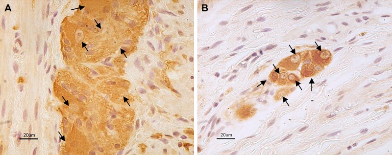
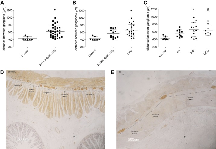
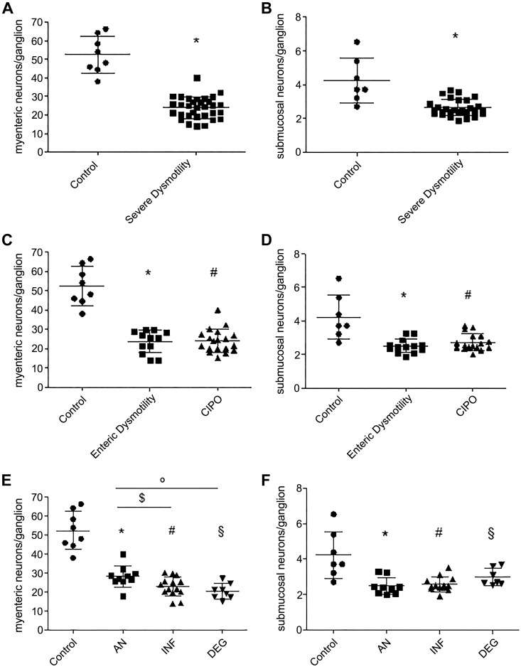
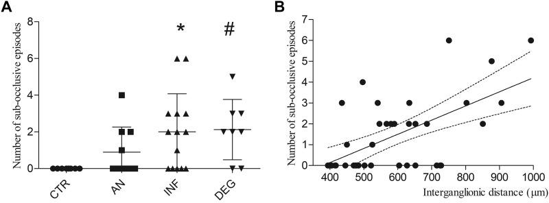
Gastrointestinal (GI) symptoms can originate from severe dysmotility due to enteric neuropathies. Current methods used to demonstrate enteric neuropathies are based mainly on classic qualitative histopathological/immunohistochemical evaluation. This study was designed to identify an objective morphometric method for paraffin-embedded tissue samples to quantify the interganglionic distance between neighboring myenteric ganglia immunoreactive for neuron-specific enolase, as well as the number of myenteric and submucosal neuronal cell bodies/ganglion in jejunal specimens of patients with severe GI dysmotility. Jejunal full-thickness biopsies were collected from 32 patients (22 females; 16-77 yr) with well-characterized severe dysmotility and 8 controls (4 females; 47-73 yr). A symptom questionnaire was filled before surgery. Mann-Whitney U test, Kruskal-Wallis coupled with Dunn's posttest and nonparametric linear regression tests were used for analyzing morphometric data and clinical correlations, respectively. Compared with controls, patients with severe dysmotility exhibited a significant increase in myenteric interganglionic distance (P = 0.0005) along with a decrease in the number of myenteric (P < 0.00001) and submucosal (P < 0.0004) neurons. A 50% reduction in the number of submucosal and myenteric neurons correlated with an increased interganglionic distance and severity of dysmotility. Our study proposes a relatively simple tool that can be applied for quantitative evaluation of paraffin sections from patients with severe dysmotility. The finding of an increased interganglionic distance may aid diagnosis and limit the direct quantitative analysis of neurons per ganglion in patients with an interganglionic distance within the control range.NEW & NOTEWORTHY Enteric neuropathies are challenging conditions characterized by a severe impairment of gut physiology, including motility. An accurate, unambiguous assessment of enteric neurons provided by quantitative analysis of routine paraffin sections may help to define neuropathy-related gut dysmotility. We showed that patients with severe gut dysmotility exhibited an increased interganglionic distance associated with a decreased number of myenteric and submucosal neurons, which correlated with symptoms and clinical manifestations of deranged intestinal motility.

Keywords: chronic intestinal pseudo-obstruction; enteric neuron cell count; interganglionic distance; severe gut dysmotility.


Conflict of interest statement

R. De Georgio has participated as a consultant for Shire, Sucampo, Coloplast, Kyowa Kirin International, and Takeda and received grant support from Shire and Takeda. These consultancies did not influence the content of this article. The other authors declare no conflict of interest.

Figures

None
Graphical abstract
Fig. 1.
Fig. 1.
Measurement of interganglionic distance. Schematic representation of the method by which interganglionic distances were defined, using the 300-μm cut-off value. Groups of immunoreactive neurons separated by less than 300 μm (distances indicated by *) were considered part of the same arbitrary ganglionic unit. Distances ≥300 μm were used as the threshold value to distinguish two different ganglia (indicated by square brackets).
Fig. 2.
Fig. 2.
Neurons identified by a pan-neuronal marker in myenteric and submucosal plexuses. Representative photomicrographs showing neuron-specific enolase (NSE) immunolabeling in the myenteric (A) and submucosal (B) plexus of a control sample. Black arrows indicate identified neuronal cell bodies.
Fig. 3.
Fig. 3.
Interganglionic distance in myenteric ganglia. Distance between ganglia using the 300-μm threshold comparing controls (622.3 ± 151.8 μm) and patients with severe dysmotility (427.4 ± 46.8 μm), *P = 0.0005 (A), enteric dysmotility (569.8 ± 117.7 μm) and CIPO (653.3 ± 164.7 μm), *P = 0.0076 and #P = 0.0001 (B), and apparently normal (AN; 562.1 ± 93.2 μm), inflammatory neuromyopathy (INF; 651.3 ± 190.6 μm), and degenerative neuromyopathy (DEG; 645.25 ± 134.2 μm), *P = 0.0099, #P = 0.0004, and §P = 0.0008 (C). Bottom: examples of interganglionic distance measurements using ImageJ software for a control sample (D) and a patient with severe dysmotility (E). Neuron-specific enolase (NSE) immunostaining; 500 μm final magnification.
Fig. 4.
Fig. 4.
Myenteric and submucosal neuronal cell counts in patients with severe dysmotility and controls. A: myenteric neurons counted in the whole severe dysmotility (24.0 ± 5.8 neurons/ganglion) group vs. controls (52.4 ± 10.0 neurons/ganglions), *P < 0.00001. B: submucosal neurons counted in the whole severe dysmotility (2.6 ± 0.5 neurons/ganglion) group vs. controls (4.2 ± 1.3 neurons/ganglion), *P = 0.0004. C: myenteric neuronal counts in enteric dysmotility (ED; 23.8 ± 5.5 neurons/ganglion) and chronic intestinal pseudo-obstruction (CIPO; 24.0 ± 6.0 neurons/ganglion) subgroups, *P < 0.00001 and #P < 0.0001. D: submucosal neuronal counts in ED (2.5 ± 0.4 neurons/ganglion) and CIPO (2.7 ± 0.5 neurons/ganglion) subgroups,*P = 0.0014 and #P = 0.0002. E: myenteric neuronal counts in apparently normal (AN; 28.3 ± 5.6 neurons/ganglion), inflammatory neuromyopathy (INF; 22.9 ± 5.6 neurons/ganglion), and degenerative neuromyopathy (DEG; 20.4 ± 4.0 neurons/ganglion) subgroups, *P = 0.0106, #§P < 0.00001, $P = 0.0390, and °P = 0.008. F: submucosal neuronal counts in AN (2.5 ± 0.5 neurons/ganglion), INF (2.6 ± 0.4 neurons/ganglion), and DEG (3.0 ± 10.5 neurons/ganglion) subgroups, *P < 0.0001, #P = 0.0003, and §P = 0.0496.
Fig. 5.
Fig. 5.
Number of subocclusive episodes distribution among immunohistochemically defined subgroups and relationship with interganglionic distance. A: number of subocclusive episodes recorded per patient before surgical intervention. Patients were clustered accordingly with the qualitative immunohistochemically defined assessment. *P = 0.0025 and #P = 0.0022. B: scatter plot illustrating the relationship between number of subocclusive episodes and interganglionic distances.

References

    1. Abalo R, José Rivera A, Vera G, Isabel Martín M. Ileal myenteric plexus in aged guinea-pigs: loss of structure and calretinin-immunoreactive neurones. Neurogastroenterol Motil 17: 123–132, 2005. doi:10.1111/j.1365-2982.2004.00612.x. - DOI - PubMed
    1. Bernard CE, Gibbons SJ, Gomez-Pinilla PJ, Lurken MS, Schmalz PF, Roeder JL, Linden D, Cima RR, Dozois EJ, Larson DW, Camilleri M, Zinsmeister AR, Pozo MJ, Hicks GA, Farrugia G. Effect of age on the enteric nervous system of the human colon. Neurogastroenterol Motil 21: 746–e46, 2009. doi:10.1111/j.1365-2982.2008.01245.x. - DOI - PMC - PubMed
    1. Bernardini N, Ippolito C, Segnani C, Mattii L, Bassotti G, Villanacci V, Blandizzi C, Dolfi A. Histopathology in gastrointestinal neuromuscular diseases: methodological and ontological issues. Adv Anat Pathol 20: 17–31, 2013. doi:10.1097/PAP.0b013e31827b65c0. - DOI - PubMed
    1. Bernardini N, Segnani C, Ippolito C, De Giorgio R, Colucci R, Faussone-Pellegrini MS, Chiarugi M, Campani D, Castagna M, Mattii L, Blandizzi C, Dolfi A. Immunohistochemical analysis of myenteric ganglia and interstitial cells of Cajal in ulcerative colitis. J Cell Mol Med 16: 318–327, 2012. doi:10.1111/j.1582-4934.2011.01298.x. - DOI - PMC - PubMed
    1. Boyer L, Ghoreishi M, Templeman V, Vallance BA, Buchan AM, Jevon G, Jacobson K. Myenteric plexus injury and apoptosis in experimental colitis. Auton Neurosci 117: 41–53, 2005. doi:10.1016/j.autneu.2004.10.006. - DOI - PubMed

Publication types

MeSH terms

Substances

LinkOut - more resources