. 2019 Sep 19;20(18):4639.
doi: 10.3390/ijms20184639.

Heightened TLR7/9-Induced IL-10 and CXCL13 Production with Dysregulated NF-ҝB Activation in CD11chiCD11b+ Dendritic Cells in NZB/W F1 Mice

Affiliations

Heightened TLR7/9-Induced IL-10 and CXCL13 Production with Dysregulated NF-ҝB Activation in CD11chiCD11b+ Dendritic Cells in NZB/W F1 Mice

Lok Yan Yim et al. Int J Mol Sci. .

Abstract

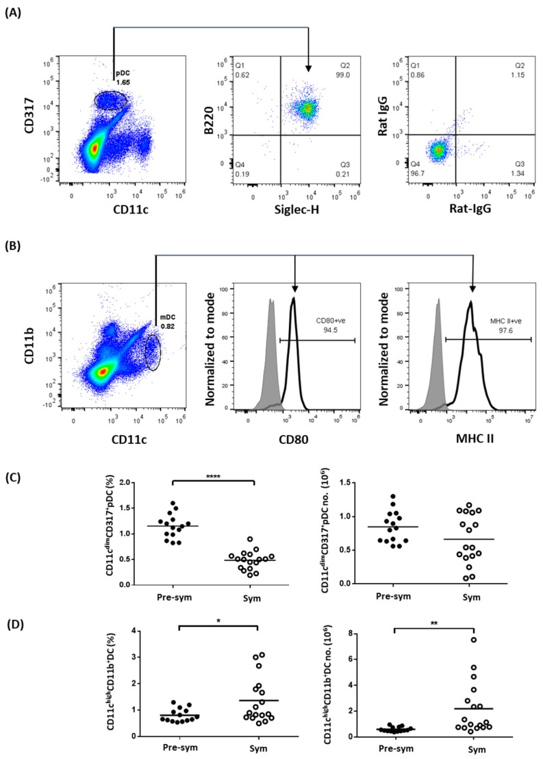
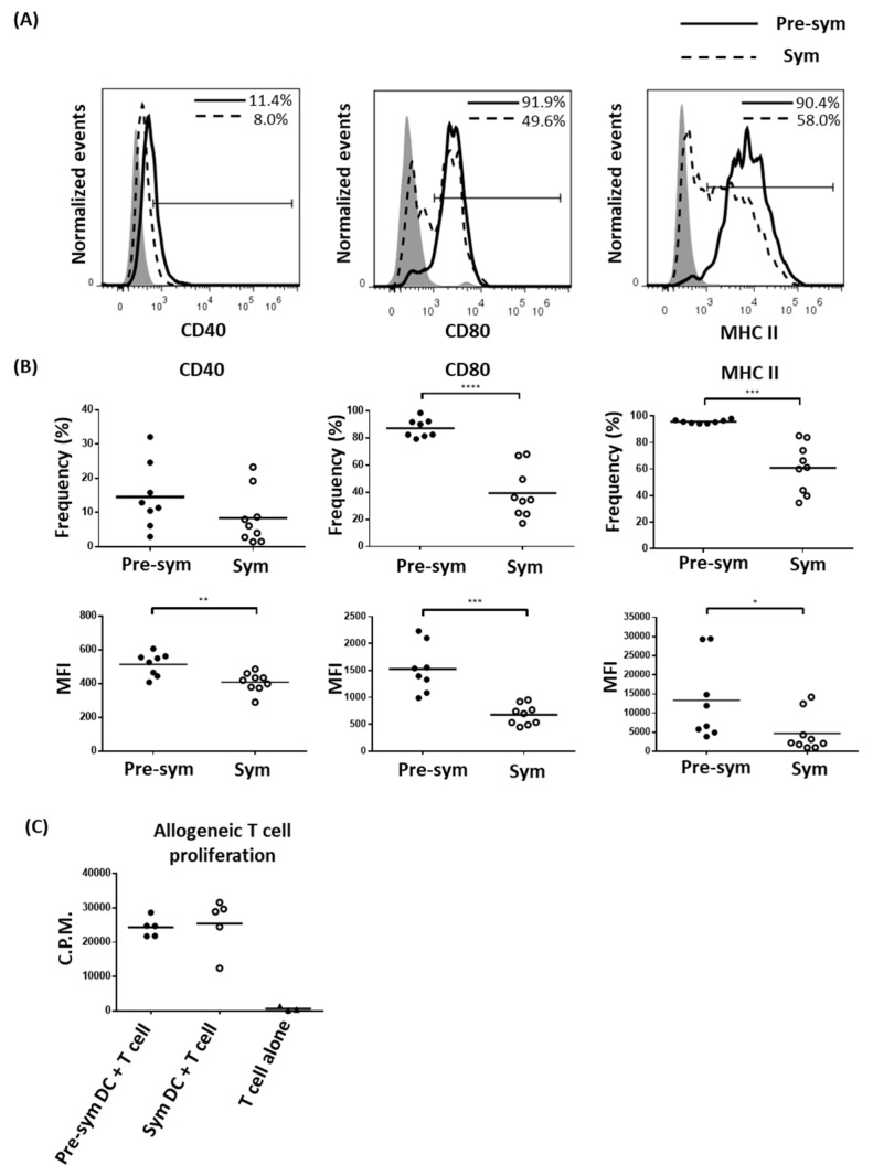
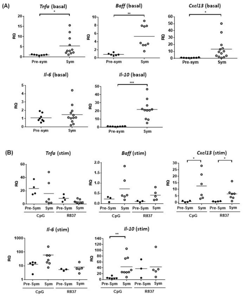
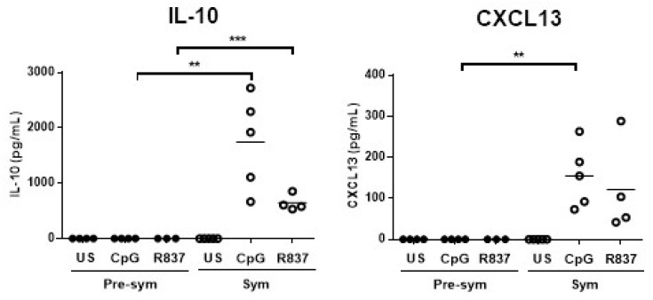
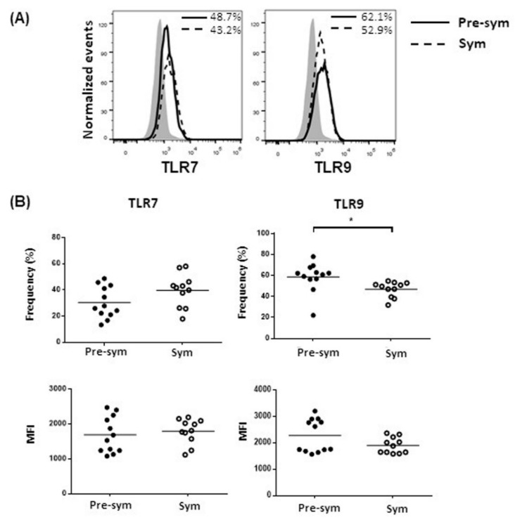
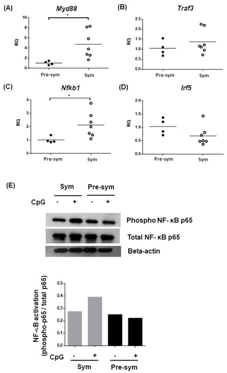
Systemic lupus erythematosus (SLE) is a chronic, multifactorial autoimmune disease that predominantly affects young females. Dysregulation of different immune cell populations leads to self-tolerance breakdown and subsequent multiple organ damage as the disease develops. Plasmacytoid dendritic cells (pDCs) are potent producers of type I interferon (IFN), while myeloid dendritic cells (mDCs) are more specialized in antigen presentations. We have previously reported that bone-marrow (BM)-derived pDCs from the murine lupus model New Zealand black/white F1 (BWF1) possess abnormalities. Therefore, this study continues to investigate what aberrant properties peripheral pDCs and mDCs possess in BWF1 and how they mediate SLE progression, by comparing their properties in pre-symptomatic and symptomatic mice. Results showed that CD11chiCD11b+ myeloid DCs expanded during the disease state with down-regulation of co-stimulatory molecules and major histocompatibility complex class II molecules (MHC II), but their capacity to stimulate T cells was not hampered. During the disease state, this subset of mDCs displayed heightened toll-like receptors 7 and 9 (TLR 7/9) responses with increased interleukin 10 (IL-10) and C-X-C motif chemokine ligand 13 (CXCL13) expressions. Moreover, the expressions of myeloid differentiation primary response 88 (Myd88) and nuclear factor kappa B subunit 1 (Nfkb1) were higher in CD11chiCD11b+ DCs at the disease stage, leading to higher nuclear factor kappa-light-chain-enhancer of activated B cells (NF-κB) p65 phosphorylation activity. In summary, we reported aberrant phenotypic properties with enhanced TLR7/9 responses of CD11chiCD11b+ DCs in SLE mediated by aberrant NF-κB signaling pathway. Our findings add additional and novel information to our current understanding of the role of DCs in lupus immunopathogenesis. Lastly, molecular candidates in the NF-κB pathway should be exploited for developing therapeutic targets for SLE.

Keywords: C-X-C motif chemokine ligand 13; Toll-like receptor 7; Toll-like receptor 9; interleukin 10; myeloid dendritic cell; systemic lupus erythematosus.


Conflict of interest statement

The authors declare no conflict of interests.

Figures

Figure 1
Figure 1
The abundance of myeloid dendritic cells (mDCs) but not plasmacytoid dendritic cells (pDCs) increases in symptomatic black/white F1 (BWF1). Total splenocytes were stained with different pDC and mDC markers to distinguish the two dendritic cell (DC) subtypes in the spleen of BWF1 using flow cytometry. (A) Splenocytes were stained with the pDC markers CD11c, CD317, B220, and Siglec-H. Expression of B220 and Siglec-H were detected on CD11cdimCD317+ gated cells (indicated by arrow). The right panel represents isotype control antibodies staining. (B) Splenocytes were stained for mDC markers CD11c and expressions of CD80 and MHC II were evaluated within the CD11chiCD11b+ gated population (indicated by arrows). (C) Summary plots comparing the frequency and total number of CD11cdimCD317+pDCs and (D) CD11chiCD11b+ DCs from pre-symptomatic (pre-sym) and symptomatic (sym) mice. Each symbol represents an individual mouse, and student’s t-test was used for statistical analysis (* p ≤ 0.05, ** p ≤ 0.01, **** p ≤ 0.0001).
Figure 2
Figure 2
Dampen expressions of co-stimulatory molecules and MHC II on symptomatic BWF1 CD11chiCD11b+ DCs does not hamper the ability of mDCs to induce T cell proliferation. Splenocytes from pre-symptomatic and symptomatic black/white F1 (BWF1) were isolated and stained for CD11c and CD11b that are also mDC markers together with the indicated activation marker. (A) Representative histograms showing the expression of different activation markers on CD11chiCD11b+ DCs from pre-symptomatic (solid line) and symptomatic BWF1 (dotted line), respectively. Shaded histogram represents the isotype control. (B) Summary plots comparing the expression of CD40, CD80, and MHC II on CD11chiCD11b+ DCs from pre-sym and sym BWF1 in terms of frequency (%) and mean fluorescence intensity (MFI). Each symbol represents an individual mouse. (C) Fluorescence-activated cell sorting (FACS)-purified splenic CD11chiCD11b+mDCs were co-cultured with C57BL/6 T cells in 1:10 ratio for two days. 3H-thymidine was then added and cultured for another 24 h to evaluate T cell proliferation using thymidine incorporation assay. A summary plot comparing the ability of mDCs from pre-symptomatic (filled circle) and symptomatic mice (open circle) to induce allogeneic T cell proliferation is shown, and each symbol represents the mean counts per minute (C.P.M.) of triplicate wells of an individual mouse. For (A) to (C), student’s t-test was used for statistical analysis (* p ≤ 0.05, ** p ≤ 0.01, *** p ≤ 0.001, and **** p ≤ 0.0001).
Figure 3
Figure 3
The basal expressions of different cytokines and chemokines in BWF1 mDCs are higher in symptomatic mice with CD11chiCD11b+ DCs displaying a heightened TLR7/9 response. Splenic CD11chiCD11b+ DCs were FACS-purified, and mRNA expressions of Tnfa, Baff, Il-6, Il-10, and Cxcl13 was determined by quantitative polymerase chain reaction (qPCR). The mRNA expression of each target was normalized with the house-keeping gene β-actin. (A) Summary plot showing the basal mRNA expression of the indicated target in CD11chiCD11b+ DCs from pre-sym and sym BWF1. The relative quantity (RQ) was the level of mRNA relative to unstimulated CD11chiCD11b+ DCs from pre-symptomatic mice. (B) The mRNA induction of the indicated target in CD11chiCD11b+ DCs from pre-symptomatic and symptomatic BWF1 upon toll-like receptor (TLR) stimulation. Cells were stimulated for 5 h in the presence or absence of the TLR9 ligand CpG (1 µM) or TLR7 ligand R837 (2.5 μg/mL). The induction of mRNA is expressed as relative quantity (RQ) relative to un-stimulated CD11chiCD11b+ DCs of the respective group. For (A) and (B), each symbol represents an individual mouse, and student’s t-test was used for statistical analysis (* p ≤ 0.05, ** p ≤ 0.01, and *** p ≤ 0.001).
Figure 4
Figure 4
Symptomatic BWF1 CD11chiCD11b+ DCs are potent in producing IL-10 and CXCL13, while their pre-symptomatic counterpart does not produce these cytokines upon TLR7/9 stimulation in vitro. Summary plots comparing IL-10 and CXCL13 protein production by TLR7- or TLR9-stimulated CD11chiCD11b+ DCs from pre-sym and sym BWF1. FACS-purified CD11chiCD11b+ DCs were stimulated with the TLR9 ligand CpG (1μM) or TLR7 ligand R837 (2.5 μg/mL) for 24 h. ELISA was used to detect the indicated cytokine in supernatant and each symbol represents an individual mouse. Student’s t-test was used for statistical analysis (** p ≤ 0.01, *** p ≤ 0.001).
Figure 5
Figure 5
Symptomatic BWF1 CD11chiCD11b+ DCs do not increase TLR7 and TLR9 expressions. Ex-vivo CD11chiCD11b+ DCs in total splenocytes from pre-sym and sym BWF1 were stained and gated for CD11chiCD11b+ cells to evaluate their intracellular TLR7 or TLR9 expressions. (A) Representative histograms showing the expressions of TLR7 and TLR9 in CD11chiCD11b+, respectively, in pre-symptomatic (solid line) and symptomatic BWF1 (dotted line). Shaded histogram represents the isotype control. (B) Summary plots comparing TLR7 and TLR9 protein expressions in CD11chiCD11b+ DCs from pre-sym and sym BWF1. Each symbol represents an individual mouse, and student’s t-test was used for statistical analysis (* p ≤ 0.05).
Figure 6
Figure 6
Expressions of different components of the TLR7 and TLR9 signaling pathway. Summary plots comparing the basal mRNA expressions of (A) Myd88, (B) Traf3, (C) Nfkb1, and (D) Irf5, in pre-sym and sym mice. Basal mRNA expressions were determined in purified CD11chiCD11b+ DCs using qPCR. The mRNA level of each target was normalized with the house-keeping gene β-actin, and relative quantity (RQ) represents the mRNA level relative to CD11chiCD11b+ DCs from pre-symptomatic BWF1. Each symbol represents an individual mouse, and student’s t-test was used for statistical analysis (* p ≤ 0.05). (E) FACS-purified CD11chiCD11b+ DCs were stimulated with 5μM CpG for 20 min and assayed for NF-ҝB activation by western blotting analysis for phosphorylated p65. Twenty micrograms of protein samples were pooled from three independent stimulation experiments for one western blot. Densitometry analysis showed signal intensity of phosphorylated p65 normalized with total p65 of the respective samples.

Similar articles

Cited by

References

    1. Petri M., Orbai A.M., Alarcon G.S., Gordon C., Merrill J.T., Fortin P.R., Bruce I.N., Isenberg D., Wallace D.J., Nived O., et al. Derivation and validation of the Systemic Lupus International Collaborating Clinics classification criteria for systemic lupus erythematosus. Arthritis Rheum. 2012;64:2677–2686. doi: 10.1002/art.34473. - DOI - PMC - PubMed
    1. Swiecki M., Colonna M. The multifaceted biology of plasmacytoid dendritic cells. Nat. Rev. Immunol. 2015;15:471–485. doi: 10.1038/nri3865. - DOI - PMC - PubMed
    1. Kumagai Y., Kumar H., Koyama S., Kawai T., Takeuchi O., Akira S. Cutting Edge: TLR-Dependent Viral Recognition Along with Type I IFN Positive Feedback Signaling Masks the Requirement of Viral Replication for IFN-α Production in Plasmacytoid Dendritic Cells. J. Immunol. 2009;182:3960–3964. doi: 10.4049/jimmunol.0804315. - DOI - PubMed
    1. Young L.J., Wilson N.S., Schnorrer P., Proietto A., ten Broeke T., Matsuki Y., Mount A.M., Belz G.T., O’Keeffe M., Ohmura-Hoshino M., et al. Differential MHC class II synthesis and ubiquitination confers distinct antigen-presenting properties on conventional and plasmacytoid dendritic cells. Nat. Immunol. 2008;9:1244–1252. doi: 10.1038/ni.1665. - DOI - PubMed
    1. Chan V.S.F., Nie Y.J., Shen N., Yan S., Mok M.Y., Lau C.S. Distinct roles of myeloid and plasmacytoid dendritic cells in systemic lupus erythematosus. Autoimmun. Rev. 2012;11:890–897. doi: 10.1016/j.autrev.2012.03.004. - DOI - PubMed

MeSH terms