Review
. 2019 Sep 19;20(18):4649.
doi: 10.3390/ijms20184649.

Retinal Ganglion Cell Death as a Late Remodeling Effect of Photoreceptor Degeneration

Affiliations
Review

Retinal Ganglion Cell Death as a Late Remodeling Effect of Photoreceptor Degeneration

Diego García-Ayuso et al. Int J Mol Sci. .

Abstract

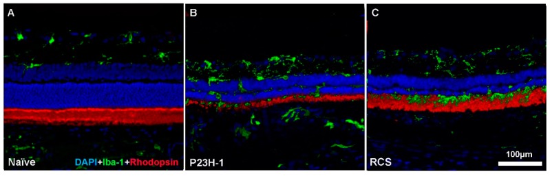
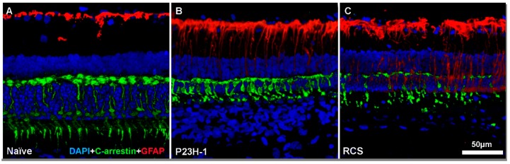
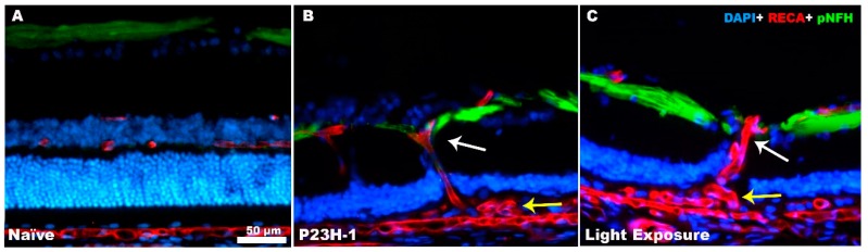
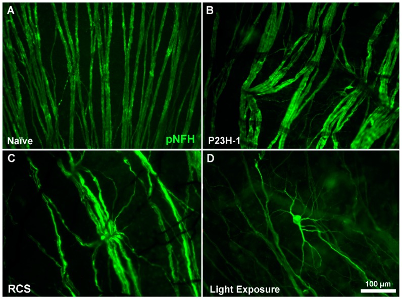
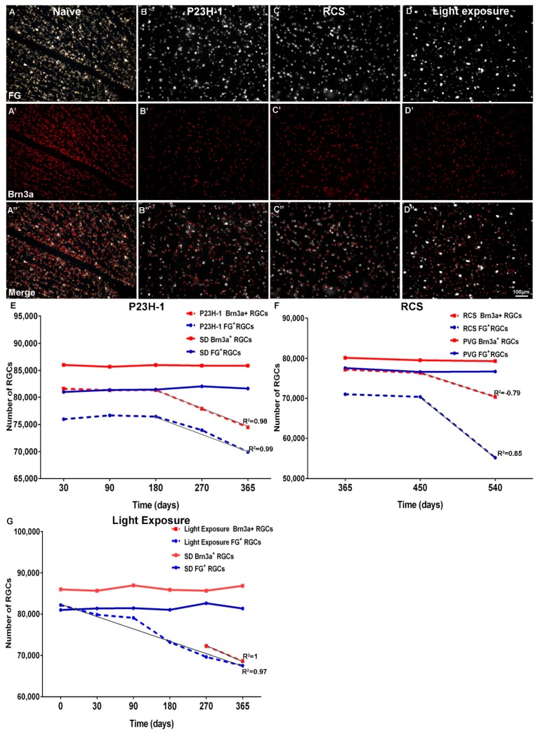
Inherited or acquired photoreceptor degenerations, one of the leading causes of irreversible blindness in the world, are a group of retinal disorders that initially affect rods and cones, situated in the outer retina. For many years it was assumed that these diseases did not spread to the inner retina. However, it is now known that photoreceptor loss leads to an unavoidable chain of events that cause neurovascular changes in the retina including migration of retinal pigment epithelium cells, formation of "subretinal vascular complexes", vessel displacement, retinal ganglion cell (RGC) axonal strangulation by retinal vessels, axonal transport alteration and, ultimately, RGC death. These events are common to all photoreceptor degenerations regardless of the initial trigger and thus threaten the outcome of photoreceptor substitution as a therapeutic approach, because with a degenerating inner retina, the photoreceptor signal will not reach the brain. In conclusion, therapies should be applied early in the course of photoreceptor degeneration, before the remodeling process reaches the inner retina.

Keywords: axonal compression; cones; neurovascular alterations; retinal degeneration; retinal ganglion cells; retinal remodeling.


Conflict of interest statement

The authors declare no conflict of interest. The funders had no role in the design of the study; in the collection, analyses, or interpretation of data; in the writing of the manuscript, or in the decision to publish the results.

Figures

Figure 1
Figure 1
Rod degeneration and microglial cell responses. Microphotographs of representative retinal cross sections from a naïve SD rat (A) showing a normal retina, a retina from a P23H-1 rat (B) and a retina from an RCS rat (C) in the early stages of retinal degeneration. Rod outer segments appear in red and microglial cells in green. In the P23H-1 rat retina, rhodopsin expression was clearly affected; however, in the RCS rat, rhodopsin expression is not altered since the retinal pigment epithelium (RPE) cells are not phagocytising the rod outer segment debris. In both experimental retinas, microglial cells were activated and migrated from the inner to the outer retinal layers.
Figure 2
Figure 2
Cone degeneration and macroglial cell response. Microphotographs of representative retinal cross sections from a naïve SD rat (A) showing a normal retina, a retina from a P23H-1 rat (B) and a retina from an RCS rat (C) in the early stages of retinal degeneration. Cone outer segments appear in green and macroglial cells (GFAP) in red. In both P23H-1 rats and RCS rats, cones degenerate and lose their typically elongated morphology. Moreover, GFAP overexpression in astrocyte and Müller cells could be seen.
Figure 3
Figure 3
Retinal remodeling causes axonal and vascular changes. Microphotographs of retinal cross sections from a naïve SD rat showing a normal retinal structure (A), the retinal structure of a P23H-1 rat (B) and the retinal structure of a light-exposed SD rat (C) after the complete loss of photoreceptors. Retinal ganglion cell (RGC) axons appear in green labelled with anti-neurofilament antibodies (pNFH), the blood vessels in red labelled with anti-rat endothelial cell antigen (RECA) and the nuclei in blue labelled with DAPI. In B and C, blood vessels are running vertically in the retina (white arrow) because they are dragged to the subretinal vascular complexes that appear between the RPE and Bruch’s membrane (yellow arrow) with the inner retinal vascular plexus. RGC axons are displaced by the vessels, which will eventually cause RGC death.
Figure 4
Figure 4
RGC axonal alterations. Magnifications from flat mounted retinas from a naïve SD rat (A), a P23H-1 rat (B), an RCS rat (C) and a light-exposed SD rat (D) after a complete loss of photoreceptors. RGC axons appear in green labelled with anti-neurofilament antibodies (pNFH). In the degenerating retina, the linear trajectory of RGC axons is disrupted and, in addition, some RGC bodies and their proximal dendrites appear labelled with pNFH, indicating RCG degeneration.
Figure 5
Figure 5
RGC loss following photoreceptor degeneration. Microphotographs from a representative naïve SD rat retina and a P23H-1, RCS and light-exposed retina showing fluorogold (FG)+RGCs (A, B, C, D), Brn3a+RGCs (A’, B’, C’, D’), merged images (A’’, B’’, C’’, D’’), and X, Y graphs showing the evolution of the RGC populations (Y axis) with age or time (X axis) in naïve SD and P23H-1 rats (E), RCS and Pievald Viro Glaxo (PVG) rats (F) and in naïve SD and light-exposed SD rats (G). Data are from García-Ayuso et al. [9,10,11].

References

    1. Marc R.E., Jones B.W., Watt C.B., Strettoi E. Neural remodeling in retinal degeneration. Prog. Retin. Eye Res. 2003;22:607–655. doi: 10.1016/S1350-9462(03)00039-9. - DOI - PubMed
    1. Jones B.W., Marc R.E., Pfeiffer R.L. Webvision: The Organization of the Retina and Visual System. University of Utah Health Sciences Center; Salt Lake City, UT, USA: 2016. Retinal Degeneration, Remodeling and Plasticity. - PubMed
    1. Villegas-Pérez M.P., Vidal-Sanz M., Lund R.D. Mechanism of retinal ganglion cell loss in inherited retinal dystrophy. Neuroreport. 1996;7:1995–1999. doi: 10.1097/00001756-199608120-00028. - DOI - PubMed
    1. Villegas-Pérez M.P., Lawrence J.M., Vidal-Sanz M., Lavail M.M., Lund R.D. Ganglion cell loss in RCS rat retina: A result of compression of axons by contracting intraretinal vessels linked to the pigment epithelium. J. Comp. Neurol. 1998;392:58–77. doi: 10.1002/(SICI)1096-9861(19980302)392:1<58::AID-CNE5>3.0.CO;2-O. - DOI - PubMed
    1. Wang S., Villegas-Pérez M.P., Holmes T., Lawrence J.M., Vidal-Sanz M., Hurtado-Montalbán N., Lund R.D. Evolving neurovascular relationships in the RCS rat with age. Curr. Eye Res. 2003;27:183–196. doi: 10.1076/ceyr.27.3.183.16053. - DOI - PubMed

MeSH terms

LinkOut - more resources