. 2019 Sep 19;11(9):1397.
doi: 10.3390/cancers11091397.

γ-H2AX Foci Persistence at Chromosome Break Suggests Slow and Faithful Repair Phases Restoring Chromosome Integrity

Affiliations

γ-H2AX Foci Persistence at Chromosome Break Suggests Slow and Faithful Repair Phases Restoring Chromosome Integrity

Michelle Ricoul et al. Cancers (Basel). .

Abstract

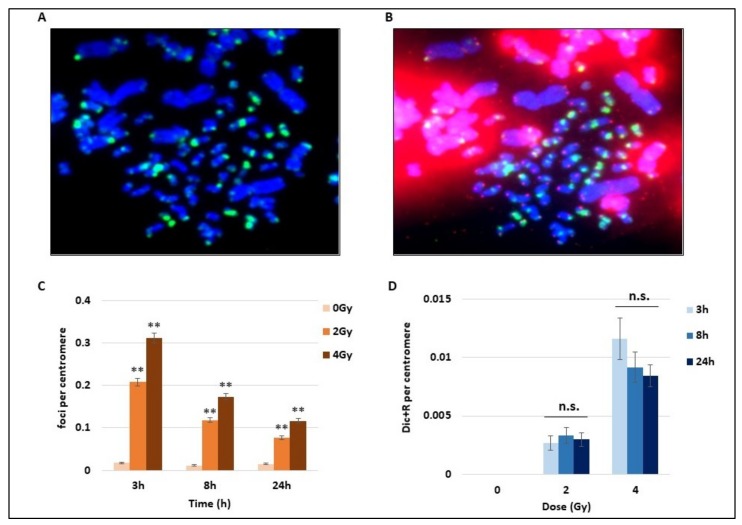
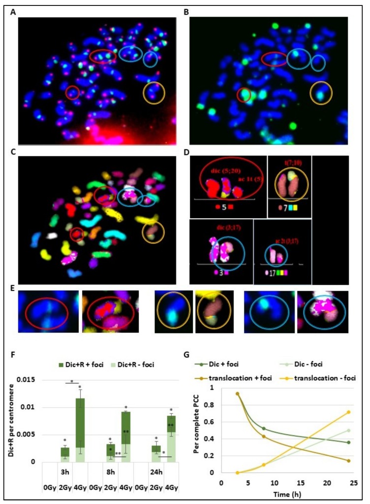
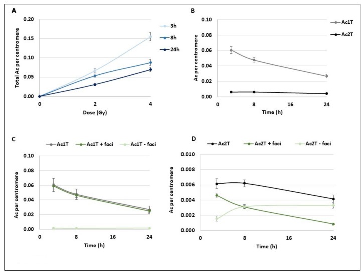
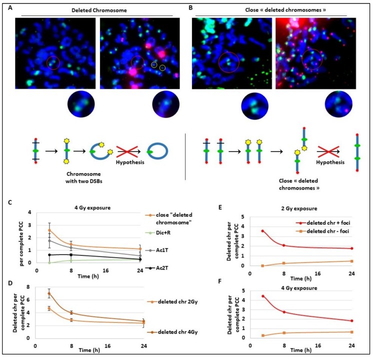
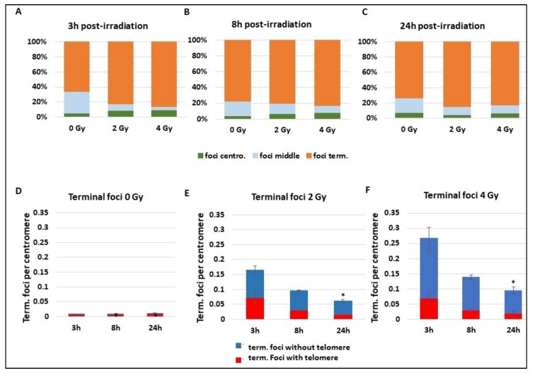
Many toxic agents can cause DNA double strand breaks (DSBs), which are in most cases quickly repaired by the cellular machinery. Using ionising radiation, we explored the kinetics of DNA lesion signaling and structural chromosome aberration formation at the intra- and inter-chromosomal level. Using a novel approach, the classic Premature Chromosome Condensation (PCC) was combined with γ-H2AX immunofluorescence staining in order to unravel the kinetics of DNA damage signalisation and chromosome repair. We identified an early mechanism of DNA DSB joining that occurs within the first three hours post-irradiation, when dicentric chromosomes and chromosome exchanges are formed. The slower and significant decrease of "deleted chromosomes" and 1 acentric telomere fragments observed until 24 h post-irradiation, leads to the conclusion that a second and error-free repair mechanism occurs. In parallel, we revealed remaining signalling of γ-H2AX foci at the site of chromosome fusion long after the chromosome rearrangement formation. Moreover there is important signalling of foci on the site of telomere and sub-telomere sequences suggesting either a different function of γ-H2AX signalling in these regions or an extreme sensibility of the telomere sequences to DNA damage that remains unrepaired 24 h post-irradiation. In conclusion, chromosome repair happens in two steps, including a last and hardly detectable one because of restoration of the chromosome integrity.

Keywords: DNA repair; chromosome; dicentric chromosome; irradiation; premature chromosome condensation; telomere; γ-H2AX.


Conflict of interest statement

The authors declare no potential conflict of interest.

Figures

Figure 1
Figure 1
Kinetics of DNA breaks signalling and unstable chromosome aberrations after radiation exposure. Blood samples from three donors were irradiated at 2 or 4 Gy or kept free from ionising radiation, analysed using the PCC technique and fixed 3 h, 8 h, or 24 h later. (A) Cells were first stained by immunofluorescence with a FITC (fluorescein isothiocyanate)-coupled γ-H2AX antibody (in green) used to detect foci on both human (1-chromatid chromosomes) and hamster chromosomes (2-chromatid chromosomes). (B) A second staining using PNA-FISH (Peptide Nucleic Acid Fluorescence In Situ Hybridisation) Telomere and Centromere probes was performed on the same Premature Chromosome Condensation (PCC) cells after the immunofluorescence to simplify the detection of chromosome unstable aberrations (Dicentric and Ring chromosomes and Acentric fragments). Centromeres are stained in green, while telomeres are in red, DNA appears in blue after Dapi staining. Dicentric chrosomes are chromosomes with two centromeres in green. (C) The number of γ-H2AX foci on human chromosomes was counted after immunofluorescence staining (cf image A) and expressed per centromere to exclude bias due to the possible loss of PCC fragments during the cytospin step. The increase of foci with 2 and 4 Gy irradiation (IR) is significant at 8 h and 24 h post-IR (** p < 0.01) and the bars represent standard error. The number of foci decreases with time. (D) Dicentric and ring chromosomes were counted after Telomere and Centromere FISH staining (as represented in B) and expressed per centromeres. The unstable chromosome aberrations increase with doses. There is no statistical difference between the different time points at 2 and 4 Gy (n.s).
Figure 2
Figure 2
γ-H2AX foci signalling of unstable and stable aberrations short time after irradiation. (AD) The same slide with PCC lymphocyte cells previously irradiated with 4 Gy was observed after telomere (in red) and centromere staining (in green) (A), γ-H2AX immunofluorescence assays have been performed with a FITC-coupled antibody (green) (B) or multi-FISH (C) and analysed with the ISIS software. Four abnormalities were detected and marked with coloured circles. The red and blue circles show dicentric chromosomes and their corresponding acentric fragments. The yellow circle shows a translocation between chromosomes 7 and 10. (D) Chromosome karyotype using false colours was built using the ISIS software. Data obtained from Telomere and Centromere staining and from multi-FISH were assembled to detect unstable and stable aberrations. (E) Zoom of CA stained for γ-H2AX (green) and visualized after multi-FISH. The foci localized at the junction between two chromosomes. (F) Blood samples from three donors were irradiated at 2 or 4 Gy or kept free from ionising radiation before analysis using the PCC technique. Dic+R frequency per centromere was scored 3 h, 6 h, and 24 h after IR and the percentage of co-staining with γ-H2AX foci was determined (dark green). The increase of “Dic+R + foci” or “Dic+R – foci” is significant (* p < 0.05, ** p < 0.001) at the different time points compared to 0 Gy. There is significant differences when indicated between 2 Gy and 4 Gy irradiation (* p < 0.05, ** p < 0.001). (G) Only complete PCC cells (between 45 and 48 chromosome fragments) irradiated with 4Gy were analysed by multi-FISH after γ-H2AX foci immunofluorescence assay. Proportions of Dic+R and translocations signalled with γ-H2AX foci (in green part A) or without any γ-H2AX foci were represented over time. Respectively 15, 21, and 14 PCC were analysed for 0, 2, and 4 Gy exposure.
Figure 3
Figure 3
Kinetics of different subtypes of acentric fragments after radiation exposure. Blood samples from three donors were irradiated at 2 or 4 Gy or kept free from ionising radiation and analysed using the PCC technique. (A) After Telomere/Centromere staining on PCC, the total number of acentric fragments per centromere was scored for each dose and 3 h, 8 h, and 24 h post-IR. The total acentric fragments increase with dose and decrease overtime. Bars represent standard deviation. (BD) Focus was made on PCC analysis after 2 Gy radiation exposure. Bars represent standard deviation. (B) Acentric fragments containing 1 telomere or resulting from fusion and containing 2 telomeres were separately represented at 3 h, 8 h, and 24 h post IR. A decrease of both types of acentric fragments is observed overtime (C,D) All acentric fragments were counted and classified in two classes, acentric fragments signalled by γ-H2AX foci or without γ-H2AX signal. Their proportions were followed at 3 h, 8 h, and 24 h post IR. Acentric fragments with 1 telomere resulting from one DNA DSB (C) and acentric fragments with 2 telomeres resulting from two DNA double strand breaks (DSBs) and fusions of two fragments (D) were represented separately.
Figure 4
Figure 4
Kinetics of late and slow DNA repair mechanisms. Blood samples from three donors were irradiated at 2 or 4 Gy or kept free from ionising radiation, analysed using the PCC technique and fixed 3 h, 8 h, or 24 h post-exposition. (A,B) Images of the same PCC exposed to 4Gy were observed after γ-H2AX immunofluorescence staining (in green on the left panels) and after TC staining (right panels). Centromeres are stained in green and telomeres in red after PNA-FISH staining. Red circles show the zones of interest and yellow circles indicate acentric fragments. A chromosome with two DSBs (A) and two chromosomes with close signalled DSBs (B) are both signalled by γ-H2AX and considered as “in repair”. In (A), the chromosome is expected to become a ring while in (B), the two chromosomes are expected to fuse and make a dicentric chromosome. Below each image, a schema represents the hypothetical mechanism of CA formation. On the schema, centromeres are represented with green circles, telomeres with red circles, and γ-H2AX foci with yellow stars. The red crosses on the hypothesis conclude that the data will not confirm the hypothesis. (CF) Scoring was performed only on complete PCC cells containing between 45 and 48 fragments. (C) The frequency of “close deleted chromosomes”, Dic+R, Ac1T, and Ac2T, per PCC were reported at 3 h, 8 h, and 24 h after 4 Gy irradiation. (D) The frequency of deleted chromosomes (with one telomere only) was followed after 2 and 4 Gy irradiation at three time points: 3 h, 8 h, and 24 h post-IR. (E,F). The proportion of deleted chromosomes containing γ-H2AX signalling or not signalled was quantified after 2 Gy (E) and 4 Gy (F) exposure.
Figure 5
Figure 5
Localization of γ-H2AX foci on chromosomes. Blood samples from three donors were irradiated at 2 or 4 Gy or kept free from ionising radiation and then analysed using the PCC technique. (AC) After immunofluorescence staining, γ-H2AX foci position on chromosome sequence was determined “close to chromosome extremities”, “close to centromere”, or “in the middle of the chromatide”. Analyses were performed at 3 h (A), 8 h (B), or 24 h (C) after radiation exposure. (DF) Only γ-H2AX foci “close to chromosome extremities” were sub-classed in two groups, γ-H2AX foci associated “with telomeres” or “without telomere” and followed at 3 h, 8 h, and 24 h post radiation exposure. The graphs show the repartition of foci without irradiation (D) and after 2 Gy exposure (E) or 4 Gy (F) radiation exposition. The decrease of terminal foci without telomere is significant at 24 h (p < 0.05).

References

    1. Rogakou E.P., Pilch D.R., Orr A.H., Ivanova V.S., Bonner W.M. DNA double-stranded breaks induce histone H2AX phosphorylation on serine 139. J. Biol. Chem. 1998;273:5858–5868. doi: 10.1074/jbc.273.10.5858. - DOI - PubMed
    1. Stiff T., O’Driscoll M., Rief N., Iwabuchi K., Löbrich M., Jeggo P.A. ATM and DNA-PK function redundantly to phosphorylate H2AX after exposure to ionizing radiation. Cancer Res. 2004;64:2390–2396. doi: 10.1158/0008-5472.CAN-03-3207. - DOI - PubMed
    1. Motoyama N., Naka K. DNA damage tumor suppressor genes and genomic instability. Curr. Opin. Genet. Dev. 2004;14:11–16. doi: 10.1016/j.gde.2003.12.003. - DOI - PubMed
    1. Blackford A.N., Jackson S.P. ATM, ATR, and DNA-PK: The Trinity at the Heart of the DNA Damage Response. Mol. Cell. 2017;66:801–817. doi: 10.1016/j.molcel.2017.05.015. - DOI - PubMed
    1. Eliezer Y., Argaman L., Rhie A., Doherty A.J., Goldberg M. The direct interaction between 53BP1 and MDC1 is required for the recruitment of 53BP1 to sites of damage. J. Biol. Chem. 2009;284:426–435. doi: 10.1074/jbc.M807375200. - DOI - PubMed

LinkOut - more resources