Review
. 2019 Sep 19;20(18):4653.
doi: 10.3390/ijms20184653.

Regulatory Effects and Interactions of the Wnt and OPG-RANKL-RANK Signaling at the Bone-Cartilage Interface in Osteoarthritis

Affiliations
Review

Regulatory Effects and Interactions of the Wnt and OPG-RANKL-RANK Signaling at the Bone-Cartilage Interface in Osteoarthritis

Béla Kovács et al. Int J Mol Sci. .

Abstract

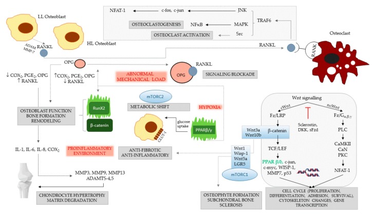
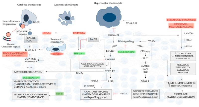
Cartilage and the bordering subchondral bone form a functionally active regulatory interface with a prominent role in osteoarthritis pathways. The Wnt and the OPG-RANKL-RANK signaling systems, as key mediators, interact in subchondral bone remodeling. Osteoarthritic osteoblasts polarize into two distinct phenotypes: a low secretory and an activated, pro-inflammatory and anti-resorptive subclass producing high quantities of IL-6, PGE2, and osteoprotegerin, but low levels of RANKL, thus acting as putative effectors of subchondral bone sclerosis. Wnt agonists, Wnt5a, Wisp-1 initiate excessive bone remodeling, while Wnt3a and 5a simultaneously cause loss of proteoglycans and phenotype shift in chondrocytes, with decreased expression of COL2A, aggrecan, and Sox-9. Sclerostin, a Wnt antagonist possesses a protective effect for the cartilage, while DKK-1 inhibits VEGF, suspending neoangiogenesis in the subchondral bone. Experimental conditions mimicking abnormal mechanical load, the pro-inflammatory milieu, but also a decreased OPG/RANKL ratio in the cartilage, trigger chondrocyte apoptosis and loss of the matrix via degradative matrix metalloproteinases, like MMP-13 or MMP-9. Hypoxia, an important cofactor exerts a dual role, promoting matrix synthesis via HIF-1α, a Wnt silencer, but turning on HIF-2α that enhances VEGF and MMP-13, along with aberrant collagen expression and extracellular matrix deterioration in the presence of pro-inflammatory cytokines.

Keywords: Wnt signaling; bone-cartilage interface; osteoarthritis.


Conflict of interest statement

The authors declare no conflict of interest.

Figures

Figure 1
Figure 1
Signaling events and their consequences in osteoarthritic subchondral bone. Three classical factors—abnormal mechanical load, hypoxia and pro-inflammatory transformation play a trigger role in osteoarthritis (represented in red). Two types of osteoarthritic osteoblasts are illustrated: high-level (HL) and low-level (LL) synthesizer cells. The first type produces high levels of Cox-2, PGE2 and OPG and less RANKL; the second type has an opposite secretory pattern. The HL synthesizers maintain pro-inflammatory conditions in the subchondral bone, and they are increased in the presence of abnormal mechanical load, when they produce OPG, which may enhance focal osteosclerosis. The LL synthesizers release important quantities of RANKL, which activate osteoclasts by linking RANK. Signal cascades are activated via TRAF6, JNK, MAP and Src kinases, resulting in osteoclast differentiation and activation. Activation of Cox-2 and PGE2 stimulates the synthesis of IL-1, IL-6, and IL-8, which turn on the production of degradative MMPs in the cartilage. In normal conditions, osteoblasts rely for energy mainly on glucose, and PPARβ/γ enhances glucose uptake via the GLUT-1 transporter; however, in hypoxic conditions, the cells undergo a strong metabolic shift to anaerobic glycolysis, driven by mTORC2 that stabilizes RunX2. PPARβ/γ cooperates with canonical signal molecules, e.g., Wnt3a and Wnt10b, to exert anti-fibrotic and anti-inflammatory effects, further inducing OPG and RunX2 and fine-tuning osteoblast function, bone formation and remodeling. RunX2 alone induces RANKL and controls osteoclast function, but when acting together with β-catenin, it stimulates mainly osteoblasts, which increase trabecular bone volume. The two linker factors of OPG/RANKL and Wnt signaling, RunX2 and PPARβ/γ, are shown in green.
Figure 2
Figure 2
Effects of OPG/RANKL and Wnt signaling on chondrocytes and cartilage. The presence of catabolic, apoptotic and hypertrophic chondrocytes is a characteristic of osteoarthritis. Chondrocytes expressing RANKL release it in the soluble form, triggering IL-1β and IL-6 overexpression in the cartilage. Senescent chondrocytes contribute to this pro-inflammatory environment, increasing the synthesis of MMP-3, -9 -13, ADAMTS-4 and -5. The pro-inflammatory activity results in degradation of cartilage extracellular matrix. OPG links to RANKL and facilitates its internalization with consequent degradation. Sclerostin is a strong inhibitor of cartilage degradation, which suspends MMPs, ADAMTs and even their inhibitors—tissue inhibitors of metalloproteinases—stimulating in parallel aggrecan and collagen type II production. Canonical Wnt signaling and β-catenin are anti-apoptotic and stimulate chondrocyte proliferation; however, TCF/LEF is a downstream activator of apoptosis and matrix degradation. Hypoxia, triggering HIF-1α, generates an inhibitory effect on β-catenin and RunX2 and enhances the homeostatic effects of Sox-9. Non-canonical Wnt activation has different pathological consequences: chondrocyte dedifferentiation and loss of phenotype, the agonist Wnt5a up-regulating MMP-1, -3 and -13, and inhibition of aggrecan along with collagen type II. Wnt3a, together with Wnt5a, suspends hypertrophic transformation, while Wnt4, 8 and 11 are stimulatory. The metabolic syndrome and abnormal mechanical load down-regulate AMPKα, glycolysis and mitochondrial respiration in an adaptive response to hypoxia, which also results in a catabolic phenotype switch of chondrocytes. Factors of chondrocyte and cartilage deterioration are shown in red, and protective mediators are drawn in green.

Similar articles

Cited by

References

    1. Hügle T., Geurts J. What drives osteoarthritis?-Synovial versus subchondral bone pathology. Rheumatology. 2017;56:1461–1471. doi: 10.1093/rheumatology/kew389. - DOI - PubMed
    1. Yuan X.L., Meng H.Y., Wang Y.C., Peng J., Guo Q.Y., Wang A.Y., Lu S.B. Bone-cartilage interface crosstalk in osteoarthritis: Potential pathways and future therapeutic strategies. Osteoarthr. Cartil. 2014;22:1077–1089. doi: 10.1016/j.joca.2014.05.023. - DOI - PubMed
    1. Madry H., Luyten F.P., Facchini A. Biological aspects of early osteoarthritis. Knee Surgery Sports Traumatol. Arthrosc. 2012;20:407–422. doi: 10.1007/s00167-011-1705-8. - DOI - PubMed
    1. Martel-Pelletier J., Boileau C., Pelletier J.P., Roughley P.J. Cartilage in normal and osteoarthritis conditions. Best Pract. Res. Clin. Rheumatol. 2008;22:351–384. doi: 10.1016/j.berh.2008.02.001. - DOI - PubMed
    1. Christgau S., Garnero P., Fledelius C., Moniz C., Ensig M., Gineyts E., Rosenquist C., Qvist P. Collagen type II C-telopeptide fragments as an index of cartilage degradation. Bone. 2001;29:209–215. doi: 10.1016/S8756-3282(01)00504-X. - DOI - PubMed

MeSH terms