. 2019 Sep 19;8(9):1109.
doi: 10.3390/cells8091109.

Dynamic Interplay between Pericytes and Endothelial Cells during Sprouting Angiogenesis

Affiliations

Dynamic Interplay between Pericytes and Endothelial Cells during Sprouting Angiogenesis

Giulia Chiaverina et al. Cells. .

Abstract

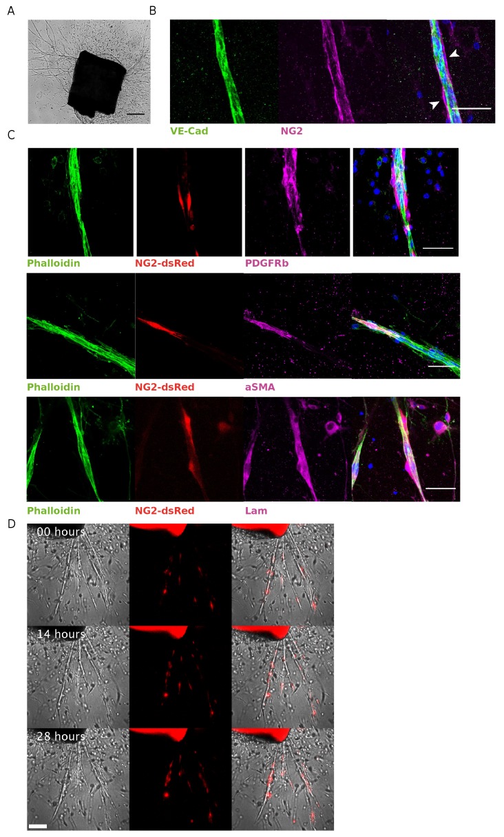
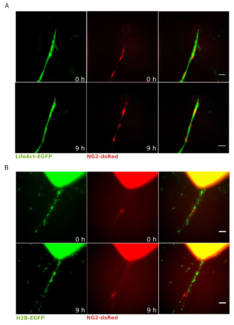
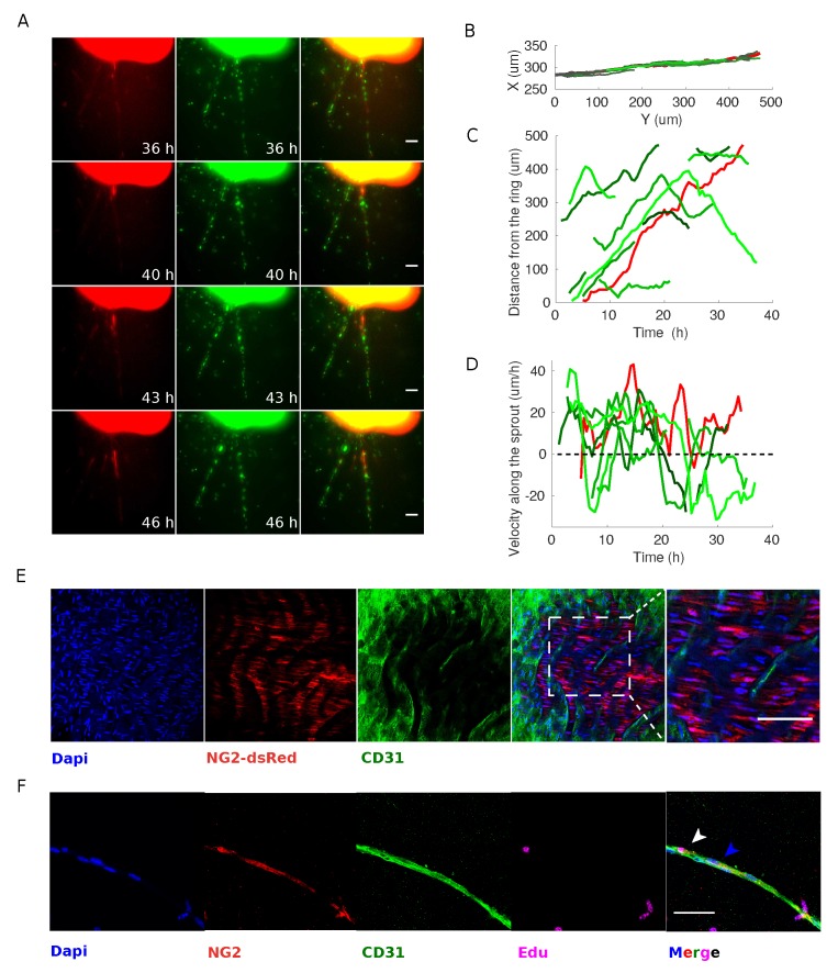
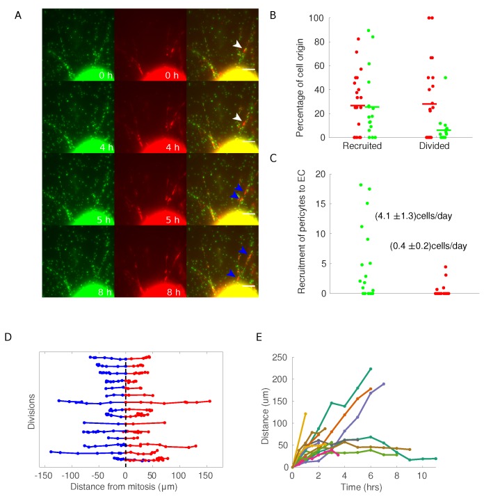
Vascular physiology relies on the concerted dynamics of several cell types, including pericytes, endothelial, and vascular smooth muscle cells. The interactions between such cell types are inherently dynamic and are not easily described with static, fixed, experimental approaches. Pericytes are mural cells that support vascular development, remodeling, and homeostasis, and are involved in a number of pathological situations including cancer. The dynamic interplay between pericytes and endothelial cells is at the basis of vascular physiology and few experimental tools exist to properly describe and study it. Here we employ a previously developed ex vivo murine aortic explant to study the formation of new blood capillary-like structures close to physiological situation. We develop several mouse models to culture, identify, characterize, and follow simultaneously single endothelial cells and pericytes during angiogenesis. We employ microscopy and image analysis to dissect the interactions between cell types and the process of cellular recruitment on the newly forming vessel. We find that pericytes are recruited on the developing sprout by proliferation, migrate independently from endothelial cells, and can proliferate on the growing capillary. Our results help elucidating several relevant mechanisms of interactions between endothelial cells and pericytes.

Keywords: NG2; aortic ring assay; cancer; endothelial cells; pericytes; sprouting angiogenesis.


Conflict of interest statement

The authors declare no conflict of interest.

Figures

Figure 1
Figure 1
Characterization of NG2-dsRed mAR. (A) Representative bright-field image of mouse aortic ring cultured for one week, showing microvessel outgrowth (scale bar represents 200 μm). (B) Whole-mount immunofluorescence staining of a mAR fixed after 6 days of culture. The mAR was stained for the endothelial marker VE-cadherin (green) and the pericyte marker NG2 (magenta) (Scale bar: 50 μm). (C) mARs obtained from NG2-dsRed mouse model were cultured for 6 days then fixed to perform a whole-mount immunofluorescence staining. mARs were stained for classical pericyte markers, like PDGFRβ (magenta, top row) and αSMA (magenta, middle row), to verify the colocalization with dsRed signal (red). mARs were also stained for laminin (magenta, bottom row), one of the main components of vBM. Phalloidin staining was performed to identify microvessel outgrowths (green) (Scale bar: 50 μm). (D) Snapshots of time-lapse microscopy of angiogenic outgrowths from NG2-dsRed mAR.
Figure 2
Figure 2
LifeAct-EGFP or H2B-EGFP NG2-dsRed mAR models for pericyte–EC interactions. (A) Time-lapse microscopy was performed on LifeAct-EGFP NG2-dsRed mARs cultured for 5 days then imaged for 72 h. This mouse model allows to clearly identify pericytes thanks to dsRed signal (red) running over the endothelial layer labeled by LifeAct-EGFP (green) (scale bar: 50 μm). (B) Time-lapse microscopy of H2B-EGFP NG2-dsRed mARs cultured for 5 days then imaged for 72 h. Thanks to this model we could identify pericytes (red) as well as track all individual cells thanks to histone H2B-EGFP (green) (scale bar: 50 μm).
Figure 3
Figure 3
Pericytes recruited on newly formed sprouting capillaries originate from the aortic ring but can proliferate. (A) Time-lapse microscopy analysis of H2B-EGFP NG2-dsRed mARs cultured for 5 days then imaged for 72 h. The observed pericyte originates from the edge of the mAR and moves along the ECs during sprouting process (scale bar: 50 μm). (B) Cell trajectory analysis shows that endothelial cells and pericytes move along the sprout. This set of trajectories is used for the following two panels. (C) The distance of each cell from the ring plotted as a function of time shows that endothelial cells (plotted in different shades of green) move at a rather constant speed and, in some cases, suddenly change direction, as witnessed by the change in slope of some trajectories. (D) The velocity of each cell shows that pericytes and endothelial cells do not proceed coherently, but conversely they move independently of each other. In particular cells proceed with bursts (accelerations and decelerations) (E) Whole-mount immunofluorescence staining of a NG2-dsRed mA-sheet embedding in a collagen gel and fixed. mA-sheet was stained for the endothelial marker CD31 (green) and DAPI (blue). The rightmost panel shows an enlarged detail. (scale bar: 50 μm). (F) mARs were cultured in standard medium supplemented with EdU for 1 week after explant in order to detect whether pericytes and/or ECs coming from the ring originate from a proliferative event or not. Images show a representative experiment. Arrows indicate pericytes Edu-positive (white) or -negative (blue). (Scale bar: 50 μm.)
Figure 4
Figure 4
H2B-EGFP NG2-dsRed mAR mouse models to study pericytes dynamics during the sprouting process. (A) Time-lapse widefield microscopy analysis of H2B-EGFP NG2-dsRed mARs. White arrow indicate a pericyte that undergoes cell division after 5 h from the beginning of the experiment, generating the two daughter cells marked with blue arrows. (Scale bar: 50 μm.) (B) For each cell in a sprout, we verified if it appeared on the sprout coming out of the ring (recruited) or if it divided on the sprout (divided). Each dot represents a single sprout, with green dots for endothelial cells and red dots for pericytes. (C) To measure the recruitment rate of cells on the sprouts, we measured the number of cells at the end of each sprouting assay (each dot represents a sprout) and measured the number of cells during the time of the experiment. (D) Each line represents a division event of a pericyte. Red and blue trajectories are projections of the trajectories of the daughter cells on the direction of the sprout, where the origin has been defined as the coordinate of the mother cells at the frame preceding mitosis. (E) Plot of the distance between daughter cells as a function of time.

Similar articles

Cited by

References

    1. Armulik A., Genove G., Betsholtz C. Pericytes: Developmental, physiological, and pathological perspectives, problems, and promises. Dev. Cell. 2011;21:193–215. doi: 10.1016/j.devcel.2011.07.001. - DOI - PubMed
    1. Van Dijk C.G., Nieuweboer F.E., Pei J.Y., Xu Y.J., Burgisser P., Van Mulligen E., El Azzouzi H., Duncker D.J., Verhaar M.C., Cheng C. The complex mural cell: Pericyte function in health and disease. Int. J. Cardiol. 2015;190:75–89. doi: 10.1016/j.ijcard.2015.03.258. - DOI - PubMed
    1. Birbrair A., Zhang T., Wang Z.M., Messi M.L., Olson J.D., Mintz A., Delbono O. Type-2 pericytes participate in normal and tumoral angiogenesis. Am. J. Physiol.-Cell Physiol. 2014;307:C25–C38. doi: 10.1152/ajpcell.00084.2014. - DOI - PMC - PubMed
    1. Ozerdem U., Monosov E., Stallcup W.B. NG2 proteoglycan expression by pericytes in pathological microvasculature. Microvasc. Res. 2002;63:129–134. doi: 10.1006/mvre.2001.2376. - DOI - PubMed
    1. Song S., Ewald A.J., Stallcup W., Werb Z., Bergers G. PDGFRbeta+ perivascular progenitor cells in tumours regulate pericyte differentiation and vascular survival. Nat. Cell Biol. 2005;7:870–879. doi: 10.1038/ncb1288. - DOI - PMC - PubMed

Publication types