. 2019 Sep 20;8(10):420.
doi: 10.3390/antiox8100420.

Combined Treatment with Three Natural Antioxidants Enhances Neuroprotection in a SH-SY5Y 3D Culture Model

Affiliations

Combined Treatment with Three Natural Antioxidants Enhances Neuroprotection in a SH-SY5Y 3D Culture Model

Pasquale Marrazzo et al. Antioxidants (Basel). .

Abstract

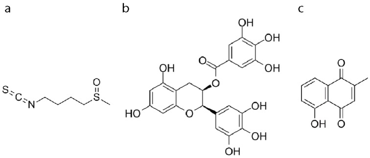
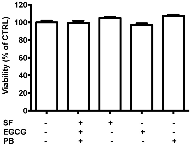
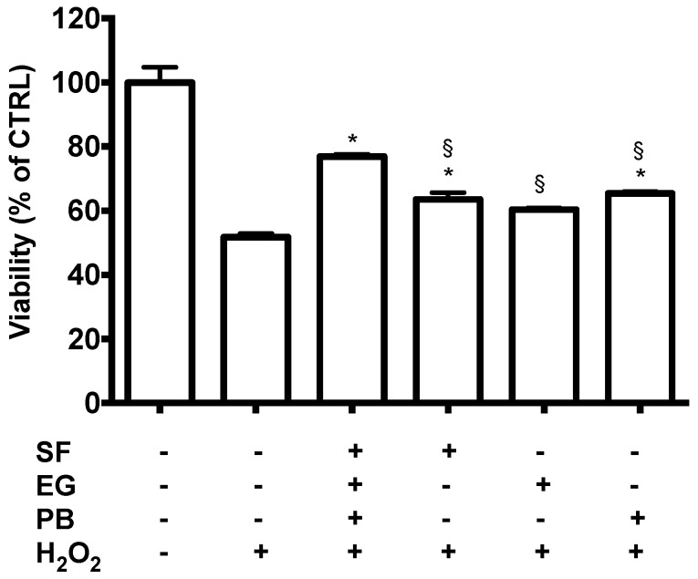
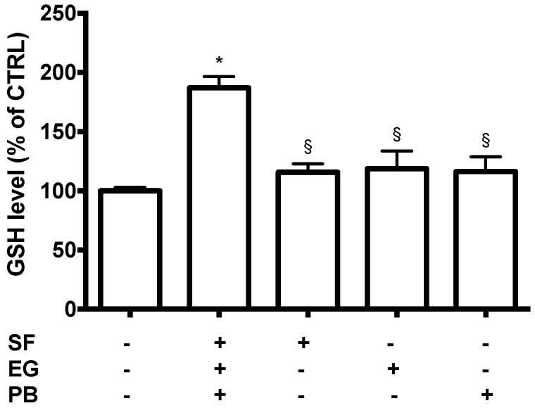
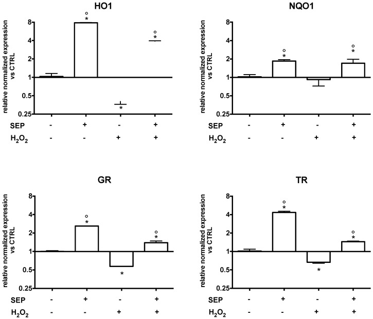
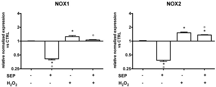
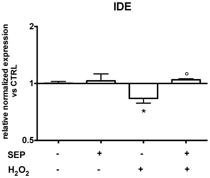
Currently, the majority of cell-based studies on neurodegeneration are carried out on two-dimensional cultured cells that do not represent the cells residing in the complex microenvironment of the brain. Recent evidence has suggested that three-dimensional (3D) in vitro microenvironments may better model key features of brain tissues in order to study molecular mechanisms at the base of neurodegeneration. So far, no drugs have been discovered to prevent or halt the progression of neurodegenerative disorders. New therapeutic interventions can come from phytochemicals that have a broad spectrum of biological activities. On this basis, we evaluated the neuroprotective effect of three phytochemicals (sulforaphane, epigallocatechin gallate, and plumbagin) alone or in combination, focusing on their ability to counteract oxidative stress. The combined treatment was found to be more effective than the single treatments. In particular, the combined treatment increased cell viability and reduced glutathione (GSH) levels, upregulated antioxidant enzymes and insulin-degrading enzymes, and downregulated nicotinamide adenine dinucleotide phosphate (NADPH) oxidase 1 and 2 in respect to peroxide-treated cells. Our data suggest that a combination of different phytochemicals could be more effective than a single compound in counteracting neurodegeneration, probably thanks to a pleiotropic mechanism of action.

Keywords: 3D cultures; Antioxidants; Neurodegeneration; Oxidative stress; Phytochemicals; SH-SY5Y cell line.


Conflict of interest statement

The authors declare that there is no conflict of interest regarding the publication of this paper.

Figures

Figure 1
Figure 1
Chemical structures of the natural compounds used in this study. (a) Sulforaphane (SF), (b) epigallocatechin gallate (EGCG), (c) plumbagin (PB).
Figure 2
Figure 2
Characterization of the 3D SH-SY5Y model. (a) Cellular adhesion to the scaffold was evaluated 24 h after cell seeding by MTT assay as reported in Materials and Methods. Data are expressed as a percentage of total viable cells and represent the mean of three independent experiments. (b) Metabolic activity of the 3D model was evaluated after 1 and 6 days from cell seeding by a Prestoblue assay as reported in Materials and Methods. Each bar represents the mean ± SEM of three independent experiments. Data were analyzed with a two-way ANOVA followed by the Fisher’s test. * p < 0.05. (c) Real time-PCR was performed in the 3D culture for neuronal markers. Each bar represents the mean ± SEM of three independent experiments, which were analyzed with an unpaired T-test. * p < 0.05.
Figure 3
Figure 3
Potential cytotoxicity of sulforaphane (SF), epigallocatechin gallate (EGCG), and plumbagin (PB) on SH-SY5Y cells. Cells were treated with 1 µM SF, 2.5, µM EGCG, and 0.5 µM PB, and after 24 h, viability was evaluated by a Prestoblue assay as reported in Materials and Methods. Results are expressed as a percentage of untreated cells. Each bar represents the mean ± SEM of three independent experiments, which were analyzed with a one-way ANOVA followed by the Fisher’s test.
Figure 4
Figure 4
Neuroprotective activity of SF, EGCG, and PB compounds against H2O2-induced damage. Cells were treated with 1 µM SF, 2.5 µM EGCG, and 0.5 µM PB, and after 24 h, were exposed to 700 µM H2O2 to induce oxidative stress. Cell viability in 3D cultures was measured by a Prestoblue assay as reported in Materials and Methods. Data are expressed as a percentage of untreated cells. Each bar represents mean ± SEM of three independent experiments. Data were analyzed with a one-way ANOVA followed by the Fisher’s test. * p < 0.05 vs. H2O2 treated cells; § p < 0.05 vs. sulforaphane, epigallocatechin gallate, and plumbagin (SEP) co-treatment.
Figure 5
Figure 5
Antioxidant activity of SF, EGCG, and PB compounds on SH-SY5Y cells. Cells were treated with 1 µM SF, 2.5 µM EGCG, and 0.5 µM PB, and after 24 h GSH levels were evaluated with an monochlorobimane (MCB) assay as reported in Materials and Methods. Data are expressed as a percentage of untreated cells (CTRL). Each bar represents mean ± SEM of three independent experiments. Data were analyzed with a one-way ANOVA followed by the Fisher’s test. * p < 0.05 vs. untreated cells; § p < 0.05 vs. SEP co-treatment.
Figure 6
Figure 6
Effect of SEP co-treatment on antioxidant enzyme expression. Cells were co-treated with 1 µM SF, 2.5 µM EGCG, and 0.5 µM PB for 6 h. Oxidative stress was induced with 700 µM H2O2 for 1 h prior to lysis. Real time-PCR was performed to detect heme oxygenase 1 (HO1), NADPH: quinone oxidoreductase 1 (NQO1), glutathione reductase (GR), and thioredoxin reductase (TR) mRNA levels. Data are expressed as relative abundance compared to untreated cells. Each bar represents mean ± SEM of three independent experiments. Data were analyzed with a one-way ANOVA followed by the Fisher’s test. * p < 0.05 vs. untreated cells, ° p < 0.05 vs. H2O2.
Figure 7
Figure 7
Effect of SEP co-treatment on NADPH oxidase 1 (NOX1) and NADPH oxidase 2 (NOX2). Cells were co-treated with 1 μM SF, 2.5 μM EGCG, and 0.5 μM PB for 6 h. Oxidative stress was induced with 700 µM H2O2 for 1 h prior to lysis. Real time-PCR was performed to detect NOX1 and NOX2 mRNA levels. Data are expressed as relative abundance compared to untreated cells. Each bar represents mean ± SEM of three independent experiments. Data were analyzed using a one-way ANOVA followed by the Fisher’s test. * p < 0.05 vs. untreated cells, ° p < 0.05 vs. H2O2.
Figure 8
Figure 8
Effect of SEP co-treatment on insulin-degrading enzyme (IDE). Cells were co-treated with 1 μM SF, 2.5 μM EGCG, and 0.5 μM PB for 6 h. Oxidative stress was induced with 700 µM H2O2 for 1 h prior to lysis. Real time-PCR was performed to detect IDE mRNA levels. Data are expressed as relative abundance compared to untreated cells. Each bar represents mean ± SEM of three independent experiments. Data were analyzed with a one-way ANOVA followed by the Fisher’s test. * p < 0.05 vs. untreated cells, ° p < 0.05 vs. H2O2.

References

    1. Barnham K.J., Masters C.L., Bush A.I. Neurodegenerative diseases and oxidative stress. Nat. Rev. Drug Discov. 2004;3:205–214. doi: 10.1038/nrd1330. - DOI - PubMed
    1. Gammon K. Neurodegenerative disease: Brain windfall. Nature. 2014;515:299–300. doi: 10.1038/nj7526-299a. - DOI - PubMed
    1. Tarozzi A., Angeloni C., Malaguti M., Morroni F., Hrelia S., Hrelia P. Sulforaphane as a Potential Protective Phytochemical against Neurodegenerative Diseases. Oxid. Med. Cell. Longev. 2013;2013:415078. doi: 10.1155/2013/415078. - DOI - PMC - PubMed
    1. Angeloni C., Malaguti M., Hrelia S. Antiglycative activity of sulforaphane: A new avenue to counteract neurodegeneration? Neural Regen. Res. 2015;10:1750. doi: 10.4103/1673-5374.169613. - DOI - PMC - PubMed
    1. Lenzi M., Fimognari C., Hrelia P. Advances in Nutrition and Cancer. Volume 159. Springer; Berlin/Heidelberg, Germany: 2014. Sulforaphane as a Promising Molecule for Fighting Cancer; pp. 207–223. Cancer Treatment and Research. - PubMed

LinkOut - more resources