. 2019 Sep 20;20(19):4663.
doi: 10.3390/ijms20194663.

Platelet-Derived Growth Factor Receptor and Ionizing Radiation in High Grade Glioma Cell Lines

Affiliations

Platelet-Derived Growth Factor Receptor and Ionizing Radiation in High Grade Glioma Cell Lines

Oana Alexandru et al. Int J Mol Sci. .

Abstract

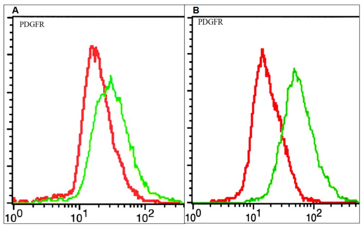
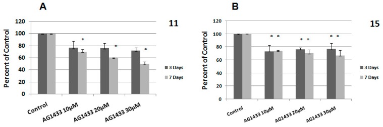
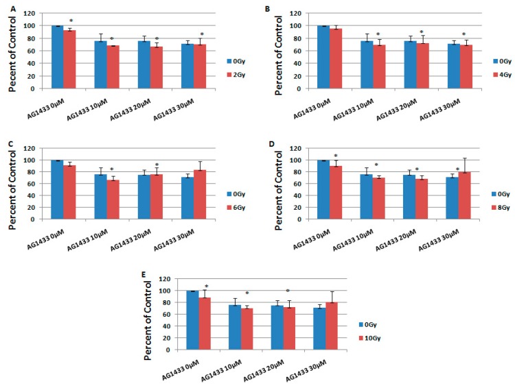
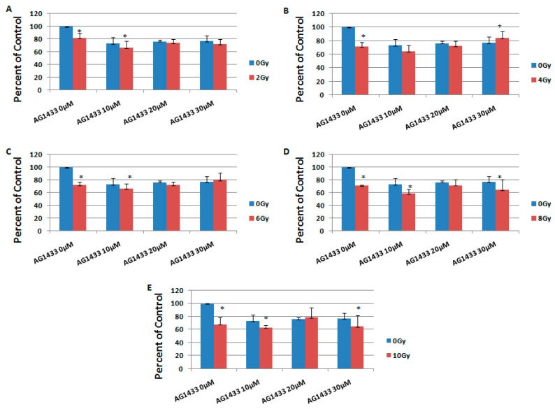
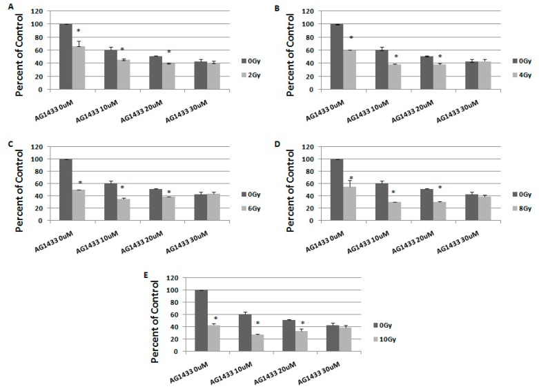
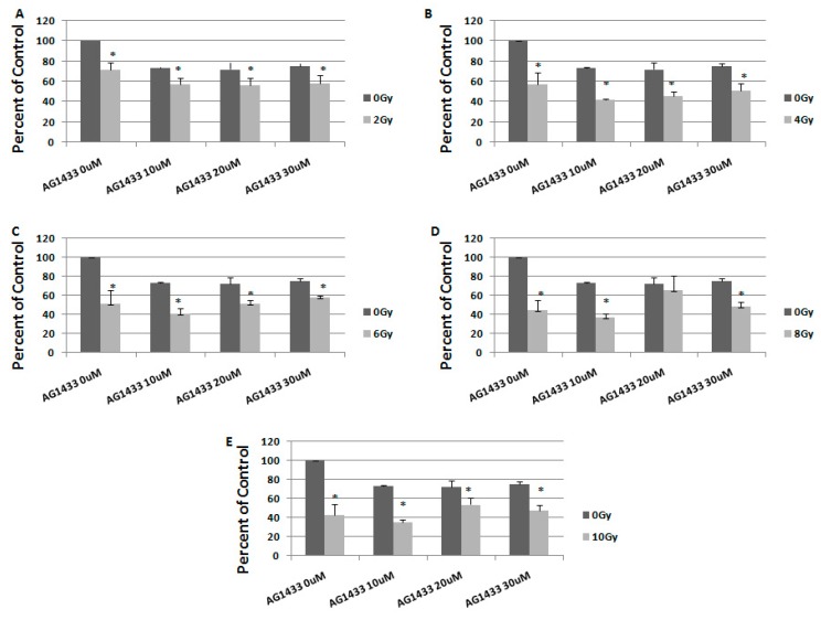
Treatment of high grade gliomas (HGGs) has remained elusive due to their high heterogeneity and aggressiveness. Surgery followed by radiotherapy represents the mainstay of treatment for HGG. However, the unfavorable location of the tumor that usually limits total resection and the resistance to radiation therapy are the major therapeutic problems. Chemotherapy with DNA alkylating agent temozolomide is also used to treat HGG, despite modest effects on survival. Disregulation of several growth factor receptors (GFRs) were detected in HGG and receptor amplification in glioblastoma has been suggested to be responsible for heterogeneity propagation through clonal evolution. Molecularly targeted agents inhibiting these membrane proteins have demonstrated significant cytotoxicity in several types of cancer cells when tested in preclinical models. Platelet-derived growth factor receptors (PDGFRs) and associated signaling were found to be implicated in gliomagenesis, moreover, HGG commonly display a Platelet-derived growth factor (PDGF) autocrine pathway that is not present in normal brain tissues. We have previously shown that both the susceptibility towards PDGFR and the impact of the PDGFR inactivation on the radiation response were different in different HGG cell lines. Therefore, we decided to extend our investigation, using two other HGG cell lines that express PDGFR at the cell surface. Here, we investigated the effect of PDGFR inhibition alone or in combination with gamma radiation in 11 and 15 HGG cell lines. Our results showed that while targeting the PDGFR represents a good means of treatment in HGG, the combination of receptor inhibition with gamma radiation did not result in any discernable difference compared to the single treatment. The PI3K/PTEN/Akt/mTOR and Ras/Raf/MEK/ERK pathways are the major signaling pathways emerging from the GFRs, including PDGFR. Decreased sensitivity to radiation-induced cell death are often associated with redundancy in these pro-survival signaling pathways. Here we found that Phosphoinositide 3-kinases (PI3K), Extracellular-signal-regulated kinase 1/2 (ERK1/2), or c-Jun N-terminal kinase 1/2 (JNK1/2) inactivation induced radiosensitivity in HGG cells.

Keywords: Platelet-derived growth factor receptor (PDGFR); high grade glioma; radiotherapy.


Conflict of interest statement

The authors state no conflict of interest.

Figures

Figure 1
Figure 1
Membrane expression of Platelet-derived growth factor receptor (PDGFR) on glioblastoma cells: 11 high grade glioma (HGG) cells (A) and 15 HGG cells (B). Cells were stained with a plating efficiency (PE)-conjugated anti-PDGFR (green line) or a PE-labelled isotype control (red line) antibody and analyzed by flow cytometry as described in materials and methods.
Figure 2
Figure 2
Effect of inhibition of PDGFR on glioblastoma (GBM) cells viability: 11 HGG cells (A) and 15 HGG cells (B) were seeded in 96-well culture plates (1–10 × 103 cells/well) and treated with AG1433 for 3 days (grey bars) and for 7 days (black bars). Results are expressed as percentage of control and each experiment was repeated at least three times. Data are mean and standard error of three separate experiments. Data are reported as the mean ± SD (error bars). * p < 0.05 vs. untreated control cells.
Figure 3
Figure 3
The effect of γ-radiation on colony-forming ability in 11 HGG (A) and 15 HGG (B) cells. Cells were plated (100–7500 cells/ 60-mm dish) and then were irradiated, using a 137Cs source. Twelve days later colonies were fixed and stained with crystal violet. Colonies that contained more than 50 cells were counted. Data are mean and standard error of two separate experiments.
Figure 4
Figure 4
The effect of PDGFR inhibition on ionizing radiation response in 11 HGG cells, 3 days after the treatment. The cells were seeded in 96-well culture plates (5000 cells/well) then irradiated with 2 Gy (A); 4 Gy (B); 6 Gy (C); 8 Gy (D); 10 Gy (E) and treated with 10, 20, or 30 μM AG556. The cells were incubated for 3 days and cell viability was determined by MTT assay that is based upon the cleavage of the yellow tetrazolium salt MTT to purple formazan crystals by metabolically active cells. All results show the mean of three independent experiments ± SD (error bars). * p < 0.05 vs. irradiated cells.
Figure 5
Figure 5
The effect of PDGFR inhibition on ionizing radiation response in 15 HGG cells, 3 days after the treatment. The cells were seeded in 96-well culture plates (5000 cells/well) then irradiated with 2 Gy (A); 4 Gy (B); 6 Gy (C); 8 Gy (D); 10 Gy (E) and treated with 10, 20, or 30 μM AG556. The cells were incubated for 3 days and cell viability was determined by MTT assay. All results show the mean of three independent experiments ± SD (error bars), * p < 0.05 vs. irradiated cells.
Figure 6
Figure 6
The effect of PDGFR inhibition on ionizing radiation response in 11 HGG cells, 7 days after treatment. The cells were seeded in 96-well culture plates (3000 cells/well) then irradiated with 2 Gy (A); 4 Gy (B); 6 Gy (C); 8 Gy (D); 10 Gy (E) and treated with 10, 20, or 30 μM AG556. The cells were incubated for 3 days and cell viability was determined by MTT assay. All results show the mean of three independent experiments ± SD, * p < 0.05 vs. irradiated cells.
Figure 7
Figure 7
The effect of PDGFR inhibition on ionizing radiation response in 15 HGG cells, 7 days after treatment. The cells were seeded in 96-well culture plates (3000 cells/well) then irradiated with 2 Gy (A); 4 Gy (B); 6 Gy (C); 8 Gy (D); 10 Gy (E) and treated with 10, 20, or 30 μM AG556. The cells were incubated for 3 days and cell viability was determined by MTT assay. All results show the mean of three independent experiments ± SD, * p < 0.05 vs. irradiated cells.
Figure 8
Figure 8
The effect of signal transduction inhibition on ionizing radiation response in 11 (A) and 15 HGG (B) cells. The cells were seeded in 96-well culture plates (5000 cells/well) then irradiated with 2 (red bar) and 10 Gy (green bar) and treated with 10 μM LY294002, PD 98059, and SP600125 and then cell growth was measured using MTT assays after 3 days. All results show the mean of three independent experiments ± SD, * p < 0.05 vs. irradiated cells.

References

    1. Louis D.N., Perry A., Reifenberger G., von Deimling A., Figarella-Branger D., Cavence W.K., Ohgaki H., Wiestler O.D., Kleihues P., Ellison D.W. The 2016 World Health Organization Classification of Tumors of the Central Nervous System. Acta Neuropathol. 2016;131:803–820. doi: 10.1007/s00401-016-1545-1. - DOI - PubMed
    1. Weller M., van den Bent M., Hopkins K., Tonn J.C., Cohen-Jonathan-Moyal E., Frappaz D., Henriksson R., Balana C. EANO Guideline for the Diagnosis and Treatment of Anaplastic Gliomas and Glioblastoma. Lancet Oncol. 2014;15:395–403. doi: 10.1016/S1470-2045(14)70011-7. - DOI - PubMed
    1. Toepoel M., Joosten P.H.L.J., Knobbe C.B., Afink G.B., Zotz R.B., Steegers-Theunissen R.P., Reifenberger G., van Zoelen E.J.J. Haplotype-Specific Expression of the Human PDGFRA Gene Correlates with the Risk of Glioblastomas. Int. J. Cancer. 2008;123:322–329. doi: 10.1002/ijc.23432. - DOI - PubMed
    1. Nister M., Heldin C.H., Wasteson A., Westmark B. A Platelet-Derived Growth Factor Analog Produced by a Human Glioma Cell Line. Ann. N.Y. Acad. Sci. 1982;397:25–33. doi: 10.1111/j.1749-6632.1982.tb43414.x. - DOI - PubMed
    1. Hermanson M., Funa K., Hartman M., Claesson-Welsh L., Heldin C.-H., Westmark B., Nister M. Platelet-Derived Growth Factor and its Receptors in Human Glioma Tissue: Expression of Messenger RNA and Protein Suggests the Presence of Autocrine and Paracrine Loops. Cancer Res. 1992;52:3213–3219. - PubMed

MeSH terms

Substances