. 2019 Sep 12;20(18):4509.
doi: 10.3390/ijms20184509.

Cx43-Gap Junctions Accumulate at the Cytotoxic Immunological Synapse Enabling Cytotoxic T Lymphocyte Melanoma Cell Killing

Affiliations

Cx43-Gap Junctions Accumulate at the Cytotoxic Immunological Synapse Enabling Cytotoxic T Lymphocyte Melanoma Cell Killing

Francisca Hofmann et al. Int J Mol Sci. .

Abstract

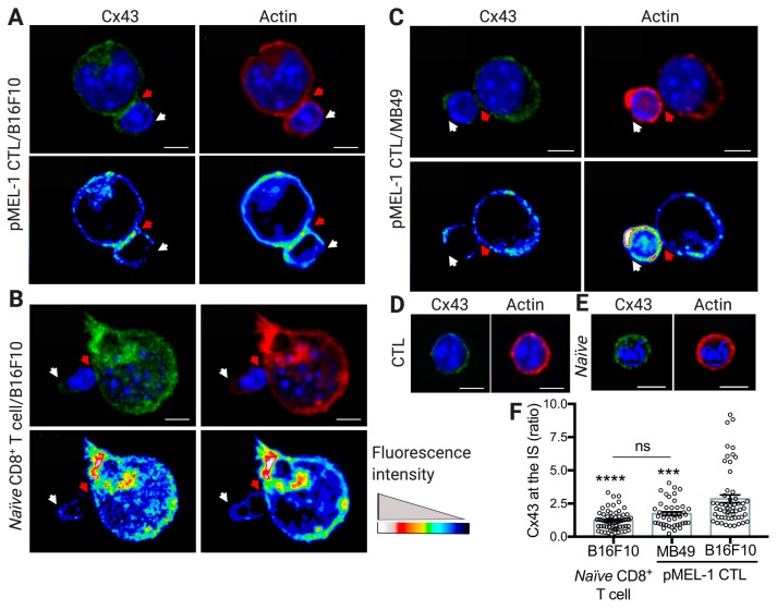
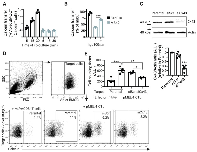
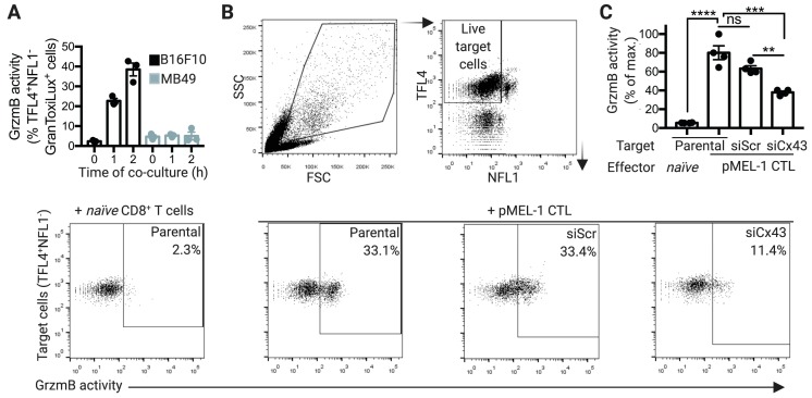
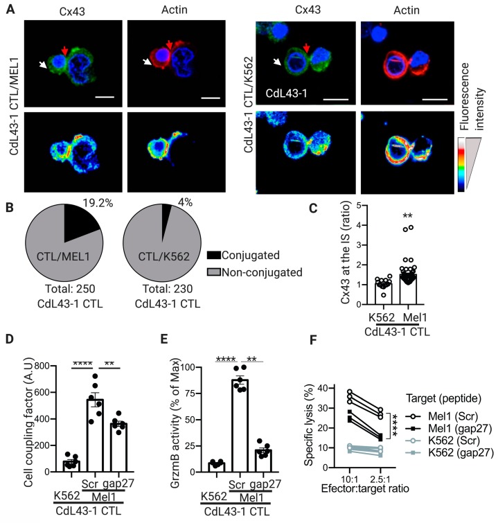
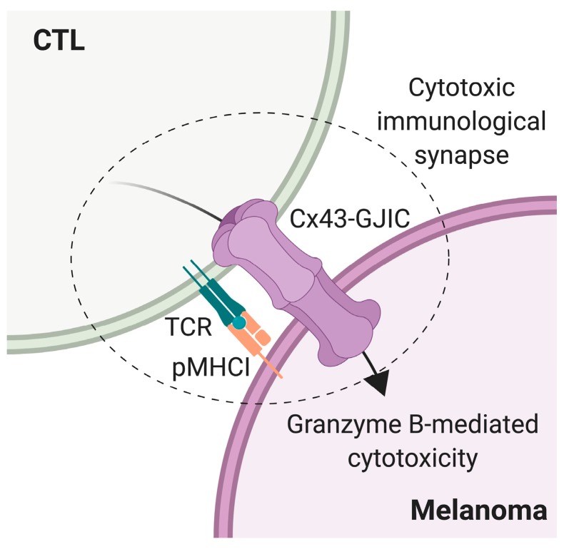
Upon tumor antigen recognition, cytotoxic T lymphocytes (CTLs) and target cells form specialized supramolecular structures, called cytotoxic immunological synapses, which are required for polarized delivery of cytotoxic granules. In previous reports, we described the accumulation of connexin 43 (Cx43)-formed gap junctions (GJs) at natural killer (NK) cell-tumor cell cytotoxic immunological synapse. In this report, we demonstrate the functional role of Cx43-GJs at the cytotoxic immunological synapse established between CTLs and melanoma cells during cytotoxicity. Using confocal microscopy, we evaluated Cx43 polarization to the contact site between CTLs isolated from pMEL-1 mice and B16F10 melanoma cells. We knocked down Cx43 expression in B16F10 cells and evaluated its role in the formation of functional GJs and the cytotoxic activity of CTLs, by calcein transfer and granzyme B activity assays, respectively. We found that Cx43 localizes at CTL/B16F10 intercellular contact sites via an antigen-dependent process. We also found that pMEL-1 CTLs but not wild-type naïve CD8+ T cells established functional GJs with B16F10 cells. Interestingly, we observed that Cx43-GJs were required for an efficient granzyme B activity in target B16F10 cells. Using an HLA-A2-restricted/MART-1-specific CD8+ T-cell clone, we confirmed these observations in human cells. Our results suggest that Cx43-channels are relevant components of cytotoxic immunological synapses and potentiate CTL-mediated tumor cell killing.

Keywords: connexin 43; cytotoxic T lymphocyte; cytotoxic immunological synapse; gap junctions; melanoma.


Conflict of interest statement

The authors declare no conflict of interest.

Figures

Figure 1
Figure 1
Connexin-43 (Cx43) accumulates at the pMEL-1 cytotoxic T lymphocyte (CTL)/B16F10 melanoma cell contact site upon cytotoxic immunological synapse (IS) formation. Representative images for cell conjugates formed after 30 min of co-culture of pMEL-1 CTLs or naïve CD8+ T cells with wheat germ agglutinin (WGA) pre-loaded (not shown) target (A,B) B16F10 cells or (C) MB49 cells. The distributions of both Cx43 (green) and F-actin (rhodamine-phalloidin, red) at the cytotoxic IS are indicated by red arrows and were analyzed by immunofluorescence and confocal microscopy. (AE) Hoechst nuclear staining is shown in blue. Representative images of Cx43 and actin staining in (D) unconjugated pMEL-1 CTL and (E) naïve CD8+ T cell. (AC) Pseudocolor images for the distribution of Cx43 and F-actin are shown at the bottom of each figure. (AE) Scale bars = 5 μm. (F) Quantification of Cx43 accumulation at the cytotoxic IS as ratio of Cx43 fluorescence intensity in the cell-cell contact site (red arrows) versus the opposite site (white arrows). *** p < 0.001, **** p < 0.0001, versus CTL/B16F10 conjugates; ns, non-significant (one-way ANOVA, Tukey’s multiple comparison test); n = approximately 60 cell conjugates by condition, two independent experiments.
Figure 2
Figure 2
pMEL-1 cytotoxic T lymphocytes (CTLs) form functional connexin-43 (Cx43)-mediated gap junction (GJ) communications with B16F10 melanoma cells. (A) B16F10 or MB49 cells were pre-loaded with the CellTracker Violet BMQC and co-cultured for different time points (as indicated) with calcein-AM pre-loaded pMEL-1 CTLs, at a 1:5 ratio. Calcein transfer from effector to target tumor cells was assessed by flow cytometry. The bar graph shows Violet BMQC+calcein+ cells. (B) Calcein transfer from pMEL-1 CTLs to target tumor cells was evaluated as described before, after 30 min of co-culture. MB49 cells were pre-loaded or not with hgp10025–33 peptide before co-culturing with pMEL-1 CTLs. The bar graph shows Violet BMQC+calcein+ cells as a percentage of the maximum calcein transfer. (C) B16F10 cells were transfected with siRNAs against Cx43 (siCx43) or control-scrambled siRNAs (siScr). The expression of Cx43 and actin was assessed three days after transfection by Western blot in transfected or non-transfected (parental) B16F10 cells, and Cx43/actin ratios were quantified by ImageJ software. The bar graph at the bottom shows the average of Cx43 expression depicted as Cx43/actin ratio relative to parental untransfected cells (n = 5 independent experiments). (D) Representative dot plots showing the strategy for Cx43-GJ communication measuring. Target (parental, siScr- or siCx43-transfected B16F10) cells were pre-loaded with the CellTracker Violet BMQC and co-cultured for 30 min with calcein-AM pre-loaded effector cells (naïve CD8+ T cells or pMEL-1 CTLs), at a 1:5 ratio. Calcein transfer from effector to target tumor cells was assessed by flow cytometry. The numbers in the dot plots represent the percentage of Violet BMQC+calcein+ cells. (E) The bar graph shows the cell coupling factor calculated as (%Violet BMQC+calcein+ cells × Calcein MFI of Calcein+ cells)/100. * p < 0.05; ** p < 0.01; *** p < 0.001; ns = non-significant (one-way ANOVA, Tukey’s multiple comparison test). A.U., arbitrary units.
Figure 3
Figure 3
Connexin-43 gap junctions (Cx43-GJs) are required for granzyme b (GrzmB)-mediated cytotoxicity of pMEL-1 cytotoxic T lymphocytes (CTLs) against B16F10 melanoma cells. (A) B16F10 or MB49 cells were pre-stained with TFL4 (CellTracker) and NFL1 (viability marker) and co-cultured for different times (as indicated) with pMEL-1 CTLs, at a 1:5 ratio, in the presence of a permeable fluorogenic substrate for GrzmB (GranToxiLux; GrzmB activity). GrzmB activity was evaluated on TFL4+NFL1 target tumor cells by flow cytometry. (B) Representative dot plots showing the strategy for measuring GrzmB activity in target tumor cells co-cultured with effector cells. Target cells (parental, siScr- or siCx43-transfected B16F10) were pre-stained with TFL4 and NFL1 and co-cultured for 2 h with effector cells (naïve CD8+ T cells or pMEL-1 CTLs), at a 1:5 ratio, in the presence of GranToxiLux substrate. The numbers in the dot plots represent the percentage of TFL4+NFL1GranToxiLux+ cells. (C) The bar graph shows the GrzmB activity in target tumor cells (TFL4+NFL1GranToxiLux+) as a percentage of the maximum (target tumor cell: parental/effector cell: pMEL-1 CTL); n = 4 independent experiments. ** p < 0.01; *** p < 0.001; **** p < 0.0001; ns = non-significant (one-way ANOVA, Tukey’s multiple comparison test).
Figure 4
Figure 4
Connexin-43 (Cx43) localizes at cytotoxic immunological synapses (IS) and contributes to cell coupling and granzyme b (GrzmB)-mediated cytotoxicity of human cytotoxic T lymphocytes (CTLs) against melanoma cells. (A) Representative images for cell conjugates formed after 30 min of co-culture of HLA-A2-restricted/MART-1-specific CD8+ T-cell clone (CdL43-1 CTL) with wheat germ agglutinin (WGA) pre-loaded (not shown) target Mel1 cells (left) or K562 cells (right, negative control). The distributions of both Cx43 (green) and F-actin (rhodamine-phalloidin, red) at the cytotoxic IS are indicated by red arrows and were analyzed by immunofluorescence and confocal microscopy. Hoechst nuclear staining is shown in blue. Pseudocolor images of the distribution for Cx43 and F-actin are shown at the bottom of each figure. Scale bars = 5 μm. (B) Quantification of the percentage of CdL43-1 CTLs conjugated to target tumor cells after co-culture with Mel1 (left) or K562 (right) cells. (C) Quantification of Cx43 accumulation at the cytotoxic IS as ratio of Cx43 fluorescence intensity in the cell-cell contact site (red arrows) versus at the opposite site (white arrows). (D) Target (K562 or Mel1) tumor cells were pre-loaded with the CellTracker Violet BMQC and co-cultured for 1 hour with calcein-AM pre-loaded CdL43-1 CTL effector cells at 1:3 ratios, in the presence of Cx43 mimetic inhibitory (gap27) or control (Scr) peptides (300 μM). Calcein transfer from effector to target tumor cells was assessed by flow cytometry. The bar graph shows the cell coupling factor calculated as (%Violet BMQC+calcein+ cells × Calcein MFI of Calcein+ cells)/100. (E) Mel1 or K562 cells were pre-stained with TFL4 (CellTracker) and NFL1 (viability marker) and co-cultured for 2 h with CdL43- CTLs, at a 1:3 ratio, in the presence of a permeable fluorogenic substrate for GrzmB (GranToxiLux; GrzmB activity), and gap27 or Scr peptides. GrzmB activity was evaluated on TFL4+NFL1 target tumor cells by flow cytometry. The bar graph shows the GrzmB activity in target tumor cells (TFL4+NFL1GranToxiLux+) as a percentage of the maximum (target tumor cell: Mel1; peptide: Scr). (F) CdL43-1 CTLs were co-cultured with Mel1 or K562 cells at different effector:target cell ratios in the presence of gap27 or Scr peptides. Cytotoxicity was assessed by conventional 51Cr release assays. The results are plotted as a percentage of specific lysis. All the results are from three independent experiments. ** p < 0.01; **** p < 0.0001 (one-way ANOVA, Tukey’s multiple comparison test, or two-tailed Student’s t-test (C)).
Figure 5
Figure 5
Model of connexin-43 (Cx43)-gap junction (GJ) intercellular communication (Cx43-GJIC) in the cytotoxic T lymphocyte (CTL)-mediated melanoma cell killing. Upon cytotoxic immunological synapse formation, initiated by T cell receptor (TCR)-cognate antigenic peptide-MHC I (pMHCI) interaction, Cx43-GJ channels polarize to the CTL-melanoma cell contact site, allowing Cx43-mediated cell coupling and the efficient activity of CTL-derived granzyme B on the target tumor cell.

Similar articles

Cited by

References

    1. Nielsen M.S., Axelsen L.N., Sorgen P.L., Verma V., Delmar M., Holstein-Rathlou N.H. Gap junctions. Compr. Physiol. 2012;2:1981–2035. - PMC - PubMed
    1. Sáez P.J., Shoji K.F., Aguirre A., Sáez J.C. Regulation of hemichannels and gap junction channels by cytokines in antigen-presenting cells. Mediat. Inflamm. 2014;2014:742734. doi: 10.1155/2014/742734. - DOI - PMC - PubMed
    1. Neijssen J., Pang B., Neefjes J. Gap junction-mediated intercellular communication in the immune system. Prog. Biophys. Mol. Biol. 2007;94:207–218. doi: 10.1016/j.pbiomolbio.2007.03.008. - DOI - PubMed
    1. Oviedo-Orta E., Hoy T., Evans W.H. Gap junctions and connexin-mediated communication in the immune system. Biochim. Biophys. Acta. 2004;1662:102–112. doi: 10.1016/j.bbamem.2003.10.021. - DOI - PubMed
    1. Oviedo-Orta E., Perreau M., Evans W.H., Potolicchio I. Control of the proliferation of activated CD4+ T cells by connexins. J. Leukoc. Biol. 2010;88:79–86. doi: 10.1189/jlb.0909613. - DOI - PubMed