Review
. 2019 Sep 23;6(4):34.
doi: 10.3390/jcdd6040034.

Ca2+ Signaling in Cardiac Fibroblasts and Fibrosis-Associated Heart Diseases

Affiliations
Review

Ca2+ Signaling in Cardiac Fibroblasts and Fibrosis-Associated Heart Diseases

Jianlin Feng et al. J Cardiovasc Dev Dis. .

Abstract

Cardiac fibrosis is the excessive deposition of extracellular matrix proteins by cardiac fibroblasts and myofibroblasts, and is a hallmark feature of most heart diseases, including arrhythmia, hypertrophy, and heart failure. This maladaptive process occurs in response to a variety of stimuli, including myocardial injury, inflammation, and mechanical overload. There are multiple signaling pathways and various cell types that influence the fibrogenesis cascade. Fibroblasts and myofibroblasts are central effectors. Although it is clear that Ca2+ signaling plays a vital role in this pathological process, what contributes to Ca2+ signaling in fibroblasts and myofibroblasts is still not wholly understood, chiefly because of the large and diverse number of receptors, transporters, and ion channels that influence intracellular Ca2+ signaling. Intracellular Ca2+ signals are generated by Ca2+ release from intracellular Ca2+ stores and by Ca2+ entry through a multitude of Ca2+-permeable ion channels in the plasma membrane. Over the past decade, the transient receptor potential (TRP) channels have emerged as one of the most important families of ion channels mediating Ca2+ signaling in cardiac fibroblasts. TRP channels are a superfamily of non-voltage-gated, Ca2+-permeable non-selective cation channels. Their ability to respond to various stimulating cues makes TRP channels effective sensors of the many different pathophysiological events that stimulate cardiac fibrogenesis. This review focuses on the mechanisms of Ca2+ signaling in fibroblast differentiation and fibrosis-associated heart diseases and will highlight recent advances in the understanding of the roles that TRP and other Ca2+-permeable channels play in cardiac fibrosis.

Keywords: Ca2+ signaling pathways; TRP channels; cardiac fibroblasts; cardiac fibrosis; ion channels.


Conflict of interest statement

The authors declare no conflict of interest.

Figures
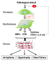
Figure 1
Figure 1
Schematic diagram illustrating cardiac fibrogenesis cascade and fibrosis-associated heart diseases. Pathological stresses stimulate fibroblasts to differentiate into myofibroblasts, during which an increase in intracellular Ca2+ plays a key role. Myofibroblasts synthesize and secrete extracellular matrix (ECM) proteins, matrix metalloproteinases (MMPs), and cytokines such as TGFβ1. Excessive deposition of ECM proteins results in cardiac fibrosis. This fibrogenesis cascade is perpetuated by TGFβ1 produced by myofibroblasts. Cardiac fibrosis is involved in a variety of pathological remodeling, which can lead to arrhythmia, hypertrophy and heart failure.
Figure 2
Figure 2
Ca2+ signaling mechanisms in cardiac fibroblasts and myofibroblasts. Intracellular Ca2+ levels are finely controlled by: (1) Ca2+ entry through Ca2+-permeable channels in the plasma membrane, including TRP channels, P2X receptors, and Orai/STIM channels; (2) Ca2+ release via IP3Rs in the ER; and (3) Ca2+ extrusion pumps, including SERCA in the ER and PMCA4 in the plasma membrane. Pathophysiological stimuli can activate Gq-coupled receptors to induce Ca2+ release, which is secondarily followed by Ca2+ entry. Receptor stimulation can also directly activate Ca2+-permeable (TRP) channels in the plasma membrane. Other ion channels, including voltage-gated K+ (Kv) channels, Kir channels, Ca2+-activated potassium (KCa) channels, and NaV channels, may influence the resting membrane potential or depolarization to indirectly influence Ca2+ entry in fibroblasts and myofibroblasts. An increase in intracellular Ca2+ activates the calcineurin/NFAT (CN/NFAT), ERK1/2, ROS/RhoA, and sFRP2 pathways to promote profibrotic gene expression.

Similar articles

  • Transient receptor potential (TRP) channels and cardiac fibrosis.
    Yue Z, Zhang Y, Xie J, Jiang J, Yue L. Yue Z, et al. Curr Top Med Chem. 2013;13(3):270-82. doi: 10.2174/1568026611313030005. Curr Top Med Chem. 2013. PMID: 23432060 Free PMC article. Review.
  • TRP Channels in the Heart.
    Freichel M, Berlin M, Schürger A, Mathar I, Bacmeister L, Medert R, Frede W, Marx A, Segin S, Londoño JEC. Freichel M, et al. In: Emir TLR, editor. Neurobiology of TRP Channels. Boca Raton (FL): CRC Press/Taylor & Francis; 2017. Chapter 9. In: Emir TLR, editor. Neurobiology of TRP Channels. Boca Raton (FL): CRC Press/Taylor & Francis; 2017. Chapter 9. PMID: 29356479 Free Books & Documents. Review.
  • Regulation and Role of Store-Operated Ca2+ Entry in Cellular Proliferation.
    Hodeify R, Yu F, Courjaret R, Nader N, Dib M, Sun L, Adap E, Hubrack S, Machaca K. Hodeify R, et al. In: Kozak JA, Putney JW Jr, editors. Calcium Entry Channels in Non-Excitable Cells. Boca Raton (FL): CRC Press/Taylor & Francis; 2018. Chapter 12. In: Kozak JA, Putney JW Jr, editors. Calcium Entry Channels in Non-Excitable Cells. Boca Raton (FL): CRC Press/Taylor & Francis; 2018. Chapter 12. PMID: 30299656 Free Books & Documents. Review.
  • Emerging role of transient receptor potential (TRP) ion channels in cardiac fibroblast pathophysiology.
    Gwanyanya A, Mubagwa K. Gwanyanya A, et al. Front Physiol. 2022 Oct 6;13:968393. doi: 10.3389/fphys.2022.968393. eCollection 2022. Front Physiol. 2022. PMID: 36277180 Free PMC article. Review.
  • Progress on role of ion channels of cardiac fibroblasts in fibrosis.
    Xing C, Bao L, Li W, Fan H. Xing C, et al. Front Physiol. 2023 Mar 9;14:1138306. doi: 10.3389/fphys.2023.1138306. eCollection 2023. Front Physiol. 2023. PMID: 36969589 Free PMC article. Review.

Cited by

References

    1. Weber K. Cardiac interstitium. In: Poole-Wilson P., Colucci W., Massie B., Chatterjee K., Coats A., editors. Heart Failure. Churchill Livingstone; New York, NY, USA: 1997. pp. 13–31.
    1. Gourdie R.G., Dimmeler S., Kohl P. Novel therapeutic strategies targeting fibroblasts and fibrosis in heart disease. Nat. Rev. Drug Discov. 2016;15:620–638. doi: 10.1038/nrd.2016.89. - DOI - PMC - PubMed
    1. Travers J.G., Kamal F.A., Robbins J., Yutzey K.E., Blaxall B.C. Cardiac Fibrosis: The Fibroblast Awakens. Circ. Res. 2016;118:1021–1040. doi: 10.1161/CIRCRESAHA.115.306565. - DOI - PMC - PubMed
    1. Ma Z.G., Yuan Y.P., Wu H.M., Zhang X., Tang Q.Z. Cardiac fibrosis: New insights into the pathogenesis. Int. J. Biol. Sci. 2018;14:1645–1657. doi: 10.7150/ijbs.28103. - DOI - PMC - PubMed
    1. Nguyen M.N., Kiriazis H., Gao X.M., Du X.J. Cardiac Fibrosis and Arrhythmogenesis. Compr. Physiol. 2017;7:1009–1049. doi: 10.1002/cphy.c160046. - DOI - PubMed

LinkOut - more resources