. 2019 Sep 23;11(10):889.
doi: 10.3390/v11100889.

Cytokine Effects on the Entry of Filovirus Envelope Pseudotyped Virus-Like Particles into Primary Human Macrophages

Affiliations

Cytokine Effects on the Entry of Filovirus Envelope Pseudotyped Virus-Like Particles into Primary Human Macrophages

Tzanko S Stantchev et al. Viruses. .

Abstract

Macrophages are one of the first and also a major site of filovirus replication and, in addition, are a source of multiple cytokines, presumed to play a critical role in the pathogenesis of the viral infection. Some of these cytokines are known to induce macrophage phenotypic changes in vitro, but how macrophage polarization may affect the cell susceptibility to filovirus entry remains largely unstudied. We generated different macrophage subsets using cytokine pre-treatment and subsequently tested their ability to fuse with beta-lactamase containing virus-like particles (VLP), pseudotyped with the surface glycoprotein of Ebola virus (EBOV) or the glycoproteins of other clinically relevant filovirus species. We found that pre-incubation of primary human monocyte-derived macrophages (MDM) with interleukin-10 (IL-10) significantly enhanced filovirus entry into cells obtained from multiple healthy donors, and the IL-10 effect was preserved in the presence of pro-inflammatory cytokines found to be elevated during EBOV disease. In contrast, fusion of IL-10-treated macrophages with influenza hemagglutinin/neuraminidase pseudotyped VLPs was unchanged or slightly reduced. Importantly, our in vitro data showing enhanced virus entry are consistent with the correlation established between elevated serum IL-10 and increased mortality in filovirus infected patients and also reveal a novel mechanism that may account for the IL-10-mediated increase in filovirus pathogenicity.

Keywords: Ebola virus (EBOV); cytokines; filoviruses; interleukin-10 (IL-10).


Conflict of interest statement

The authors declare no conflict of interest. The funders had no role in the design of the study; in the collection, analyses, or interpretation of data; in the writing of the manuscript, or in the decision to publish the results. This work represents the views of the authors and does not necessarily reflect those of the Food and Drug Administration, the National Institute of Allergy and Infectious Diseases/National Institutes of Health and/or the Uniformed Services University.

Figures
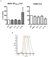
Figure 1
Figure 1
(A) IL-10, but not TNF-α, IL-4 or IL-13, significantly enhance EBOVKikwit glycoprotein(s) (GP) pseudotyped virus-like particles (VLP) entry in primary human monocyte-derived macrophages (MDM). MDM from three healthy donors were pre-treated with TNF-α, IL-4, IL-13 or IL10 (all at concentrations of 20 ng/mL) for 48 h prior to incubation with EBOVKikwit GP or influenza hemagglutinin/neuraminidase (HA/NA) pseudotyped VLP for 3.5 h at 37 °C. Subsequently, the cells were processed at room temperature, washed twice, loaded with CCF2/AM fluorescent dye for 1.5 h, washed again (2 times), incubated overnight to allow the cleavage of CCF2/AM by the VLP introduced β-lactamase (BlaM) and fixed in 1.6% paraformaldehyde. The extent of CCF2/AM cleavage, resulting in the change of the emission spectrum from 530 nm (green) to 460 nm (blue), was assessed by Laser Scanning Cytometry. The levels of VLP fusion with mock-treated MDM (treated with an equivalent volume of cytokine re-suspension buffer) were assumed to be 100%. Each treatment condition was performed in triplicate wells and the mean value(s) was used to calculate “% of control”. The data from the individual experiments, used to generate Figure 1A, are included in Table S1A through Table S1H (donor #1, donor #2 and donor #17). (B) TNF-α, IL-4, IL-13 or IL-10 affect CD206 expression in a cytokine-specific manner. Aliquots of MDM from one of the donors presented in Figure 1A were pre-incubated with TNF-α, IL-4, IL-13 or IL10 (20 ng/mL), detached by incubation in EDTA/EGTA buffer at 4°C and gentle scraping, and then labeled with phycoerythrin-conjugated anti-CD206 monoclonal antibody. CD206 cell surface expression was evaluated using BD FACSCanto II cell analyzer. The gray, yellow, red, green and purple lines represent control (mock treated), TNF-α, IL-10, IL-4 and/or IL-13 pre-incubated cells, respectively. The dash lines with the same colors represent the relevant isotype controls.
Figure 2
Figure 2
(A) IL-10 enhances entry of full-length EBOVKikwit GP pseudotyped VLPs in MDM from multiple donors (n = 25). MDM were pre-incubated with IL10 (20 ng/mL) for 48 h prior to infection. The cells were processed and the EBOVKikwit VLP (solid diamonds) entry/fusion was analyzed by Laser Scanning Cytometry as described in the Materials and Methods and Figure 1. BlaM-containing virus particles, pseudotyped with influenza HA/NA (white triangles), served as controls. The levels of virus entry in mock-treated MDM (with an equivalent volume of cytokine re-suspension buffer) were assumed to be 100%. The graph summarizes data from multiple experiments and includes the results for the IL-10 effect on EBOVKikwit VLP entry at a concentration of 20 ng/mL in the MDM of donors also used to generate the data presented in Figure 1, Figure 2B, Figure 3, Figure 4A–C, Supplemental Figure S1 and Supplemental Figure S2. The data, including statistical analysis, from the multiple experiments, used to generate Figure 2A, are summarized in Table S1A,B. (B) Representative fluorescent images, generated by the iCys Laser Scanning Cytometer using MDM from one of the donors, show increased numbers of cells fused with the EBOV GP VLPs (blue cells) after IL-10 treatment. No significant difference was observed in the background fluorescence of uninfected IL-10 or mock treated cells. (C) IL-10 induced enhancement is independent of the EBOVKikwit GP Δmucin domain. MDM from 4 healthy donors were infected in parallel with VLPs pseudotyped with either full length EBOVKikwit GP (solid diamonds) or EBOVKikwit Δ mucin GP (black and white diamonds). The cells were processed and the “% of control” was calculated as described for Figure 1. The data from the individual experiments, used to generate this figure, are included in Table S1A,B,I (donors #10, #11, #13, #15 and #29). (D) Donor-dependent effect of TNF-α on EBOVKikwit GP VLP entry into primary human MDM. In several different experiments, cells from a total of seven healthy donors were incubated with TNF-α (20 ng/mL) for 48 h, infected with EBOVKikwit GP VLP (solid circles) for 3.5 h, loaded with CCF2/AM fluorescent dye and processed as described in Materials and Methods. The subsequent evaluation of VLP fusion was performed either by Laser Scanning Cytometry (6 donors) or Flow cytometry (1 donor). The levels of VLP fusion with mock-treated MDM (equivalent volume of cytokine re-suspension buffer) were assumed to be 100%. VLPs pseudotyped with influenza HA/NA served as controls (open triangles). The data from the individual experiments, used to generate Figure 2D are summarized in Table S1C,F.
Figure 2
Figure 2
(A) IL-10 enhances entry of full-length EBOVKikwit GP pseudotyped VLPs in MDM from multiple donors (n = 25). MDM were pre-incubated with IL10 (20 ng/mL) for 48 h prior to infection. The cells were processed and the EBOVKikwit VLP (solid diamonds) entry/fusion was analyzed by Laser Scanning Cytometry as described in the Materials and Methods and Figure 1. BlaM-containing virus particles, pseudotyped with influenza HA/NA (white triangles), served as controls. The levels of virus entry in mock-treated MDM (with an equivalent volume of cytokine re-suspension buffer) were assumed to be 100%. The graph summarizes data from multiple experiments and includes the results for the IL-10 effect on EBOVKikwit VLP entry at a concentration of 20 ng/mL in the MDM of donors also used to generate the data presented in Figure 1, Figure 2B, Figure 3, Figure 4A–C, Supplemental Figure S1 and Supplemental Figure S2. The data, including statistical analysis, from the multiple experiments, used to generate Figure 2A, are summarized in Table S1A,B. (B) Representative fluorescent images, generated by the iCys Laser Scanning Cytometer using MDM from one of the donors, show increased numbers of cells fused with the EBOV GP VLPs (blue cells) after IL-10 treatment. No significant difference was observed in the background fluorescence of uninfected IL-10 or mock treated cells. (C) IL-10 induced enhancement is independent of the EBOVKikwit GP Δmucin domain. MDM from 4 healthy donors were infected in parallel with VLPs pseudotyped with either full length EBOVKikwit GP (solid diamonds) or EBOVKikwit Δ mucin GP (black and white diamonds). The cells were processed and the “% of control” was calculated as described for Figure 1. The data from the individual experiments, used to generate this figure, are included in Table S1A,B,I (donors #10, #11, #13, #15 and #29). (D) Donor-dependent effect of TNF-α on EBOVKikwit GP VLP entry into primary human MDM. In several different experiments, cells from a total of seven healthy donors were incubated with TNF-α (20 ng/mL) for 48 h, infected with EBOVKikwit GP VLP (solid circles) for 3.5 h, loaded with CCF2/AM fluorescent dye and processed as described in Materials and Methods. The subsequent evaluation of VLP fusion was performed either by Laser Scanning Cytometry (6 donors) or Flow cytometry (1 donor). The levels of VLP fusion with mock-treated MDM (equivalent volume of cytokine re-suspension buffer) were assumed to be 100%. VLPs pseudotyped with influenza HA/NA served as controls (open triangles). The data from the individual experiments, used to generate Figure 2D are summarized in Table S1C,F.
Figure 3
Figure 3
IL-10 enhances fusion of primary MDM with VLPs pseudotyped with envelope glycoproteins from all clinically significant filovirus species. Cells were pre-incubated with 20 ng/mL IL-10 or DPBS supplemented with 0.5% HSA (mock treated), infected, loaded with CCF2/AM, processed and analyzed by Flow cytometry as described in Materials and Methods. The figure summarizes the results from several different experiments, utilizing MDM from a total of seven different donors. In each experiment, MDM were infected with EBOVKikwit GP VLP (solid diamonds) in parallel with one or more different VLP types, pseudotyped with the surface glycoprotein of one of the indicated filovirus species. Data from one of the individual donors are presented in Supplemental Figure S3. Influenza HA/NA pseudotyped virus particles served as a control (open triangles).
Figure 4
Figure 4
IL-10 enhances fusion of primary MDM with VLPs pseudotyped with EBOV GP in a dose-dependent manner, but has no significant effect or slightly inhibits entry of VLPs pseudotyped with influenza HA/NA. MDM from three different healthy donors were pre-incubated for 48 h with increasing concentrations of IL-10 and infected for 3.5 h with VLPs pseudotyped with the surface glycoproteins of the Kikwit (Panel A), Mayinga (Panel B) or Makona (Panel C) strains, respectively. Subsequently, the cells were washed, loaded with the fluorescent dye CCF2/AM and prepared for analysis by Laser Scanning Cytometry as described in Material and Methods. * p ≤ 0.05; ** p ≤ 0.01; *** p ≤ 0.001.
Figure 4
Figure 4
IL-10 enhances fusion of primary MDM with VLPs pseudotyped with EBOV GP in a dose-dependent manner, but has no significant effect or slightly inhibits entry of VLPs pseudotyped with influenza HA/NA. MDM from three different healthy donors were pre-incubated for 48 h with increasing concentrations of IL-10 and infected for 3.5 h with VLPs pseudotyped with the surface glycoproteins of the Kikwit (Panel A), Mayinga (Panel B) or Makona (Panel C) strains, respectively. Subsequently, the cells were washed, loaded with the fluorescent dye CCF2/AM and prepared for analysis by Laser Scanning Cytometry as described in Material and Methods. * p ≤ 0.05; ** p ≤ 0.01; *** p ≤ 0.001.
Figure 5
Figure 5
Increased binding of EBOV GP to IL-10 treated primary human MDM. (A) Cells were pre-treated for 48 h with IL-10 and incubated for 1.5 h at 4 °C in binding buffer (0.5% HSA in Ca2+ and Mg2+ containing DPBS) with a recombinant protein, consisting of the EBOV GP receptor binding domain (GP1 residues 57 to 149), fused to the Fc portion of rabbit IgG. MDM incubated with the binding deficient construct 4merRBR-Fc served as a negative control (red line). Subsequently, the MDM were washed, labeled with FITC conjugated polyclonal goat anti-rabbit antibody, fixed with paraformaldehyde and analyzed by Flow Cytometry. The staining indexes for the mock and IL-10 treated pairs of negative and positive samples are 0.3593 and 0.7086, respectively (for details regarding the staining indexes calculation please refer to Material and Methods). (B) Alternatively, MDM were incubated for 2.5 h at 4 °C with GFP-containing (VP40-GFP) VLPs pseudotyped with full length EBOVKikwit GP (blue line). Full length EBOV GP- pseudotyped VLPs, which do not contain GFP, served as a negative control (red line). The staining indexes for the mock and IL-10 treated pairs negative and positive samples are 0.8444 and 1.8228, respectively.
Figure 6
Figure 6
The enhancing effect of IL-10 on EBOV VLP entry is preserved in the presence of pro-inflammatory cytokines. (A) MDM were pre-treated with IL-10, TNF-α or a combination of both for 48 h (IL-10 and TNF-α were used at a concentration of 20 ng/mL) and infected with EBOVKikwit GP VLP. The first columns from the left represent mock infected cells. There was no difference in the background fluorescence of mock-infected MDM incubated with cytokines or in medium alone. (B) MDM were pre-incubated with IL-10 (0.5 ng/mL or 1 ng/mL) or IL-10 plus a cocktail of pro-inflammatory cytokines (TNF-α, 0.1ng/mL; MIP-1α, 0.1ng/mL; MIP-1β and MCP-1, 2ng/mL; IL-8, 0.5ng/mL) for 48 h before infection. After the 3.5 h infection period, the cells were prepared for analysis by Laser Scanning Cytometry as described in Materials and Methods. * p ≤ 0.05; ** p ≤ 0.01; *** p ≤ 0.001; ns = not significant.
Figure 6
Figure 6
The enhancing effect of IL-10 on EBOV VLP entry is preserved in the presence of pro-inflammatory cytokines. (A) MDM were pre-treated with IL-10, TNF-α or a combination of both for 48 h (IL-10 and TNF-α were used at a concentration of 20 ng/mL) and infected with EBOVKikwit GP VLP. The first columns from the left represent mock infected cells. There was no difference in the background fluorescence of mock-infected MDM incubated with cytokines or in medium alone. (B) MDM were pre-incubated with IL-10 (0.5 ng/mL or 1 ng/mL) or IL-10 plus a cocktail of pro-inflammatory cytokines (TNF-α, 0.1ng/mL; MIP-1α, 0.1ng/mL; MIP-1β and MCP-1, 2ng/mL; IL-8, 0.5ng/mL) for 48 h before infection. After the 3.5 h infection period, the cells were prepared for analysis by Laser Scanning Cytometry as described in Materials and Methods. * p ≤ 0.05; ** p ≤ 0.01; *** p ≤ 0.001; ns = not significant.
Figure 7
Figure 7
TGF-β1 enhances fusion of primary MDM with VLPs pseudotyped with EBOVKikwit GP. MDM were pre-incubated for 72 h in macrophage medium supplemented with TGF-β1 (2.5 or 5 ng/mL) or cytokine resuspension buffer (0.5% HSA DPBS) prior to infection with EBOVKikwit VLP or HA/NA VLP, respectively. The cells were processed and the VLP entry/fusion was analyzed by Laser Scanning Cytometry, as described in Materials and Methods. No significant difference was observed in the background fluorescence of uninfected TGF-β1 or mock treated cells. ** p ≤ 0.01; *** p ≤ 0.001; ns = not significant.

Similar articles

Cited by

References

    1. Villinger F., Rollin P.E., Brar S.S., Chikkala N.F., Winter J., Sundstrom J.B., Zaki S.R., Swanepoel R., Ansari A.A., Peters C.J. Markedly elevated levels of interferon (ifn)-gamma, ifn-alpha, interleukin (il)-2, il-10, and tumor necrosis factor-alpha associated with fatal ebola virus infection. J. Infect. Dis. 1999;179(Suppl. 1):S188–S191. doi: 10.1086/514283. - DOI - PubMed
    1. Baize S., Leroy E.M., Georges A.J., Georges-Courbot M.C., Capron M., Bedjabaga I., Lansoud-Soukate J., Mavoungou E. Inflammatory responses in ebola virus-infected patients. Clin. Exp. Immunol. 2002;128:163–168. doi: 10.1046/j.1365-2249.2002.01800.x. - DOI - PMC - PubMed
    1. Ruibal P., Oestereich L., Ludtke A., Becker-Ziaja B., Wozniak D.M., Kerber R., Korva M., Cabeza-Cabrerizo M., Bore J.A., Koundouno F.R., et al. Unique human immune signature of ebola virus disease in guinea. Nature. 2016;533:100–104. doi: 10.1038/nature17949. - DOI - PMC - PubMed
    1. Leroy E.M., Baize S., Debre P., Lansoud-Soukate J., Mavoungou E. Early immune responses accompanying human asymptomatic ebola infections. Clin. Exp. Immunol. 2001;124:453–460. doi: 10.1046/j.1365-2249.2001.01517.x. - DOI - PMC - PubMed
    1. Leroy E.M., Baize S., Volchkov V.E., Fisher-Hoch S.P., Georges-Courbot M.C., Lansoud-Soukate J., Capron M., Debre P., McCormick J.B., Georges A.J. Human asymptomatic ebola infection and strong inflammatory response. Lancet. 2000;355:2210–2215. doi: 10.1016/S0140-6736(00)02405-3. - DOI - PubMed

Publication types

MeSH terms