. 2019 Sep 23;50(1):69.
doi: 10.1186/s13567-019-0687-2.

Haemophilus parasuis VtaA2 is involved in adhesion to extracellular proteins

Affiliations

Haemophilus parasuis VtaA2 is involved in adhesion to extracellular proteins

Mar Costa-Hurtado et al. Vet Res. .

Abstract

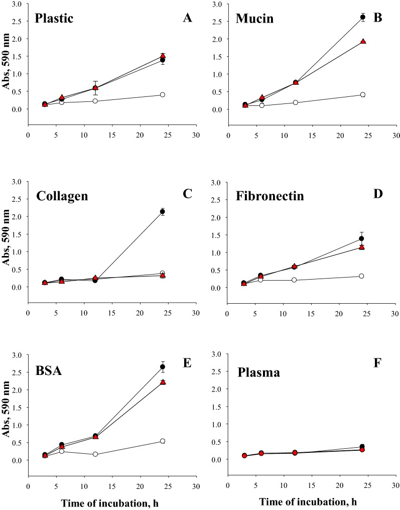
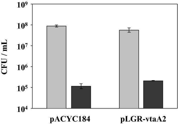
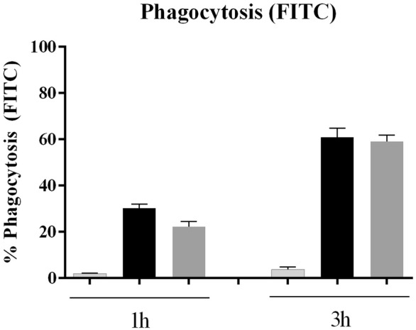
Haemophilus parasuis is part of the microbiota of the upper respiratory tract in swine. However, virulent strains can cause a systemic disease known as Glässer's disease. Several virulence factors have been described in H. parasuis including the virulence-associated trimeric autotransporters (VtaAs). VtaA2 is up-regulated during infection and is only found in virulent strains. In order to determine its biological function, the vtaA2 gene was cloned with its native promotor region in pACYC184, and the transformed Escherichia coli was used to perform functional in vitro assays. VtaA2 was found to have a role in attachment to plastic, mucin, BSA, fibronectin and collagen. As other VtaAs from H. parasuis, the passenger domain of VtaA2 contains collagen domains. In order to examine the contribution of the collagen repeats to VtaA2 function, a recombinant vtaA2 without the central collagen domains was obtained and named vtaA2OL. VtaA2OL showed similar capacity than VtaA2 to adhere to plastic, mucin, BSA, fibronectin and plasma but a reduced capacity to adhere to collagen, suggesting that the collagen domains of VtaA2 are involved in collagen attachment. No function in cell adhesion and invasion to epithelial alveolar cell line A549 or unspecific binding to primary alveolar macrophages was found. Likewise VtaA2 had no role in serum or phagocytosis resistance. We propose that VtaA2 mediates adherence to the host by binding to the mucin, found in the upper respiratory tract mucus, and to the extracellular matrix proteins, present in the connective tissue of systemic sites, such as the serosa.


Conflict of interest statement

The authors declare that they have no competing interests.

Figures

Figure 1
Figure 1
Kinetics of attachment to different substrates of E. coli clones expressing VtaA2 and VtaA2OL. Wells were coated with mucin (B), collagen (C), fibronectin (D), BSA (E), or plasma (F), or were left uncoated (plastic A). Plates were inoculated and incubated under static conditions. Attachment was measured at different time points. Black circles: BL21 (pLGR-vtaA2); red triangles: BL21 (pLGR-vtaA2OL); and white circles BL21 (pACY184) (control). Bars represent the standard deviation of the mean Abs590 from triplicate wells.
Figure 2
Figure 2
VtaA2 has no role in the adhesion to lung epithelial cells A549 cells. Adhesion of the clones E. coli BL21 (pLGR-vtaA2) and (pACYC184) to lung epithelial cells A549 after 1 h of incubation (black bars). Concentration of bacteria in the wells at the beginning of the assay are shown in gray bars. Bars represent the mean ± standard deviation of the concentration of bacteria from duplicate wells. Results are representative of 2 independent experiments.
Figure 3
Figure 3
VtaA2 has no role in the bacterial interaction with PAMs; bacteria stained with FITC. E. coli BL21 clones (pLGR-vtaA2; dark grey bars) and empty vector pACYC184 (black bars) were stained with FITC, incubated for 1 h or 3 h with PAMs and the interaction with the macrophages was analyzed by flow cytometry. Results are shown as the percentage of fluorescent macrophages. Macrophages without bacteria were included as negative (light grey bars). Bars represent the mean ± standard deviation.
Figure 4
Figure 4
VtaA2 has no role in the bacterial interaction with PAMs; bacteria stained with SYTO9. E. coli BL21 clones (pLGR-vtaA2; dark grey bars) and empty vector pACYC184 (black bars) were stained with SYTO9, incubated for 1 h with PAMs and the interaction with the macrophages was analyzed by flow cytometry. Results are shown as the percentage of fluorescent macrophages. Macrophages without bacteria were included as negative (light grey bars). Bars represent the mean ± standard deviation.
Figure 5
Figure 5
VtaA2 and VtA2OL are produced under in vitro conditions. Detection by immunoblot of VtaA2 and its truncated version VtaA2OL from E. coli BL21 (pLGR-vtaA2) and E. coli BL21 (pLGR-vtaA2OL) respectively. Both proteins were extracted from bacterial pellets grown in LB-Cm30 or incubated with CDMEM. Molecular size standards in kDa are shown on the left. Protein extraction from E. coli BL21 transformed with pACYC184 empty plasmid was used as negative control of expression.

References

    1. Møller K, Kilian M. V factor-dependent members of the family Pasteurellaceae in the porcine upper respiratory tract. J Clin Microbiol. 1990;28:2711–2716. - PMC - PubMed
    1. Vahle JL, Haynes JS, Andrews JJ. Experimental reproduction of Haemophilus parasuis infection in swine: clinical, bacteriological, and morphologic findings. J Vet Diagn Invest. 1995;7:476–480. doi: 10.1177/104063879500700409. - DOI - PubMed
    1. Holtkamp D, Rotto H, Garcia R. Economic cost of major health challenges in large US swine production systems. Swine News Newslett. 2007;30:4.
    1. Bouchet B, Vanier G, Jacques M, Auger E, Gottschalk M. Studies on the interactions of Haemophilus parasuis with porcine epithelial tracheal cells: limited role of LOS in apoptosis and pro-inflammatory cytokine release. Microb Pathog. 2009;46:108–113. doi: 10.1016/j.micpath.2008.10.008. - DOI - PubMed
    1. Aragon V, Bouchet B, Gottschalk M. Invasion of endothelial cells by systemic and nasal strains of Haemophilus parasuis. Vet J. 2009;186:264–267. doi: 10.1016/j.tvjl.2009.08.013. - DOI - PubMed

MeSH terms

Substances