Review
. 2020 Mar 2;10(3):a037259.
doi: 10.1101/cshperspect.a037259.

Liver Tropism in Cancer: The Hepatic Metastatic Niche

Affiliations
Review

Liver Tropism in Cancer: The Hepatic Metastatic Niche

Ainhoa Mielgo et al. Cold Spring Harb Perspect Med. .

Abstract

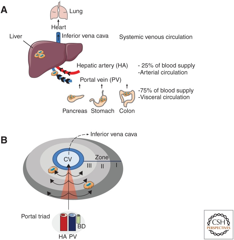
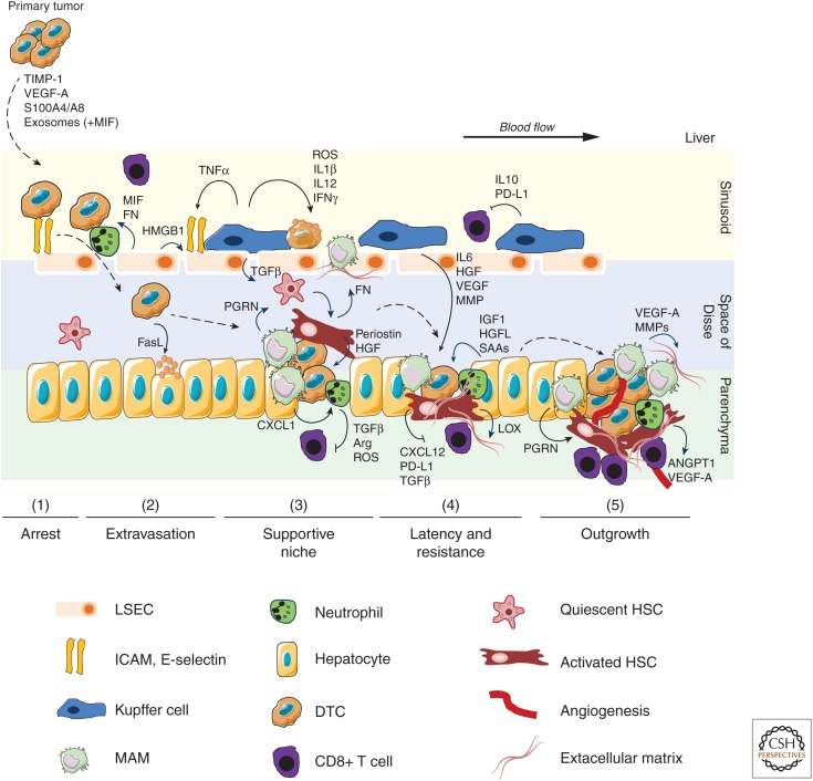
The liver is the largest organ in the human body and is prone for cancer metastasis. Although the metastatic pattern can differ depending on the cancer type, the liver is the organ to which cancer cells most frequently metastasize for the majority of prevalent malignancies. The liver is unique in several aspects: the vascular structure is highly permeable and has unparalleled dual blood connectivity, and the hepatic tissue microenvironment presents a natural soil for the seeding of disseminated tumor cells. Although 70% of the liver is composed of the parenchymal hepatocytes, the remaining 30% is composed of nonparenchymal cells including Kupffer cells, liver sinusoidal endothelial cells, and hepatic stellate cells. Recent discoveries show that both the parenchymal and the nonparenchymal cells can modulate each step of the hepatic metastatic cascade, including the initial seeding and colonization as well as the decision to undergo dormancy versus outgrowth. Thus, a better understanding of the molecular mechanisms orchestrating the formation of a hospitable hepatic metastatic niche and the identification of the drivers supporting this process is critical for the development of better therapies to stop or at least decrease liver metastasis. The focus of this perspective is on the bidirectional interactions between the disseminated cancer cells and the unique hepatic metastatic niche.


Figures

Figure 1.
Figure 1.
Anatomy of the liver and hemodynamic flow. (A) The liver is the largest organ of the human body and is the only organ which is connected to two blood circulation systems. Disseminated tumor cells (DTCs) in the arterial and visceral circulation are drained to the liver by the hepatic artery and the portal vein, respectively. Gastrointestinal cancers (pancreas, colon, stomach) are directly connected to the visceral circulation and show high-liver tropism for metastases. (B) General microanatomy of the liver showing the location of the portal triads [consisting of the hepatic artery (HA), portal vein (PV), bile duct (BD)], the central vein (CV), and the direction of the blood flow across the three different zones (I, II, III). Owing to extensive branching of portal vessels into liver sinusoids, and the accompanying increase of vascularization, the hepatic microcirculation is characterized by low pressure and slow blood flow.
Figure 2.
Figure 2.
Tumor cell interactions with parenchymal and nonparenchymal cells during liver colonization by disseminated tumor cells. The colonization of the liver is a multistep process including arrest (1), extravasation (2), supportive niche formation (3), latency and resistance (4), and finally outgrowth (5). Major intercellular interactions and factors involved in this process are depicted. Primary tumors release factors involved in the generation of a premetastatic niche. On entry of disseminated tumor cells (DTC) into the sinusoidal vessels, DTCs first interact with LSEC and KC. Arrest of DTC is increased by cell adhesion molecules expressed by inflamed LSEC and via neutrophil interaction. Depending on the activation state, KCs release tumoricidal factors, suppress cytotoxic CD8+ T cells, or secrete growth and survival factors for DTCs. On extravasation, tumor secreted FasL induces apoptosis of hepatocytes, which facilitates colonization of the parenchyma. Metastasis-associated macrophages (MAMs), mainly monocyte-derived, rapidly accumulate in high numbers and MAM-released factors, including PGRN, CXCL1, MMPs, TGFβ, and VEGF-A promote the generation of a hospitable hepatic niche. Key events are activation of hepatic stellate cells (HSC), recruitment of immunosuppressive neutrophils, and remodeling of extracellular matrix (ECM). Hepatocyte-derived factors such as IGF1, HGFL, and SAAs contribute to the generation of a supportive niche. The supportive niche also protects neoplastic cells during potential latency and against anticancer therapies. Metastatic expansion and outgrowth requires the formation of new blood vessels (angiogenesis), sustained suppression of an antitumoral immune response, and continuous ECM remodeling. (ARG) arginase, (ANGPT1) angiopoietin 1, (FN) fibronectin, (HGF) hepatocyte growth factor, (HMGB1) high mobility group box 1, (IGF1) insulin-like growth factor 1, (IL) interleukin, (LOX) lysil oxidase, (MIF) macrophage migration inhibitory factor, (MMP) matrix metalloproteinase, (PD-L1) programmed death ligand 1, (PGRN) progranulin, (ROS) reactive oxygen species, (SAA) serum amyloid A1/2, (TGFβ) transforming growth factor β, (TNFα) tumor necrosis factor α, (VEGF-A) vascular endothelial growth factor A.

References

    1. Amaro A, Gangemi R, Piaggio F, Angelini G, Barisione G, Ferrini S, Pfeffer U. 2017. The biology of uveal melanoma. Cancer Metastasis Rev 36: 109–140. 10.1007/s10555-017-9663-3 - DOI - PMC - PubMed
    1. Aychek T, Miller K, Sagi-Assif O, Levy-Nissenbaum O, Israeli-Amit M, Pasmanik-Chor M, Jacob-Hirsch J, Amariglio N, Rechavi G, Witz IP. 2008. E-selectin regulates gene expression in metastatic colorectal carcinoma cells and enhances HMGB1 release. Int J Cancer 123: 1741–1750. 10.1002/ijc.23375 - DOI - PubMed
    1. Bao S, Ouyang G, Bai X, Huang Z, Ma C, Liu M, Shao R, Anderson RM, Rich JN, Wang XF. 2004. Periostin potently promotes metastatic growth of colon cancer by augmenting cell survival via the Akt/PKB pathway. Cancer Cell 5: 329–339. 10.1016/S1535-6108(04)00081-9 - DOI - PubMed
    1. Barker HE, Cox TR, Erler JT. 2012. The rationale for targeting the LOX family in cancer. Nat Rev Cancer 12: 540–552. 10.1038/nrc3319 - DOI - PubMed
    1. Barth RJ Jr, Mule JJ, Spiess PJ, Rosenberg SA. 1991. Interferon γ and tumor necrosis factor have a role in tumor regressions mediated by murine CD8+ tumor-infiltrating lymphocytes. J Exp Med 173: 647–658. 10.1084/jem.173.3.647 - DOI - PMC - PubMed

Publication types