. 2019 Nov;181(3):1096-1113.
doi: 10.1104/pp.19.00919. Epub 2019 Sep 23.

K+ Channel-SEC11 Binding Exchange Regulates SNARE Assembly for Secretory Traffic

Affiliations

K+ Channel-SEC11 Binding Exchange Regulates SNARE Assembly for Secretory Traffic

Sakharam Waghmare et al. Plant Physiol. 2019 Nov.

Abstract

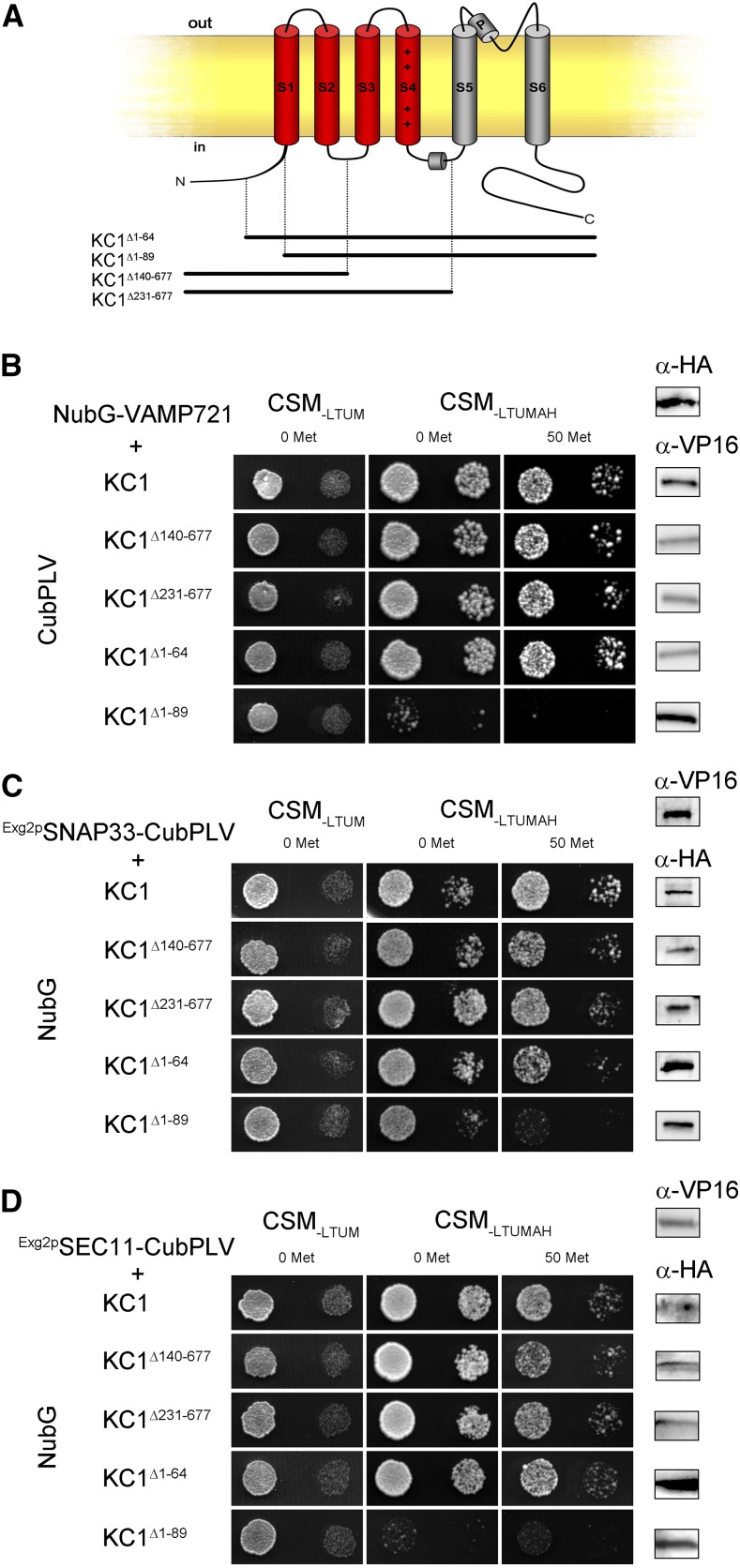
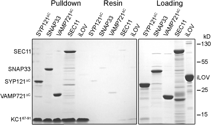
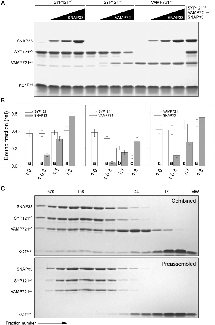
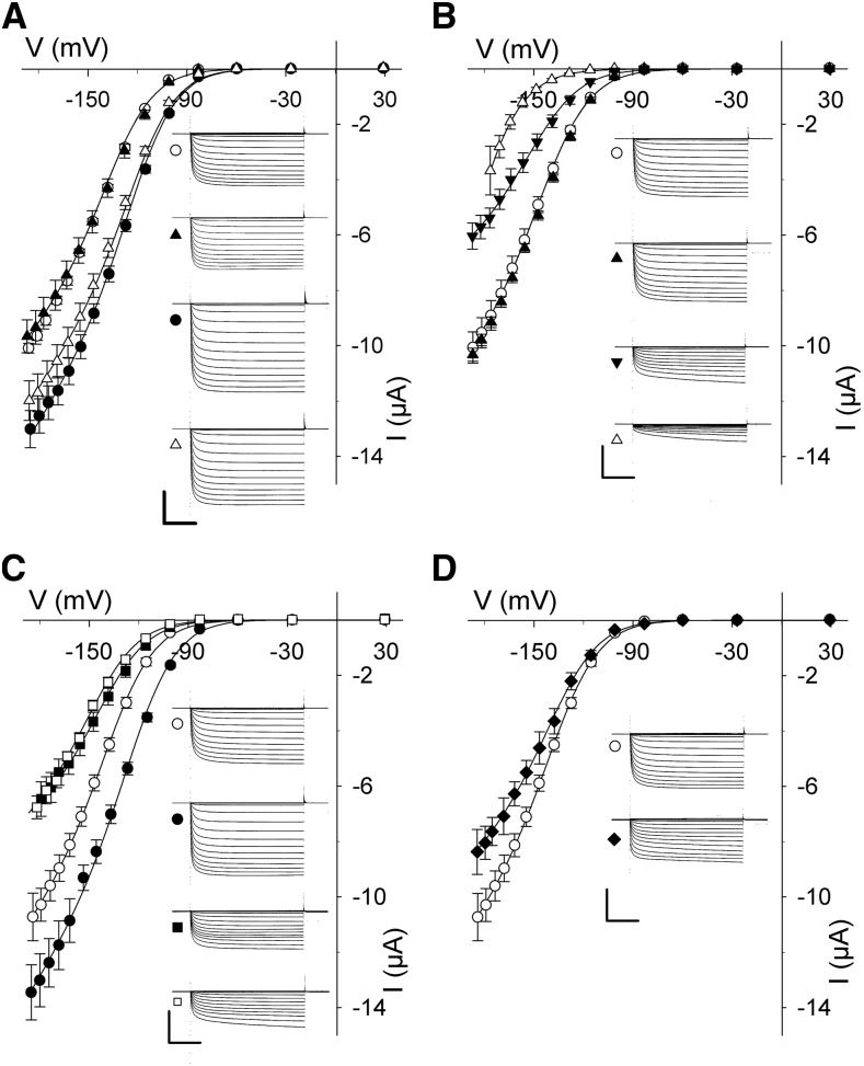
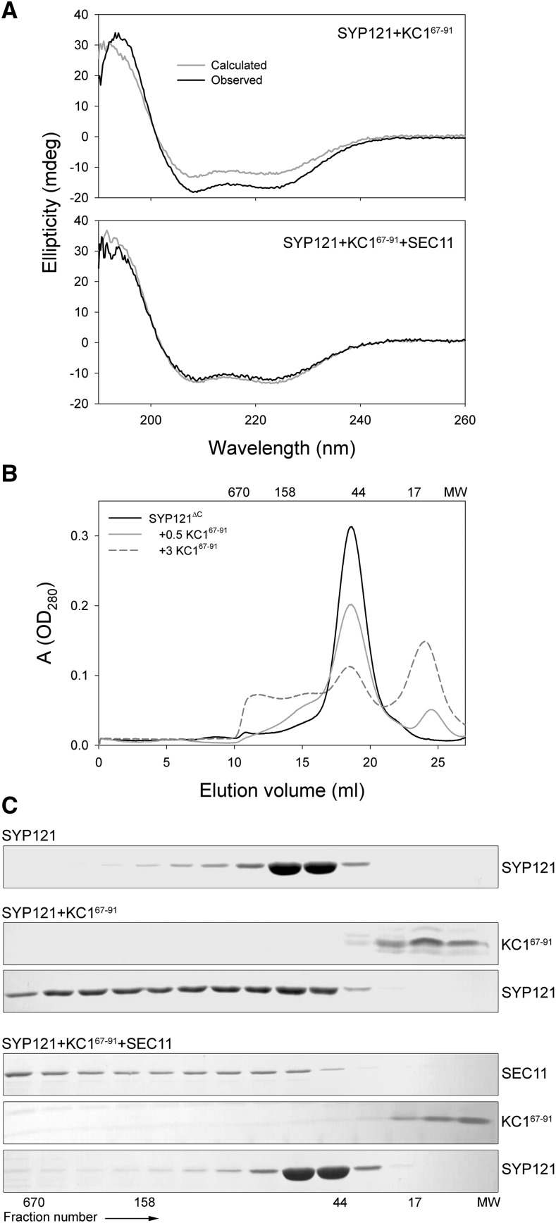
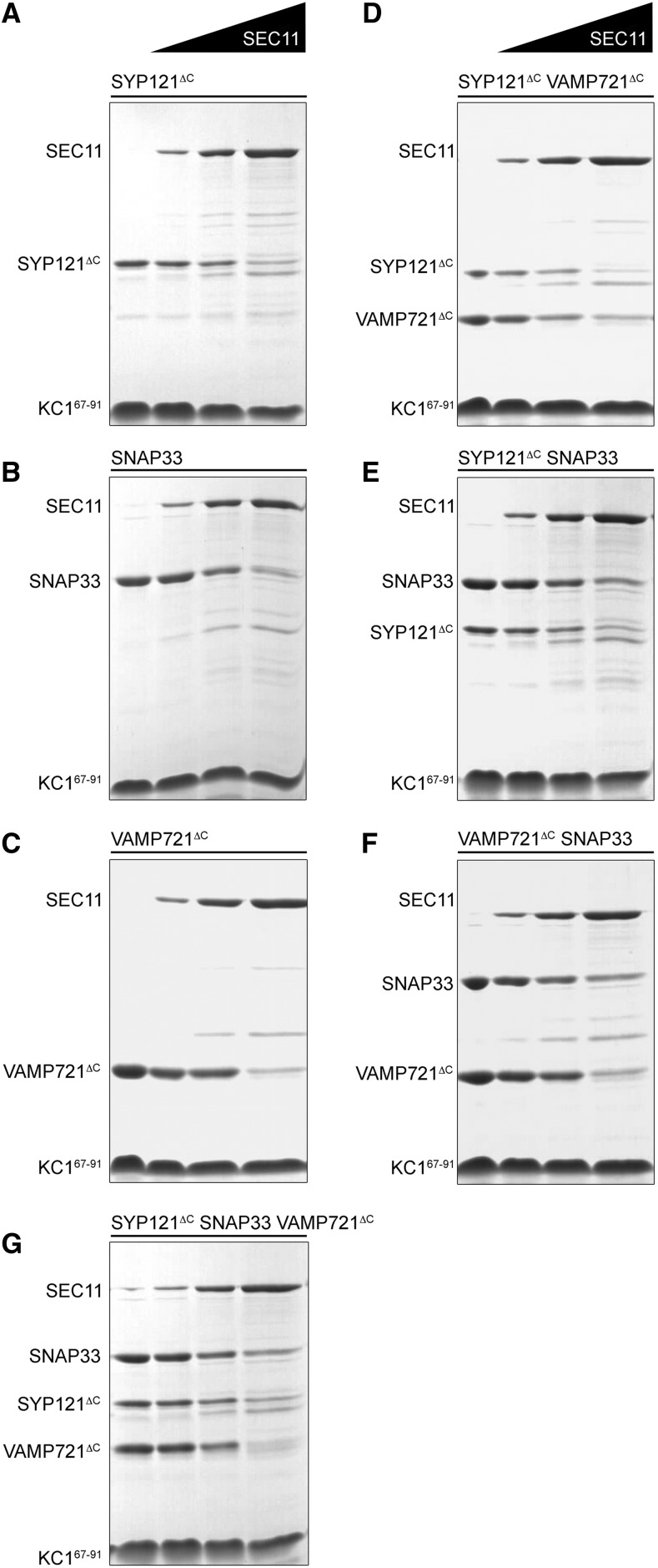
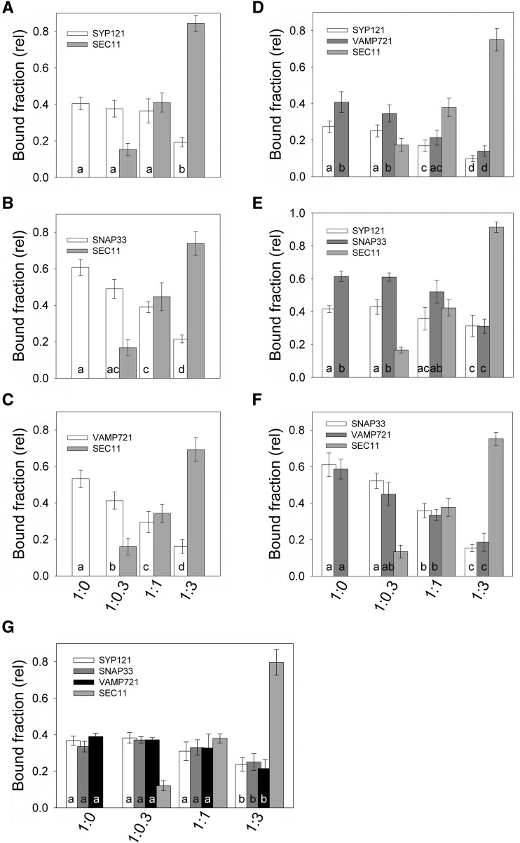
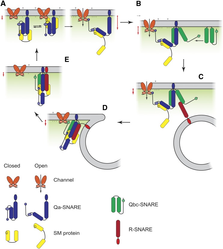
Cell expansion requires that ion transport and secretory membrane traffic operate in concert. Evidence from Arabidopsis (Arabidopsis thaliana) indicates that such coordination is mediated by physical interactions between subsets of so-called SNARE (soluble N-ethylmaleimide-sensitive factor attachment protein receptor) proteins, which drive the final stages of vesicle fusion, and K+ channels, which facilitate uptake of the cation to maintain cell turgor pressure as the cell expands. However, the sequence of SNARE binding with the K+ channels and its interweaving within the events of SNARE complex assembly for exocytosis remains unclear. We have combined protein-protein interaction and electrophysiological analyses to resolve the binding interactions of the hetero-oligomeric associations. We find that the RYxxWE motif, located within the voltage sensor of the K+ channels, is a nexus for multiple SNARE interactions. Of these, K+ channel binding and its displacement of the regulatory protein SEC11 is critical to prime the Qa-SNARE SYP121. Our results indicate a stabilizing role for the Qbc-SNARE SNAP33 in the Qa-SNARE transition to SNARE complex assembly with the R-SNARE VAMP721. They also suggest that, on its own, the R-SNARE enters an anomalous binding mode with the channels, possibly as a fail-safe measure to ensure a correct binding sequence. Thus, we suggest that SYP121 binding to the K+ channels serves the role of a primary trigger to initiate assembly of the secretory machinery for exocytosis.


Figures

Figure 1.
Figure 1.
KC1 K+ channel interacts with VAMP721, SNAP33, and SEC11 via a cytosolic N-terminal region of its voltage sensor domain. A, Schematic of Kv channel structure with the VSD comprising α-helices S1 to S4 identified in red and the pore-lining α-helices S5 and S6 in gray (adapted from Grefen et al. [2015]). Segments expressed for the KC1 deletions are indicated below. B to d, Yeast mbSUS assay for interaction of the VAMP721, SNAP33, and SEC11 fusions with KC1 and its deletion constructs (A) as bait with Y-Cub fusions. VAMP721, SNAP33, and SEC11 served as NubG-X prey fusions and SNAP33 and SEC11 were anchored via the GPI signal peptide (Zhang et al., 2018). Positive and negative controls are included in Supplemental Figure S1. Similar results were obtained in three independent experiments. Growth on CSM-LTUM was used to verify the presence of both bait and prey expression. CSM-LTUMAH was used to verify Ade- and His-independent growth of the yeast diploids. The addition of 50 μm Met to CSM-LTUMAH suppressed bait expression as a test for interaction specificity. Yeast was dropped at 1.0 and 0.1 OD600 in each case. Incubation time was 24 h for the CSM-LTUM plate and 72 h for CSM-LTUMAH plates. Immunoblot analysis (5 μg total protein/lane) of the haploid yeast used in mating (right) used the αHA antibody for the prey fusions and the αVP16 antibody for the bait fusions.
Figure 2.
Figure 2.
KC1 K+ channel interacts with VAMP721, SNAP33, and SEC11 via a common N-terminal motif, RYxxWE, at the base of its VSD. A to C, Yeast mating-based split-ubiquitin assay for interaction of the VAMP721, SNAP33, and SEC11 fusions with KC1 and with Ala substitutions of key residues at the cytosolic face of the VSD as bait with Y–Cub fusions. VAMP721, SNAP33, and SEC11 as NubG-X prey fusions and with SNAP33 and SEC11 anchored via the GPI signal peptide (Zhang et al., 2018). Positive and negative controls are included in Supplemental Figure S1. Similar results were obtained in each of three independent experiments. Growth on CSM-LTUM was used to verify the presence of both bait and prey expression. CSM-LTUMAH was used to verify Ade- and His-independent growth of the yeast diploids. The addition of 50 μm Met to CSM-LTUMAH suppressed bait expression as a test for interaction specificity. Yeast was dropped at 1.0 and 0.1 OD600 in each case. Incubation time was 24 h for CSM-LTUM plate and 72 h for CSM-LTUMAH plates. Immunoblot analysis (5 μg total protein/lane) of the haploid yeast used in mating (right), used the αHA antibody for the prey fusions and the αVP16 antibody for the bait fusions.
Figure 3.
Figure 3.
Pulldown analysis of the SNAREs and SEC11 with the KC167–91 cytosolic domain. Tagged proteins were expressed and purified from Escherichia coli using His-affinity and size-exclusion chromatography before incubation in vitro overnight at 4°C. Shown is one of three independent pulldown experiments with Coomassie-stained SDS-PAGE analysis following separation with Strep-Tactin resin-immobilized KC167–91-Flag6-StrepII. Lane sets are the SDS-PAGE results of (left to right) the pulldown, the resin control, and the loading. Incubations were carried out individually with SYP121ΔC-Flag-6His, VAMP721ΔC-Flag-6His, Flag3-SNAP33-6His, and SEC11-Flag6-6His. The unrelated iLOV-6His protein (Christie et al., 1998) was included as a control. Note the lower-molecular weight fractions that are common for purified SEC11 (Karnik et al., 2013b).
Figure 4.
Figure 4.
The cytosolic domain KC167–91 associates differentially with the cognate SNAREs in binary combination and in complex. A, Tagged proteins were expressed and purified from E. coli using His-affinity and size-exclusion chromatography before incubation in vitro overnight at 4°C. Shown is one of three independent pulldown experiments with Coomassie-stained SDS-PAGE analysis following separation with Strep-Tactin resin-immobilized KC167–91-Flag3-StrepII. Lane sets are the SDS-PAGE results of pulldowns with a 3-fold excess of SYP121ΔC and VAMP721ΔC with competing additions of 0, 0.3-, 1-, and 3-fold excess of VAMP721ΔC SNAP33, as indicated (above). Also shown are the pulldown results on incubation of all three SNAREs with the K+ channel peptide (right). SNAREs were incubated at 4°C overnight with Strep-Tactin-immobilized K+ channel peptide-Flag3-StrepII. Fixed concentrations (5 μm) of SYP121ΔC and VAMP721ΔC were used in each case. Similar results for KAT11–63 are shown in Supplemental Figure S4. B, Bound fraction analysis of KC161–97 and KAT11–63 pulldowns from all six independent experiments. Data are means ± se of the ratios of SNAREs to K+ channel peptide. Lowercase letters indicate statistical differences at P < 0.05. C, Comigration of KC167–91 peptide-SNARE complex on gel filtration. The KC167–91 peptide-SNARE complex assembly reactions were started either by comixing the channel peptide with SYP121ΔC, SNAP33, and VAMP721ΔC (above) or by adding the channel peptide following incubation of SYP121ΔC, SNAP33, and VAMP721ΔC to preform the SNARE complex. The SNARE complex was formed by incubation overnight at 4°C and purified by gel filtration chromatography, and the homo-oligomeric peak of SNARE complex was then mixed with KC167–91 peptide. Shown are the SDS-PAGE analyses of gel filtration fractions with the SNAREs and channel peptides, with molecular weights (MW) of standards indicated above each column. Note that the channel peptide remained associated with the SNARE complex when comixed with the SNAREs (Combined) but not with the preformed SNARE complex (Preassembled).
Figure 5.
Figure 5.
SNAP33 coordinates with its cognate SNAREs in regulating K+ channel activity. Mean steady-state voltage curves recorded under voltage clamp from oocytes coexpressing the KAT1 channel alone and with combinations of the cognate SNAREs SYP121, VAMP721, and SNAP33. Data in every case are measurements from at least five independent experiments and are fitted jointly by the nonlinear least-squares method to the Boltzmann function of Equation 1. Representative current traces cross-referenced by symbol are included for each set of curves, as are data for KAT1 alone, for reference. Representative immunoblot analyses and fitting results are included in Supplemental Figure S5. Water-injected controls yielded currents of <300 nA at all voltages and are omitted for clarity. Scales represent 1 s (horizontal), 6 μA (vertical). A, KAT1 alone (○), and coexpressed with SNAP33 (▲), SYP121 (●), and SNAP33 and SYP121 (△). B, KAT1 alone (○), and coexpressed with SNAP33 (▲), VAMP721 (▼), and SNAP33 and VAMP721 (△). C, KAT1 alone (○), and coexpressed with SYP121 (●), VAMP721 (▪), and SYP121 and VAMP721 (□). D, KAT1 alone (○), and coexpressed with SNAP33, VAMP721, and SYP121 (♦).
Figure 6.
Figure 6.
The K+ channel binding domain alters SYP121 structure to promote the open conformation. A, Far-UV CD spectra of KC167–91 with SYP121ΔC-2PA mixed and incubated overnight in equimolar ratio shows roughly a 24% increase in spectral peak amplitudes between 200 and 230 nm above the arithmetical sum of individual spectra for the two proteins (top). This shift in peak amplitudes is suppressed on coincubation with SEC11 (bottom). Data are from one of three independent experiments, all of which yielded equivalent results with a mean of 28 ± 5% in peak amplitude above the spectral sum. Similar results were obtained with KAT11–63 (see Supplemental Fig. S6). B, Size-exclusion chromatography of SYP121ΔC-2PA incubated overnight at 4°C alone and on coincubation with KC167–91. In the closed conformation, SYP121ΔC-2PA eluted as a single peak corresponding to a molecular weight around 48 kD. When mixed with increasing amounts of the channel peptide, this peak was reduced in favor of a higher-molecular weight shoulder and peak spanning to near 650 kD, consistent with transition to the Qa-SNARE open conformation and formation of multimers. Molecular weights of standards are indicated above the columns. C, SDS-PAGE analysis of the gel filtration eluate collected in 0.75-mL fractions for fractions 11 to 24 collected after overnight incubations of SYP121ΔC-2PA alone, with KC167–91, and with KC167–91 together with SEC11. Note the shift of the SYP121ΔC-2PA to higher-molecular weight fractions in the presence of the channel peptide and its suppression when SEC11 is included in the incubation mix. Molecular weights of standards are indicated below the columns.
Figure 7.
Figure 7.
SEC11 competes differentially with KC167–91 for cognate SNARE binding. Pulldown assays with KC167–91 alone and with 0.3-, 1-, and 3-fold molar excess of SEC11 together with each of the cognate SNAREs, as indicated (top). Binding was tested with the SNAREs singly (A–C), in binary (d–F), and in tertiary (G) combinations, as indicated. Incubations were carried out overnight at 4°C before separation. Shown are Coomassie-stained SDS-PAGE gels of the bound proteins from one of three independent experiments in each case. Similar results were obtained with the KAT11–63 peptide (Supplemental Fig. S7), and the results of all experiments are summarized in Figure 8.
Figure 8.
Figure 8.
Bound fraction analysis of KC161-97 and KAT11-63 pulldowns from all six independent experiments alone and with SEC11 additions. Binding was with the SNAREs singly (A–C), in binary (D–F) and in tertiary (G) combinations as indicated and includes the results of experiments shown in Figure 7 and Supplemental Figure S7. Data are means ±SE of the ratios of SNAREs and SEC11 bound to K+ channel peptide. Letters indicate statistical differences at P<0.05.
Figure 9.
Figure 9.
SEC11 promotes KAT1 activity with SYP121, but not with VAMP721 or SNAP33. Mean steady-state voltage curves recorded under voltage clamp from oocytes coexpressing the KAT1 channel alone and with combinations of the cognate SNAREs SYP121, VAMP721, and SNAP33 with SEC11. Data in every case are measurements from at least five independent experiments and are fitted jointly by nonlinear least-squares regression to the Boltzmann function of Equation 1. Each panel includes representative current traces cross-referenced by symbol. Representative immunoblot analyses and fitting results are included in Supplemental Figure S5. Data for KAT1 alone and with the individual SNAREs are reproduced in selected panels for reference. Water-injected controls yielded currents of <300 nA at all voltages and are omitted for clarity. Scales represent 1 s (horizontal) and 6 μA (vertical). A, KAT1 alone (○) and coexpressed with SYP121 (●) and SYP121 and SEC11 (♢). B, KAT1 alone (○) and coexpressed with VAMP721 (▼) and VAMP721 and SEC11 (♢). C, KAT1 alone (○) and coexpressed with SNAP33 (∆) and SNAP33 and SEC11 (▲). D, KAT1 alone (○) and coexpressed with SYP121 (●), SYP121 and SNAP33 (∆), and SYP121, SNAP33, and SEC11 (▼). E, KAT1 alone (○) and coexpressed with VAMP721 (▼), VAMP721 and SNAP33 (♢), and VAMP721, SNAP33, and SEC11 (▲). (F) KAT1 alone (○) and coexpressed with VAMP721 (▪), SYP121 and VAMP721 (□), and SYP121, VAMP721, and SEC11 (♦). G, KAT1 alone (○), and coexpressed with SYP121, VAMP721, SNAP33, and SEC11 (♦).
Figure 10.
Figure 10.
K+ channels KAT1 and KC1 facilitate a binding exchange with SEC11 to promote SNARE assembly for vesicle fusion. The SM protein SEC11 (A) holds SYP121 in the closed conformation through its major cleft and also binds via L128 (circled L) with the Qa-SNARE N-terminal motif centered on residue F9 (circled F; Karnik et al., 2013b, 2015). SYP121 harbors a second SEC11-binding site centered on its R20R21 motif (circled RR) that does not affect channel binding (Zhang et al., 2019). SEC11 interacts with the K+ channels through the RYxxWE motif (circled RE) and, with membrane hyperpolarization (+, −), undergoes a three-way binding exchange between SEC11 and SYP121 that culminates with binding through the two motifs, the channel RYxxWE motif (RE) and the Qa-SNARE FxRF motif (F), thereby conferring an apparent voltage dependence on the channel-SM protein interaction with SYP121 (Supplemental Fig. S8) and enhancing channel gating and activity (Honsbein et al., 2009; Grefen et al., 2010, 2015). The Qbc-SNARE SNAP33 (B) stabilizes the complex of SYP121, SEC11, and the K+ channel to moderate the open channel, Qa-SNARE, and SM protein conformations (Fig. 9). Recruiting the R-SNARE VAMP721 (C) facilitates final assembly of the SNARE core complex and transfer of channel binding (D) to SNAP33 (unknown site [circled U]) while relaxing channel gating and conductance (Fig. 9). Finally, disengaging channel binding with the cis SNARE complex (E) is followed by SNARE complex disassembly. Red arrows by each step in the cycle indicate the nominal channel activity for K+ uptake and its anticipated enhancement with the open conformation of SYP121 prior to assembly with VAMP721. Note that binding motifs on SEC11 and SNAP33 for the K+ channels are yet to be determined. The binding motif on SEC11 associated with the SYP121 R20R21 motif is also unknown. The time-averaged K+ flux needed to support cell expansion, for example during stomatal opening, is equivalent to a current of 1 to 3 μA cm−2 on a cell surface basis and is readily accommodated by KAT1 at voltages near −100 to −120 mV in vivo (Jezek and Blatt, 2017).

References

    1. Archbold JK, Whitten AE, Hu SH, Collins BM, Martin JL (2014) SNARE-ing the structures of Sec1/Munc18 proteins. Curr Opin Struct Biol 29: 44–51 - PubMed
    1. Assaad FF, Huet Y, Mayer U, Jürgens G (2001) The cytokinesis gene KEULE encodes a Sec1 protein that binds the syntaxin Knolle. J Cell Biol 152: 531–543 - PMC - PubMed
    1. Baker RW, Hughson FM (2016) Chaperoning SNARE assembly and disassembly. Nat Rev Mol Cell Biol 17: 465–479 - PMC - PubMed
    1. Bassham DC, Blatt MR (2008) SNAREs: Cogs and coordinators in signaling and development. Plant Physiol 147: 1504–1515 - PMC - PubMed
    1. Brunger AT, Weninger K, Bowen M, Chu S (2009) Single-molecule studies of the neuronal SNARE fusion machinery. Annu Rev Biochem 78: 903–928 - PMC - PubMed

Publication types

MeSH terms