. 2019 Nov 18;87(12):e00740-19.
doi: 10.1128/IAI.00740-19. Print 2019 Dec.

Antibodies Specific to Membrane Proteins Are Effective in Complement-Mediated Killing of Mycoplasma bovis

Affiliations

Antibodies Specific to Membrane Proteins Are Effective in Complement-Mediated Killing of Mycoplasma bovis

Yun-Ke Zhang et al. Infect Immun. .

Abstract

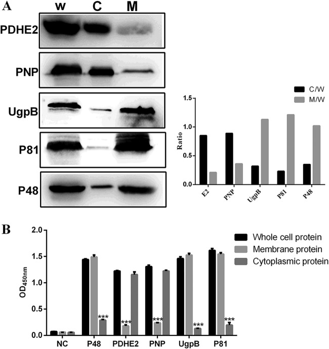
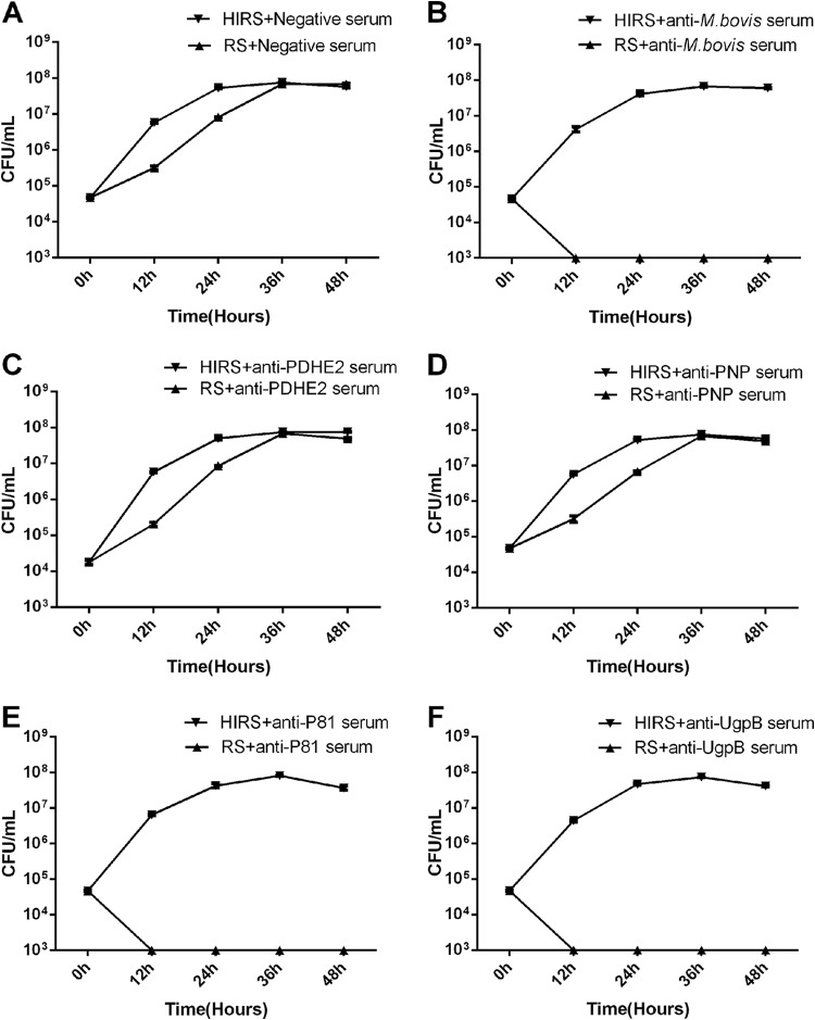
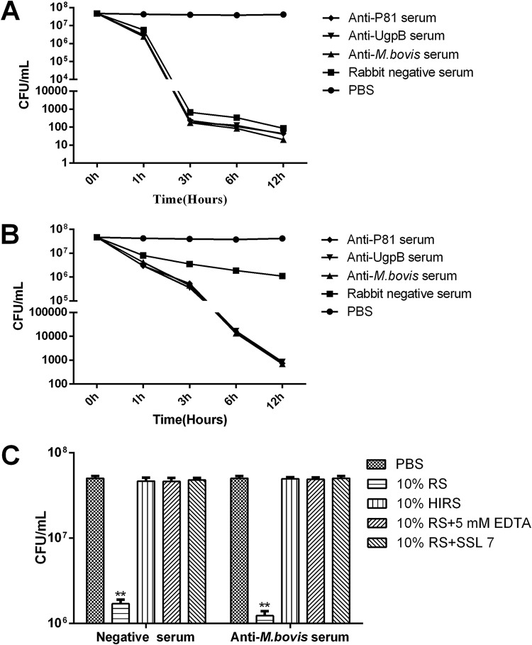
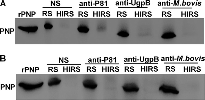
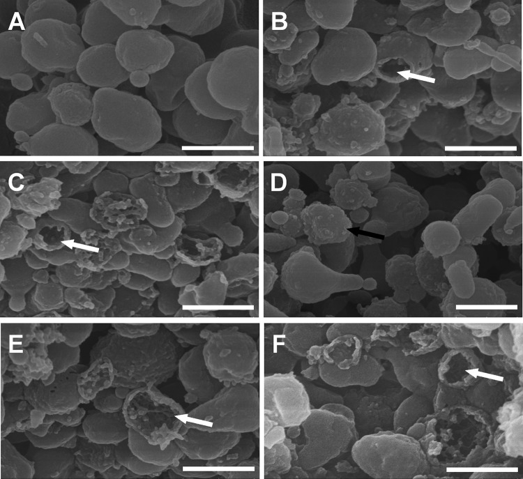
The metabolic inhibition (MI) test is a classic test for the identification of mycoplasmas, used for measuring the growth-inhibiting antibodies directed against acid-producing mycoplasmas, although their mechanism still remains obscure. To determine the major antigens involved in the immune killing of Mycoplasma bovis, we used a pulldown assay with anti-M. bovis antibodies as bait and identified nine major antigens. Among these antigens, we performed the MI test and determined that the growth of M. bovis could be inhibited effectively in the presence of complement by antibodies against specifically membrane protein P81 or UgpB in the presence of complement. Using a complement killing assay, we demonstrated that M. bovis can be killed directly by complement and that antibody-dependent complement-mediated killing is more effective than that by complement alone. Complement lysis and scanning electron microscopy results revealed M. bovis rupture in the presence of complement. Together, these results suggest that the metabolic inhibition of M. bovis is antibody-dependent complement-mediated killing. This study provides new insights into mycoplasma killing by the complement system and may guide future vaccine development studies for the treatment of mycoplasma infection. Furthermore, our findings also indicate that mycoplasmas may be an appropriate new model for studying the lytic activity of membrane attack complex (MAC).

Keywords: M. bovis; MAC; MI; bacterial lysis; complement.


Figures

FIG 1
FIG 1
Pulldown of major antigens of M. bovis. To identify the major antigens of M. bovis, a pulldown assay was performed using rabbit anti-M. bovis antibodies or normal rabbit antibodies (negative control) on extracts of M. bovis or M. bovis LAMPs. The resulting pellets were examined by SDS-PAGE.
FIG 2
FIG 2
Purification and identification of M. bovis recombinant proteins. Purified recombinant proteins were identified by SDS-PAGE (A) or Western blotting with rabbit anti-M. bovis antibodies (B). Lane M, molecular weight marker; lane 1, purified recombinant P81; lane 2, purified recombinant UgpB; lane 3, purified recombinant PNP; lane 4, purified recombinant PDHE2.
FIG 3
FIG 3
Subcellular localizations of PDHE2, PNP, UgpB, and P81 in M. bovis. (A) Western blotting with the listed specific antibodies. W, whole-cell protein; C, cytoplasmic protein; M, membrane protein. (Left) Representative blot; (right) graph showing the ratio of the protein amount in the cytoplasmic or membrane fractions to the total protein in whole-cell lysate. (B) For indirect ELISA, 96-well ELISA plates were coated with whole bacterial proteins, membrane proteins, or cytoplasmic proteins. Rabbit anti-PDHE2 serum, rabbit anti-PNP serum, rabbit anti-P81 serum, and rabbit anti-UgpB serum were used as primary antibodies, and HRP-conjugated goat anti-rabbit IgG was used as the secondary antibody. The optical density at 450 nm (OD450) was read. The whole-bacterial protein group was the control group. Data represent means ± SD from 3 independent experiments. Statistical analysis was done using a ratio-paired two-tailed t test and displayed only when significant. ***, P ≤ 0.001.
FIG 4
FIG 4
MI was detectd by CFU determination. All antisera were heated at 56°C for 30 min before use; fresh rabbit serum (RS) served as the source of complement, and heat-inactivated rabbit serum (HIRS) served as a control. Shown is growth of M. bovis in PPLO broth containing negative serum (A), anti-M. bovis serum (B), anti-PDHE2 serum (C), anti-PNP serum (D), anti-P81 serum (E), or anti-UgpB serum (F) at a dilution of 1:40 along with RS or HIRS (as the control group) at a dilution of 1:20. These data are presented as the means ± SD from three separate experiments.
FIG 5
FIG 5
Complement killing assay. All antisera were heated at 56°C for 30 min before use, and fresh rabbit serum served as the source of complement. Complement killing assays were performed on M. bovis under the following conditions: antiserum was used at a 1:20 dilution and RS at a 1:10 dilution (A), antiserum was used at a 1:20 dilution and RS at a 1:40 dilution (B), or M. bovis was incubated with fresh rabbit serum in the presence of EDTA (5 mM) or SSL7 (40 μg/ml) (C). EDTA is the Mg2+ and Ca2+ chelator, which inhibits complement activation of all three pathways. The SSL7 protein from Staphylococcus aureus binds to C5 to inhibit complement-mediated hemolytic and bacterial activity. The PBS group was the control group. These data are presented as the means ± SD from three separate experiments. Statistical analysis was done using a ratio-paired two-tailed t test and displayed only when significant. **, P ≤ 0.01.
FIG 6
FIG 6
Complement lysis. All antisera were heated at 56°C for 30 min before use, and fresh RS served as the source of complement. Western blotting to detect the level of cytoplasmic protein PNP was performed to monitor the lysis of M. bovis following treatment with normal RS (NS), anti-P81 serum, anti-UgpB serum, or anti-M. bovis serum. M. bovis was incubated with RS at a 1:10 dilution as the source of complement or with HIRS for 1 h (A) or 3 h (B).
FIG 7
FIG 7
Scanning electron microscopy of M. bovis treated with various rabbit antisera. All antisera were heated at 56°C for 30 min before use, and fresh RS served as the source of complement. Shown are representative scanning electron microscopy images of M. bovis incubated with PBS (A), normal rabbit serum (B), rabbit anti-M. bovis serum (C), rabbit anti-M. bovis serum in the presence of heat-inactivated RS (D), rabbit anti-P81 serum (E), and rabbit anti-UgpB serum (F). The white arrows indicate large holes in ghost-like structures. The black arrow indicates a mycoplasma with a rough surface. Bar, 500 nm.

References

    1. Razin S, Yogev D, Naot Y. 1998. Molecular biology and pathogenicity of mycoplasmas. Microbiol Mol Biol Rev 62:1094–1156. - PMC - PubMed
    1. Citti C, Blanchard A. 2013. Mycoplasmas and their host: emerging and re-emerging minimal pathogens. Trends Microbiol 21:196–203. doi:10.1016/j.tim.2013.01.003. - DOI - PubMed
    1. Burki S, Frey J, Pilo P. 2015. Virulence, persistence and dissemination of Mycoplasma bovis. Vet Microbiol 179:15–22. doi:10.1016/j.vetmic.2015.02.024. - DOI - PubMed
    1. Perez-Casal J, Prysliak T, Maina T, Suleman M, Jimbo S. 2017. Status of the development of a vaccine against Mycoplasma bovis. Vaccine 35:2902–2907. doi:10.1016/j.vaccine.2017.03.095. - DOI - PubMed
    1. Purcell RH, Wong D, Chanock RM, Taylor-Robinson D, Canchola J, Valdesuso J. 1967. Significance of antibody to mycoplasma as measured by metabolic-inhibition techniques. Ann N Y Acad Sci 143:664–675. doi:10.1111/j.1749-6632.1967.tb27712.x. - DOI - PubMed

Publication types

MeSH terms

Substances

LinkOut - more resources